Understanding Peripheral Nerve Injuries and Intervention Strategies
Peripheral nerves play a vital role in connecting the central nervous system to various parts of the body. Injuries to these nerves can result from different causes, leading to significant challenges. Early intervention is crucial in preventing irreversible damage and improving outcomes. The process involves a detailed approach to patient history, clinical examination, electrophysiology, and imaging. Primary repair through urgent surgery, especially in cases of sharp transection, offers benefits such as scar-free field and minimal intraneural scarring. However, determining the extent of stump resection remains a challenge.

Download Presentation

Please find below an Image/Link to download the presentation.
The content on the website is provided AS IS for your information and personal use only. It may not be sold, licensed, or shared on other websites without obtaining consent from the author. Download presentation by click this link. If you encounter any issues during the download, it is possible that the publisher has removed the file from their server.
E N D
Presentation Transcript
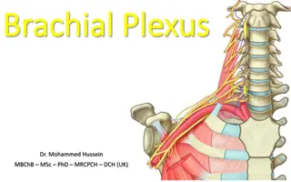
Introduction Peripheral nerves are the neural structures that connect CNS to the end organs PNS consists of: 12 pairs of cranial nerves 31 pairs of spinal nerves Unique power of regeneration
Etiology of nerve injury Three major causes: Medium to high energy nerve injuries Low energy compressive or ischemic lesions Complex injuries
Approach to the patient History: pain, sensory loss, weakness Clinical examination: general, inspection, joint mobility, motor & sensory testing, autonomic testing & special tests Electrophysiology: NCV, EMG Imaging
Time of intervention Changes following nerve injury: Central cell death, ischemia & fibrosis Target organ changes: muscle atrophy & disappearance of motor end plates- irreversible with time Proximal injury- worse outcome
Time of intervention: early Early nerve repair prevents neuronal loss & improves outcome Ma J, Novikov LN, Kellerth JO, Wiberg M: Early nerve repair after injury to the postganglionic plexus: an experimental study of sensory and motor neuronal survival in adult rats. Scand J Plast Reconstr Surg Hand Surg 2003; 37:1-9. With exception of spinal accessory improved results of early repair are found in median, ulnar, radial, musculocutaneous, sciatic, CPN & closed traction BPI Merle M, Amend P, Cour C, et al: Microsurgical repair of peripheral nerve lesions: a study of 150 injuries of the median and ulnar nerves. Peripher Nerve Repair Regen 1986; 2:17-26. Birch R, Raji AR: Repair of median and ulnar nerves. Primary suture is best. J Bone Joint Surg Br 1991; 73:154-157. Kato N, Htut M, Taggart M, et al: The effects of operative delay on the relief of neuropathic pain after injury to the brachial plexus: a review of 148 cases. J Bone Joint Surg Br 2006; 88:756-759. Limiting factor for early repair: difficult to determine the extent of stump resection
Primary repair: urgent surgery Operations done within 3-5 days of injury Indication: sharp transection Contraindication: poor clinical condition Adv: Scar free field Minimal intraneural scarring-less distortion of intraneural architecture- proper fascicular matching Disadvantage : EPS may not be available or feasible
Delayed primary repair Done after 2-3 weeks Good outcome Advantages of primary surgery disappears
Secondary repair Performed between 3 weeks to 3 months Indications: neuroma in continuity Adv: 40% of BPI recovers spontaneously- prevents unnecessary surgery Disadvantage : exploration in scarred tissue & intraneural scarring & distortion
Indications for surgery Paralysis after trauma over the course of a major nerve- including iatrogenic injuries Paralysis following closed traction BPI Associated vascular or orthopedic injury requiring treatment Worsening or failure to improve within expected time period Persistent pain
Contraindications Poor general condition of the patient Uncertainty about viability of the nerve trunks Local & systemic sepsis Early signs of recovery
Types of surgery Primary procedures Alternative methods Secondary procedures
Alternative procedures Direct muscular neurotization Nerve conduits Interposed freeze-thawed muscle Nerve allograft repair Central repair
Secondary procedures Tendon transfer Functioning free muscle transfer Arthrodesis Tenodesis Corrective osteotomy Amputation
Principles of nerve repair Environment: generous operative field, good illumination, microscope or loupe Anesthesia: Short acting paralyzing agent Flexibility regarding the position of surgeon & limb
Principles of nerve repair Wide exposure Sharp dissection in anatomic planes starting from virgin tissues & progressing towards the lesion Meticulous hemostasis- bipolar cautery Preserving fat & synovium planes- nerve s gliding planes- The gliding apparatus of peripheral nerve and its clinical significance. Millesi H, Zoch G, Rath T. Ann Chir Main Memb Super 1990;9(2):87-97.
Principles of nerve repair Preparing nerve stumps: Circumferential exposure Generous proximal & distal mobilization External neurolysis Use of intra-operative electro-physiology Placement of lateral stay sutures (6-0)- to maintain topographic alignment
Debridement of nerve stumps proximally & distally to remove scar tissue- scar > scar with some fascicles > pure healthy fascicles (fascicles appear to pout, glossy surface & fine bleeding from vessels)
Principles of nerve repair Proper alignment & positioning of nerve stumps & grafts: Longitudinal vessel alignment in epineurium Fascicular alignment
Principles of nerve repair Proper suturing: Material: 8-0, 9-0 or 10-0 monofilament nylon Two lateral sutures 1800 apart Three to four more sutures may also be placed Tensionless Avoid overzealous suturing- every suture induces fibrosis
Principles of nerve repair Use of fibrin glue: Secures the position of anastomosis When used alone: does not provide tensile strength or permit to fish-mouth Clump formation to be avoided
Decompression Release of a nerve from external compression Types: Open Endoscopic
Neurolysis Release of nerve or its part from organized scar Types: External internal External neurolysis: Nerve is set free from scar, organized hematoma or bony fragments Released in circumferential manner Epineurium is minimally breached
Neurolysis Internal neurolysis: Opening or resection the external epineurium to lyse internal scar Plain of dissection: internal epineurium Not to damage perineurium Used for preparation of nerve ends for grafting, dissection of neuroma in continuity & benign nerve sheath tumor
Direct repair Possible in most clean lacerating injuries & when co-aptation can be done without undue tension Types: 1. Epineural repair 2. Grouped fascicular repair 3. Fascicular or perineural repair Combination of epineural & grouped fascicular repair- most commonly used
Epineural repair Traditional method Appropriate for monofascicular & diffusely grouped polyfascicular nerve Goal: tensionless coaptation of proximal & distal fascicular anatomy
Epineural repair Small bite taken from internal & external epineurium Perineurium avoided Tied with mild to moderate tension Disadvantage: precise matching of proximal & distal fascicles may not be possible
Grouped fascicular repair Indication: Group of fascicles with specific functions- sensory or motor Nerve requiring split repair Debridement & alignment Inter-fascicular dissection- within internal epineurium Suturing through internal epineurium and perineurium
Fascicular repair Indication: Lacerated nerve with identifiable individual motor & sensory fascicles Partial injury to 1-2 fascicles Repair under high magnification with 10-0 nylon Sutures placed through perineurium Avoid endoneurium Maximum 2 sutures for each fascicle Strengthening by addition of epineural sutures
Epineural vs perineural sutures Perineural suture is better & epineural suture is the main source of infiltration- Millesi H: Interfascicular nerve grafting. Orthop Clin North Am 1981; 12:287-301. Epineural suture is easier & faster- Orgell M: Epineurial versus perineurial repair of peripheral nerves. In: Tertzis J, ed. Microreconstruction of Nerve Injuries, London: Saunders; 1987:97-100. Restriction of perineural sutures to oligofascicular nerves: Kline D, Hudson A, Spinner R, et al: Kline & Hudson's Nerve Injuries: Operative Results for Major Nerve Injuries, Entrapments and tumours. 2nded. Philadelphia, Saunders, 2008. No discernable difference- Urbaniak J R. Fascicular nerve suture. Clin Orthop Relat Res. 1982 Mar;(163):57-64.
Nerve auto graft repair Indication: direct repair not possible without undue tension Principles: Harvest as much of graft as possible Extremity to be in full extension Proper alignment: proximal nerves- spatial matching & distal nerves- anatomic matching Cable grafting Epineural dissection to create group of fascicles
Nerve auto graft repair Graft sutured in epineural & interfascicular epineural technique Fish mouth configuration 1-2 sutures reinforced with fibrin glue
Nerve LACN Location Terminal sensory branch of MCN. Located just lateral to biceps tendon in subcutaneous tissue. Deficit Loss of sensation over lateral aspect of forearm Contraindication Median nerve injury- significant loss of sensation over dorsolateral thumb Ulnar nerve injury MACN Derived from medial cord. Closely follows brachial vein. Loss of sensation over medial forearm Anatomical snuff box SSRN Terminal sensory part of radial nerve. Lies deep to brachioradialis muscle in proximal forearm. Good graft for proximal radial nerve recon. Most commonly used donor. Lies deep to deep fascia at proximal leg. Emerge to subcutaneous tissue at midcalf level. Significant contribution from lateral sural branch of peroneal nerve. Nil Sural Lateral order of the foot Nil
Harvesting the graft Methods: Open Endoscopic Incision: Longitudinal Step wise Proximal division: deep to deep fascia Cut to produce appropriate length
Nerve transfer Involves re-assigning an expendable or redundant nerve or its part or branch to a more important nonfunctioning nerve Indications: Nerve avulsion Rapid & reliable recovery of motor function in post- ganglionic injury To power free- functioning muscle transfer
Nerve transfer Contraindications: Absence of donor nerve Fibrosed, atrophic recipients Repairable rupture or neuroma Poor quality donor Principles: Accurate preop documentation & fall- back planning- Selection of ideal donor nerve
Nerve transfer Transection of recipient as proximal as possible Dissection of donor distal to the recipient- to gain length Selective neurotization based on fascicular anatomy Maintaining orientation Tension free repair
Alternative methods Direct muscular neurotization: Used when distal nerve stump not available Spreading out fascicle in a fan like manner and burying them in intermysial folds Becker M, Lassner F, Fansa H, et al: Refinements in nerve to muscle neurotization. Muscle Nerve 2002; 26:362-366.
Interposed freeze-thawed muscle Basal lamina of muscle acts as scaffold for axonal growth Problem: axonal growth not target oriented but diffusely over the muscle- Schlosshauer B, Dreesmann L, Schaller HE, Sinis N: Synthetic nerve guide implants in humans: a comprehensive survey. Neurosurgery 2006; 59:740- 747. Promising results for sensory nerve repair- Pereira JH, Palande DD, Narayanakumar TS, et al: Nerve repair by denatured muscle autografts promotes sustained sensory recovery in leprosy. J Bone Joint Surg Br 2008; 90:220-224.
Nerve conduits Tissue engineered bio-artificial tube placed between nerve stumps Appropriate directional & trophic cues from migrating Schwann cells & soluble growth factors Inner diameter of tube- 20% larger than that of stumps
Nerve conduits Placement of single microsuture in U fashion Reinforced with glue Tube is filled with saline Good results for defects <3cm in small nerves- Weber RA, Breidenbach WC, Brown RE, et al: A randomized prospective study of polyglycolic acid conduits for digital nerve reconstruction in humans. Plast Reconstr Surg 2000; 106:1036-1045
Nerve allograft & vascularized nerve grafts Risk of immunosuppression prevents wide spread use of allografts- Larsen M, Habermann TM, Bishop AT, et al: Epstein-Barr virus infection as a complication of transplantation of a nerve allograft from a living related donor. J Neurosurg 2007; 106:924-928.Case report Vascularized nerve graft is useful only in contralateral C7 transfer with interposition ulnar vascularized nerve graft- Birch R, Dunkerton M, Bonney G, Jamieson AM: Experience with the free vascularized ulnar nerve graft in repair of supraclavicular lesions of the brachial plexus. Clin Orthop Relat Res 1988; 237:96-104.
Central repair Central repair: reimplantation of avulsed spinal nerve- Birch R, Bonney G, Parry CW: Reimplantation of avulsed spinal nerves. Surgical Disorder of the Peripheral Nerves, London: Churchill Livingstone; 1998:201-207. Functional benefits have been observed in some cases- Carlstedt T, Grane P, Hallin RG, Noren G: Return of function after spinal cord implantation of avulsed spinal nerve roots. Lancet 1995; 346:1323-1325. Carlstedt T, Anand P, Hallin R, et al: Spinal nerve root repair and reimplantation of avulsed ventral roots into the spinal cord after brachial plexus injury. J Neurosurg 2000; 93(suppl):237-247. Carlstedt T: Central Nerve Plexus Injury. London, Imperial College Press, 2007. Carlstedt T, Hultgren T, Nyman T, et al: Cortical activity and hand function restoration in a patient after spinal cord surgery. Nat Rev Neurol 2009; 5:571-574. Should be done within 6 weeks of injury- anterior horn cells become dead after 6 weeks of avulsion- Fournier HD, Mercier P, Menei P: Repair of avulsed ventral nerve roots by direct ventral intraspinal implantation after brachial plexus injury. Hand Clin 2005; 21:109-118. Fournier HD, Mercier P, Menei P: [Spinal repair of ventral root avulsions after brachial plexus injuries: Towards new surgical strategies?]. Neurochirurgie 2006; 52:357-366.
Secondary procedures Indications: To provide additional function Delay between injury & presentation Improvement following previous procedure is less than satisfactory Unlike primary procedures these are time- independent
Tendon transfers Principles: Maintenance of tissue equilibrium- correction of contractures, joint stiffness etc Availability: removal of donor should not compromise existing function Muscle strength: >85% of normal power or 4/5 power Excursion: amplitude of motion should match & direction of action should match Synergy: transfer of synergistic muscle facilitate rehab Tension: transferred tendon should be at its resting length
Tendon transfer Shoulder function: Trapezius transfer to prox humerus- abduction Combined LD & teres major transfer- external rotation Elbow function: Modified Steindler s flexorplasty: flexor- pronator mass from medial humerus epicondyle transferred 4cm above elbow to anterior cortex of humerus Steindler A: Orthopaedic reconstruction work on hand and forearm. N Y Med J 1918; 108:1117- 1119 Chen WS: Restoration of elbow flexion by modified Steindler flexorplasty. Int Orthop 2000; 24:43-46. Pec major flexorplasty: insertion sutured to coracoid process & origin to biceps tendon Wahegaonkar AL, Doi K, Hattori Y, et al: Surgical technique of pedicled bipolar pectoralis major transfer for reconstruction of elbow flexion in brachial plexus palsy. Tech Hand Up Extrem Surg 2008; 12:12-19 Lat dorsi transfer: flexorplasty with soft tissue coverage Wrist & hand function: PT to ECRB transfer, opponensplasty
Functioning free muscle transfer Involves micro-neurovascular repair of a transplanted muscle To restore elbow flexion, shoulder abduction, elbow extension, finger flexion & extension Muscles used: gracilis, rectus femoris, LD, pec major, TFL, adductor longus