Standard of Care for Peripheral Lesion Assessment with Normal Hilar and Mediastinal Appearances
Assessing patients with peripheral lesion on staging CT with normal hilar and mediastinal findings. Further investigation with contrast-enhanced CT is recommended for fitness evaluation and treatment planning. Biopsy methods, diagnostic criteria, and considerations for nodules and nodal staging are outlined, along with steps for supportive care and prehabilitation.
Download Presentation

Please find below an Image/Link to download the presentation.
The content on the website is provided AS IS for your information and personal use only. It may not be sold, licensed, or shared on other websites without obtaining consent from the author. Download presentation by click this link. If you encounter any issues during the download, it is possible that the publisher has removed the file from their server.
E N D
Presentation Transcript
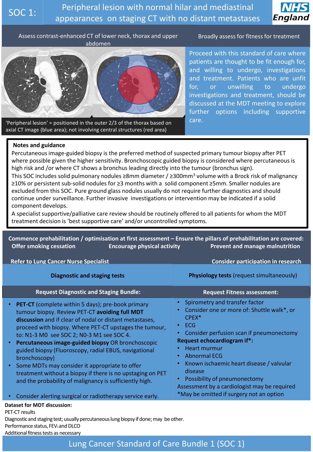
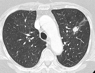
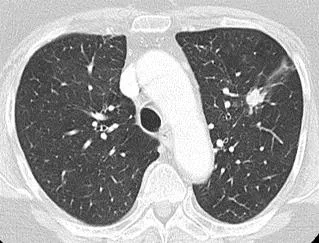
Peripheral lesion with normal hilar and mediastinal appearances on staging CT with no distant metastases SOC 1: Assess contrast-enhanced CT of lower neck, thorax and upper abdomen Broadly assess for fitness for treatment Proceed with this standard of care where patients are thought to be fit enough for, and willing to undergo, investigations and treatment. Patients who are unfit for, or unwilling investigations and treatment, should be discussed at the MDT meeting to explore further options including supportive care. to undergo Peripheral lesion = positioned in the outer 2/3 of the thorax based on axial CT image (blue area); not involving central structures (red area) Notes and guidance Percutaneous image-guided biopsy is the preferred method of suspected primary tumour biopsy after PET where possible given the higher sensitivity. Bronchoscopic guided biopsy is considered where percutaneousis high risk and /or where CT shows a bronchus leading directly into the tumour (bronchus sign). This SOC includes solid pulmonary nodules 8mm diameter / 300mm3 volume with a Brock risk of malignancy 10% or persistent sub-solid nodules for 3 months with a solid component 5mm. Smaller nodules are excluded from this SOC. Pure ground glass nodules usually do not require further diagnostics and should continue under surveillance. Further invasive investigations or intervention may be indicated if a solid component develops. A specialist supportive/palliative care review should be routinely offered to all patients for whom the MDT treatment decision is best supportive care and/or uncontrolled symptoms. Commence prehabilitation / optimisation at first assessment Ensure the pillars of prehabilitation are covered: Offer smoking cessation Encourage physical activity Prevent and manage malnutrition Refer to Lung Cancer Nurse Specialist Consider participation in research Physiology tests (request simultaneously) Physiology tests (request simultaneously) Diagnostic and staging tests Request Diagnostic and Staging Bundle: Request Fitness assessment: Spirometry and transfer factor Consider one or more of: Shuttle walk*, or CPEX* ECG Consider perfusion scan if pneumonectomy Request echocardiogram if*: Heart murmur Abnormal ECG Known ischaemic heart disease / valvular disease Possibility of pneumonectomy Assessment by a cardiologist may be required *May be omitted if surgery not an option Request Fitness assessment: PET-CT (complete within 5 days); pre-book primary tumour biopsy. Review PET-CT avoiding full MDT discussion and if clear of nodal or distant metastases, proceed with biopsy. Where PET-CT upstages the tumour, to: N1-3 M0 see SOC 2; N0-3 M1 see SOC 4. Percutaneous image-guided biopsy OR bronchoscopic guided biopsy (Fluoroscopy, radial EBUS, navigational bronchoscopy) Some MDTs may consider it appropriate to offer treatment without a biopsy if there is no upstaging on PET and the probability of malignancy is sufficiently high. Consider alerting surgical or radiotherapy service early. Dataset for MDT discussion: PET-CT results Diagnostic and staging test; usually percutaneous lung biopsy if done; may be other. Performance status, FEV1 and DLCO Additional fitness tests as necessary Lung Cancer Standard of Care Bundle 1 (SOC 1)
Lesion with mediastinal / hilar lymphadenopathy without distant metastases on staging CT SOC 2: Assess contrast-enhanced CT of lower neck, thorax and upper abdomen Broadly assess for fitness for treatment Proceed with this standard of care where patients are thought to be fit enough for, and willing to undergo, investigations and treatment. Patients who are unfit for, or unwilling to undergo investigations and treatment, should be discussed at the MDT meeting to explore further options including supportive care. Notes and guidance Staging EBUS EUS should be performed where there are enlarged nodes, including isolated N1 hilar nodes and where there is FDG avidity in normal sized nodes. PET-CT has a significant false negative rate, so sampling of normal sized, PET negative nodes is recommended when nodal appearances are not typically benign on CT or endosonography. Where staging EBUS EUS is performed there should be a systematic examination of mediastinal and hilar lymph nodes beginning with N3 stations, followed by N2 and finally N1. Any accessible lymph node based on CT ( 10mm), PET-CT (FDG avidity above the mediastinal blood pool) or sonographic assessment, is sampled. A specialist supportive/palliative care review should be routinely offered to all patients for whom the MDT treatment decision is best supportive care and/or uncontrolled symptoms. Commence prehabilitation / optimisation at first assessment Ensure the pillars of prehabilitation are covered: Offer smoking cessation Encourage physical activity Prevent and manage malnutrition Refer to Lung Cancer Nurse Specialist Consider participation in research Physiology tests (request simultaneously) Diagnostic and staging tests Request Fitness assessment: Request Diagnostic and Staging Bundle: Spirometry and transfer factor Consider one or more of: Shuttle walk*, or CPEX* ECG Consider perfusion scan if pneumonectomy PET-CT (complete within 5 days); pre-book staging EBUS EUS . Review PET-CT avoiding full MDT discussion and proceed as below. Where PET-CT upstages the tumour to M1 see SOC 4. Proceed with staging EBUS EUS where no SCN seen. US guided nodal biopsy where CT or PET-CT show enlarged or FDG avid supraclavicular nodes (SCN) Biopsy of the primary lesion where nodes negative on EBUS EUS . Reflex predictive biomarker testing is preferred Contrast-enhanced CT brain for stage II (or if known small cell). Contrast-enhanced MR brain for stage III Request echocardiogram if*: Heart murmur Abnormal ECG Known ischaemic heart disease / valvular disease Possibility of pneumonectomy Assessment by a cardiologist may be required *May be omitted if surgery not an option Dataset for MDT discussion: PET-CT and CT or MR brain results Bronchoscopy / EBUS EUS / other pathology Performance status, FEV1 and DLCO Additional fitness tests as required Lung Cancer Standard of Care Bundle 2 (SOC 2)
Contiguous or conglomerate invasive mediastinal lymphadenopathy without distant metastases on staging CT SOC 3: Assess contrast-enhanced CT of lower neck, thorax and upper abdomen Broadly assess for fitness for treatment Proceed with this standard of care where patients are thought to be fit enough for, and willing to undergo, investigations and treatment. Patients who are unfit for, or unwilling to undergo investigations and treatment, should be discussed at the MDT meeting to explore further options including supportive care. Notes and guidance This category of patients may be suitable for treatment with curative intent using radiotherapy or chemoradiotherapy. Mediastinal nodes contiguous with the primary tumour or conglomerate are almost always involved and sampling may proceed to confirm diagnosis. There is a high chance of metastatic disease. Diagnostic EBUS refers to the targeted sampling of nodal disease for pathological confirmation, tumour sub- typing and molecular pathology. Invasive mediastinal lymphadenopathy has poorly defined borders and cannot be easily measured. It forms conglomerate disease with other nodal stations. A specialist supportive/palliative care review should be routinely offered to all patients for whom the MDT treatment decision is best supportive care and/or uncontrolled symptoms. Commence prehabilitation / optimisation at first assessment Ensure the pillars of prehabilitation are covered: Offer smoking cessation Encourage physical activity Prevent and manage malnutrition Refer to Lung Cancer Nurse Specialist Consider participation in research Diagnostic and staging tests Physiology tests (request simultaneously) Request Fitness assessment: Request Diagnostic and Staging Bundle: Spirometry and transfer factor Renal function PET-CT (complete within 5 days); pre-book Bronchoscopy / EBUS EUS / SCN node biopsy. Review PET-CT; where no upstaging patient is potentially appropriate for curative treatment. Where PET-CT upstages the tumour: to N0-3 M1 see SOC 4. Proceed with EBUS EUS or where no SCN or US negative (staging EBUS may be required to define tumour extent) US guided nodal biopsy where CT or PET-CT show enlarged or FDG avid supraclavicular nodes (SCN) transfer factor may be omitted if does not influence treatment Contrast-enhanced MR brain. (CT if known small cell) Reflex predictive biomarker testing is preferred Dataset for MDT discussion: PET-CT and MR brain results Bronchoscopic / EBUS / other pathology Performance status, FEV1 and DLCO Renal function Lung Cancer Standard of Care Bundle 3 (SOC 3)
SOC 4: Distant metastases on staging CT Assess contrast-enhanced CT of lower neck, thorax and upper abdomen Broadly assess for fitness for treatment Proceed with this standard of care where patients are thought to be fit enough for, and willing to undergo, investigations and treatment. Patients who are unfit for, or unwilling to undergo investigations and treatment, should be discussed at the MDT meeting to explore further options including supportive care. Notes and guidance Follow this algorithm in cases where there is clear evidence of distant metastases on CT. Sometimes this may need to be clarified with additional tests e.g. liver US/MR/CT or PET-CT. A specialist Supportive/Palliative care review should be routinely offered to all patients, irrespective of any other treatment offered and/or uncontrolled symptoms. Diagnostic EBUS refers to the targeted sampling of nodal disease for pathological confirmation. It is essential that pathological samples are adequate to guide targeted treatment. Staging EBUS may be required to clarify tumour volume. Synchronous brain metastases may be suitable for stereotactic radiosurgery or surgery and should be discussed at the brain metastases MDT. See separate notes for metachronous oligometastatic disease. Commence prehabilitation / optimisation at first assessment Ensure the pillars of prehabilitation are covered: Offer smoking cessation Encourage physical activity Prevent and manage malnutrition Refer to Lung Cancer Nurse Specialist Consider participation in research Physiology tests (request simultaneously) Diagnostic and staging tests Request Fitness assessment: Request Diagnostic and Staging Bundle: Spirometry optional Renal function Choose the least invasive and safest sampling technique to yield adequate pathology for tumour sub-typing and targeted therapy assessment. Options include: Diagnostic bronchoscopy ( TBNA) Diagnostic EBUS US or CT guided biopsy of any target area Pleural aspiration medical thoracoscopy if pleural effusion. Reflex predictive biomarker testing is preferred Bone biopsy should be avoided where there is no significant soft tissue component because of decalcification time and inability to do molecular pathology. Consider PET-CT and contrast enhanced CT brain for oligometastatic disease (see separate notes). Dataset for MDT discussion: Bronchoscopic / EBUS / other pathology Performance status, Renal function Lung Cancer Standard of Care Bundle 4 (SOC 4)
Notes for all Lung Cancer SOCs EBUS EUS: The majority of assessments will involve EBUS only but EUS or EUSB may be added where nodes are inaccessible by EBUS. Staging EBUS EUS: Patients may need to be referred to a specialist centre for this. There should be a mechanism for rapid e-referral and prompt testing in line with the National Optimal Lung Cancer Pathway and the NHSE EBUS service specification. Reflex testing: refers to the block testing of pathological samples to assess for suitability for targeted therapy. The specific tests depend on the drugs available so will change as new drugs are approved for use. Oligometastatic Disease Synchronous brain metastases may be treated by surgery or stereotactic radiosurgery. MDTs may also elect to treat other synchronous oligometastatic sites by surgery on an individual basis (no current guidance). Oligometastatic disease and the Commissioning through Evaluation (CtE). Patients are eligible if: 1-3 sites of metastatic disease (defined after appropriate imaging) which can be treated with stereotactic radiotherapy to a radical radiation dose. A maximum of two sites of spinal metastatic disease Maximum size of any single metastasis 6cm (5 cm for lung or liver metastases) Disease free interval > 6 months; (exception: synchronous liver metastases from colorectal primary). Not more than three oligometastatic sites treated in total per patient Expected life expectancy > 6 months Performance status 2 All patients to be discussed at stereotactic MDT with presence of, or prior discussion with a disease site specific oncologist All patients willing to attend follow up and have details collected on prospective database for a minimum of two years Abbreviations CT: computed tomography PET-CT: Positron emission tomography and computed tomography US: Ultrasound MRI: Magnetic resonance imaging EBUS: Endobronchial ultrasound with needle sampling. Here refers to linear EBUS unless radial US specified EUS / EUSB: Endoscopic ultrasound / Endoscopic ultrasound with EBUS scope CPEX: Cardiopulmonary exercise test ECG: Electrocardiogram
