Understanding Image Processing Artifacts in Radiography
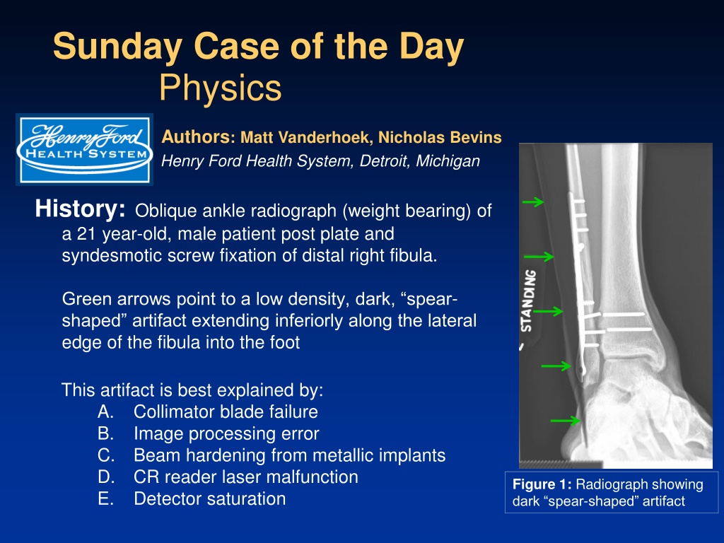
An oblique ankle radiograph of a patient showed a dark spear-shaped artifact extending from the leg to the foot, which was identified as an image processing error due to a black border imaging algorithm issue. The presence of metal in the leg caused the algorithm to fail, resulting in the artifact. This case highlights the importance of recognizing and addressing image processing failures in radiography to ensure accurate diagnosis and interpretation.

Download Presentation

Please find below an Image/Link to download the presentation.
The content on the website is provided AS IS for your information and personal use only. It may not be sold, licensed, or shared on other websites without obtaining consent from the author. Download presentation by click this link. If you encounter any issues during the download, it is possible that the publisher has removed the file from their server.
E N D
Presentation Transcript
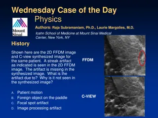
Sunday Case of the Day Physics Authors: Matt Vanderhoek, Nicholas Bevins Henry Ford Health System, Detroit, Michigan History: Oblique ankle radiograph (weight bearing) of a 21 year-old, male patient post plate and syndesmotic screw fixation of distal right fibula. Green arrows point to a low density, dark, spear- shaped artifact extending inferiorly along the lateral edge of the fibula into the foot This artifact is best explained by: A. Collimator blade failure B. Image processing error C. Beam hardening from metallic implants D. CR reader laser malfunction E. Detector saturation Figure 1: Radiograph showing dark spear-shaped artifact
Discussion: Image processing failure Figure 2: A black border was incorrectly applied to the image. The border detection image processing algorithm failed due to the presence of metal in the leg. Note that the black border runs along the edge of the metal plate and continues along the imaginary extension of that line into the foot (green arrows). Figure 3: The black border image processing has been removed and the artifact is gone. Figure 2: Image with black border image processing Figure 3: Image without black border image processing
Discussion: Incorrect answers A. Collimator blade failure Collimator blades are highly attenuating. Consequently, the presence of a collimator blade in the image would result in a high density (white) artifact. C. Beam hardening from metallic implants Metallic implants cause beam hardening which leads to artifacts in computed tomographic (CT) imaging. Beam hardening typically does not cause artifacts in standard planar radiography. D. CR reader laser malfunction A CR reader laser performs a raster scan of the imaging plate. As such, laser malfunction would likely result in degradation of the entire image, individual lines, or individual pixels. The resulting artifact is unlikely to follow the shape of an anatomical structure or implant. E. Detector saturation Digital detectors saturate when they are exposed to too much radiation. The artifact appears under patient tissue, including bone, where the x-ray intensity is significantly lower than in air.