Understanding Breast Imaging Artifacts in Digital Mammography
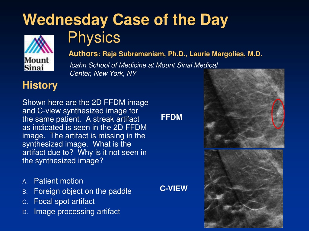
The case study discusses a streak artifact in a 2D FFDM image, caused by the patient's hair. The artifact is absent in the synthesized C-view image due to the way tomosynthesis displays breast tissue, excluding areas above the paddle. References and discussions on mammographic artifacts and quality assurance are also provided.

Download Presentation

Please find below an Image/Link to download the presentation.
The content on the website is provided AS IS for your information and personal use only. It may not be sold, licensed, or shared on other websites without obtaining consent from the author. Download presentation by click this link. If you encounter any issues during the download, it is possible that the publisher has removed the file from their server.
E N D
Presentation Transcript
Wednesday Case of the Day Physics Authors: Raja Subramaniam, Ph.D., Laurie Margolies, M.D. Icahn School of Medicine at Mount Sinai Medical Center, New York, NY History Shown here are the 2D FFDM image and C-view synthesized image for the same patient. A streak artifact as indicated is seen in the 2D FFDM image. The artifact is missing in the synthesized image. What is the artifact due to? Why is it not seen in the synthesized image? FFDM A. Patient motion B. Foreign object on the paddle C. Focal spot artifact D. Image processing artifact C-VIEW
Diagnosis Correct Answer: B - Patients Hair is the cause of Artifact in the 2D-FFDM image. FFDM C-VIEW
Discussion Tomosynthesis displays several slices equal to the measured thickness of the breast plus a few extra processing slices. For example, if the measured thickness was 42 mm there would be 42 slices + 4 or 5 more. Thickness is calculated by the area between the bottom of the paddle (pink) and the top of the detector (white) during compression. C-view and tomosynthesis images would display the content between the detector and the paddle i.e the breast tissue (beige) and not display any area above the paddle. Therefore, hair would be visible on a FFDM image, but not on C-view (nor on tomosynthesis). In this schematic, green is the patient s hair.
References/Bibliography: Choi JJ, Kim SH, Kang BJ, Choi BG, Song B, Jung H. Mammographic Artifacts on Full-Field Digital Mammography. Journal of Digital Imaging. 2014; 27(2) :231-236. 1. 2. Quality assurance in mammography: artifact analysis. Hogge JP, Palmer CH, Muller CC, Little ST, Smith DC, Fatouros PP, de Paredes ES. Radiographics. 1999 Mar-Apr;19(2):503-22. Review. 3. Gold, BM, American Journal of Roentgonology, 1992 Aug;159(2):430.