Cholera Diagnosis and Control Methods Overview
Cholera diagnosis involves various laboratory tests such as specimen analysis, smears, and cultures. Slide agglutination tests and the oxidase test can also aid in the identification of Vibrio cholerae bacteria. Understanding these diagnostic methods is crucial for effective control and management of cholera outbreaks.

Download Presentation

Please find below an Image/Link to download the presentation.
The content on the website is provided AS IS for your information and personal use only. It may not be sold, licensed, or shared on other websites without obtaining consent from the author. Download presentation by click this link. If you encounter any issues during the download, it is possible that the publisher has removed the file from their server.
E N D
Presentation Transcript
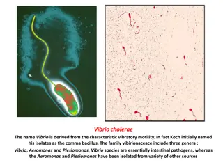
Cholera diagnosis and control
DIAGNOSTIC LABORATORY TESTS a) Specimens:- specimens from culture consist of mucus flecks from stool. b) Smears:- Dark field or phase contrast microscopy may show the rapidly motile vibrios. c) Culture:- growth is rapid in peptone agar , on blood agar with a pH near 9.0 or on TCBS agar, and typical colonies can be picked in 18 hours. For enrichment , a few drops of stool can be incubated for 6-8 hours in TAUROCHOLATEPEPTONE BROTH(pH 8.0-9.0) ; organisms from this culture can be stained or subcultured. Transport media: alkaline peptone water, sea water etc
SLIDE SLIDE AGGLUTINATION AGGLUTINATION Agglutination tests for V. cholerae somatic O antigens may be carried out in a petri dish or on a clean glass slide. Emulsify the growth in a small drop of saline and mix thoroughly by tilting back and forth for about 30 seconds. Mix the anti O serum and look for agglutination.
Figure Antisera to the O1 serogroup of Figure Antisera to the O1 serogroup of V. cholerae will agglutinate homologous organisms (left). A normal serum or agglutinate homologous organisms (left). A normal serum or saline control (right) does not show agglutination . saline control (right) does not show agglutination . V. cholerae will
OXIDASE TEST Conduct the oxidase test with fresh growth from an HIA slant or any non-carbohydrate-containing medium. Place 2 to 3 drops of oxidase reagent (1% tetramethyl-p- phenylenediamine ) on a piece of filter paper in a petri dish. Smear the culture across the wet paper with a platinum (not nichrome ) loop, a sterile wooden applicator stick, or toothpick. In a positive reaction, the bacterial growth becomes dark purple within 10 seconds Color development after 10 seconds should be disregarded. Positive and negative controls should be tested at the same time.
A positive oxidase test (as shown here) results in the A positive oxidase test (as shown here) results in the development of a dark purple color within 10 seconds. development of a dark purple color within 10 seconds. V. cholerae is oxidase cholerae is oxidase- -positive, which differentiates it from positive, which differentiates it from oxidase oxidase- -negative organisms . negative organisms . V.
Voges Voges- -Proskauer test Proskauer test The Voges-Proskauer test has been used to differentiate between the El Tor and classical biotype of V. cholerae O1. Classical biotypes usually give negative results; El Tor isolates are generally positive. V. cholerae produces acetoin, which is detected in the Voges-Proskauer test, giving a red (positive) reaction (left). A negative reaction is on the right.
HEMOLYSIS TESTING the classical and El Tor biotypes were differentiated by the ability of the El Tor group to lyse erythrocytes. However, by 1972 almost all isolates worldwide were nonhemolytic. The two exceptions to this trend have been the U.S. Gulf Coast and the Australia clones of V. cholerae O1, which are strongly hemolytic when assayed by either the plate or tube hemolysis assay (For this reason, hemolysis continues to be a useful phenotypic characteristic for differentiating the Gulf Coast and Australia clones of V cholerae O1 from El Tor strains from the rest of the world, including Latin America.
Tube hemolysis, shown in the tube on the left, is Tube hemolysis, shown in the tube on the left, is demonstrated by the absence of a button of demonstrated by the absence of a button of sedimented cells and the presence of free hemoglobin sedimented cells and the presence of free hemoglobin in the tube. in the tube.
EPIDEMIOLOGY Six pandemic of cholera occurred between 1857 and 1923, caused mostly by V. cholerae 01 of the classic biotype and largely originating in ASIA, usually the INDIAN subcontinent. Seventh pandemic began in the CELEBES ISLAND. Starting in 1991,the seventh pandemic (El Tor biotype) spread to PARIS and then to other countries of SOUTH AMERICA and CENTRAL AMERICA . Millions of people have had cholera in this pandemic . Some consider the cholera caused by the serotype O 139 strain to be the eight pandemic that begin in the INDIAN SUCONTINENT in the 1992-1993, which spread to ASIA . Cholera is endemic in INDIA and SOUTHEAST ASIA. From these centers ,it is carried along shipping lane ,trade routes and pilgrim migration routes.
The disease is spread by contact involving individuals with mild or early illness and by water, food and flies. V.cholerae lives in the aquatic environments and such environments are the vibrios natural reservoir. It can survive for year and grow , but when conditions are not suitable for growth it can become dormant.
TREATMENT Therapy consist of water and electrolyte replacement to correct the severe dehydration and salt depletion. Oral tetracycline tends to reduces stool output in cholera and shortens the period of excretion of vibrios. In some endemic areas , tetracycline resistant of V.cholerae has emerged , due to the transmissible plasmids.
Ciprofloxacin can also be used 500 mg every 12 hours for 3 days or erythromycin at 500 mg every 6 hours for 3 days is also effective In Kadoma, a child brought in a cart to an MSF cholera treatment center .
CONTROL Control rests on education and on improvement of sanitation particularly of food and water. Patients should be isolated . Repeated injections of a vaccine containing either lipopolysaccharides extracted from vibrios or dense vibrio suspension can confer limited protection to limited healthy exposed persons but not effective in epidemic control.


 results in the")