Laboratory Diagnosis of Fungal Infections: Specimen Collection and Transport

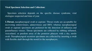
In the laboratory diagnosis of fungal infections, proper collection and transportation of specimens are crucial for accurate diagnosis and treatment. Different sites and types of specimens require specific collection techniques to avoid contamination and ensure viability. From superficial to systemic mycoses, appropriate sampling methods are essential to confirm clinical suspicion, choose the right therapeutic agent, and monitor disease progression. Early transport of specimens to the lab is vital to prevent contaminant overgrowth and obtain reliable results.

Download Presentation

Please find below an Image/Link to download the presentation.
The content on the website is provided AS IS for your information and personal use only. It may not be sold, licensed, or shared on other websites without obtaining consent from the author. Download presentation by click this link. If you encounter any issues during the download, it is possible that the publisher has removed the file from their server.
E N D
Presentation Transcript
Laboratory Diagnosis of Fungal Infections MBI 531
Introduction To confirm clinical suspicion to establish fungal cause of disease. To help in - Choosing a therapeutic agent Monitoring the course of disease Confirming mycological cure
Sites & Types of Specimens Specimen collection depends on the corresponding disease. Very important to proceed for a final diagnosis.
(a) Superficial Mycosis Clean the part with 70% alcohol Collect the material in a sterile paper or a sterile petridish to - Allow drying of the specimen Reduce bacterial contamination Maintain viability
(a) Superficial Mycosis Dermatophytic lesion spreads outward in a concentric fashion with healing in the center scrape outwards from the edge of the lesion with a scalpel blade or use Cellophane tape Scalp lesion scraping with a blunt scalpel, including hair stubs, scales & contents of plugged follicles.
(a) Superficial Mycosis Scalp lesion Woodlamp s examination of infected hair fluorescence Hairbrush sampling technique Onychomycosis stop antifungals one week prior to collection Mucosal infections mucosal scrapings
(b) Subcutaneous Mycosis Scrapings or crusts from the superficial parts of lesions Pus aspirates Biopsy
(c) Systemic Mycosis CSF Blood Scrapings or swabs from the edge of lesions. Pus Biopsy Feces Urine Sputum
Collection & Transport of specimen Proper collection of specimen and in adequate quantity. Early transport to the lab to avoid overgrowth of contaminant Respiratory specimens Sputum early morning sample, after mouth wash, flakes to be used for culturing Bronchoscopy if non productive cough Bronchial brushings or lung biopsy to rule out invasion or colonisation
Collection & Transport of specimen Blood In biphasic Brain Heart Infusion agar Inoculated in 2 bottles for dimorphic fungi Cerebrospinal fluid Should be immediately processed else stored at RT or at 30 C in an incubator Centrifuge & use sediment for culture
Collection & Transport of specimen Skin, Hair & Nail Taken for dermatophytic infections Hair plucked with forceps Tissue, BM & Body fluids Tissues grind or mince before culturing Body fluids centrifuge & use sediment for culture Urine centrifuge & use sediment for culture
Laboratory Diagnosis Direct examination Fungal culture Serological tests Skin tests PCR & other molecular methods
Direct Examination Very decisive in the diagnosis of fungal infections Wet mounts Slide & tube KOH mounts 10 to 20% KOH digests protein debris, dissolves keratin. DMSO can be added to KOH to hasten clearing in skin scrapings & nail clippings Calcofluor white fluorescent stain excellent morphology of pathogenic fungi India ink capsulated fungi
CFW yeast form of Blastomyces KOH -Aspergillus India ink - Cryptococcus
Direct Examination Gram stain fungi are Gram +ve Histopathology - yeast cells, hyphae, pseudohyphae, arthrospores, chlamydospores, and spherules. Routine stain Hematoxylin & Eosin (HE) - very usefulto visualize the host's response Superficial infection acute, subacute or chronicdermatitis with folliculitis Subcutaneous & systemic infections granulomatousreaction with fibrosis or pyogenic inflammation 1. 2.
Direct Examination Histopathology Special stains PAS (Per Iodic acid), GMS (Grocott Gomori Methanamine Silver), Mayer s mucicarmine, Gridley s stain GMS is more advantageous sinceit stains old and nonviable fungal elements more efficiently than the others Mucin stains, like Mayer's mucicarmine stain the mucopolysaccharide capsule of Cryptococcusneoformans
Direct Examination Fluorescent- antibody staining To detect fungal Ag in clinical specimen such as pus, blood, CSF, tissue sections Adv can detect fungus even when few organisms are present
Fungal Culture Sabouraud Dextrose Agar (SDA) Contains 2% dextrose, antibiotics (gentamicin, chloramphenicol) and cycloheximide Selective media Corn meal agar (CMA) sporulation, chlamydospore formation Bird seed agar cryptococcus, forms brown colonies Brain Heart Infusion (BHI) agar dimorphic & other fastidious fungi
Corn MealAgar Bird SeedAgar
Fungal Culture Temperature requirement Majority of fungi 37 C Superficial mycosis 30 C Dimorphic fungi 25 C & 37 C Incubation time At least 4 weeks Usually positive cultures are obtained in 7-10 days Candida & Aspergillus - 24 to 72 hrs
Fungal Culture Specimens should be cultured on agar slants: Safe Require less space More resistant to drying during prolonged incubation Blood cultures should be inoculated in to biphasic blood culture bottles
Interpretation of Fungal Culture Isolation of an established pathogen like H. capsulatum or C. neoformans evidence of infection Isolation of commensal or opportunistic fungi like Candida or Aspergillus consider following points: 1. Isolation of same strain in all culture tubes Repeated isolation of same strain in multiple specimens Isolation of same strain from different sites Immune status Serological evidence 2. 3. 4. 5.
Identification of fungal cultures Colony morphology colour, texture, pigment
Identification of fungal cultures Fungal morphology under microscope using Lactophenol Cotton Blue (LPCB) stain Composition of LPCB Lactic acid - preservesfungal structure Phenol kills any live organism Glycerol preventsdrying Cotton blue imparts blue color to structures
Identification of fungal cultures Special culture techniques Slide culture to see sporing structures & spore arrangement, CHROM agar for candida sps. Biochemicals ability to assimilate carbon & nitrogen, sugar fermentation
C.tropicalis C.krusei C.albicans CHROMAgar
Serology & Immunology Detection of Ag or Ab in serum or body fluids Ab detection: Diagnosis of systemic & subcutaneousmycoses Assess prognosis of the disease Assess response to treatment Ag detection: Early stages of infection In patients with impaired immunity Delayed hypersensitivity tests with Ags like candidin, histoplasmin, etc.
Serological tests used in Medical Mycology Agglutination Whole cell agglutination Latex particle agglutination Passive haemagglutination Immunodiffusion most widely used Counter immunoelectrophoresis (CIEP) Indirect fluorescent Ab detection ELISA, RIA
Other Methods PCR Polymerase Chain Reaction RFLP - Restriction fragment length polymorphism Protein electrophoresis Nucleic acid probes Serotyping Karyotyping


 Superficial Mycosis")
 Superficial Mycosis")
 Superficial Mycosis")
 Subcutaneous Mycosis")
 Systemic Mycosis")