Understanding Vibrio Cholerae: Biology and Impact on Human Health

Vibrio cholerae is a significant bacterium responsible for causing cholera, a life-threatening diarrheal disease. With over 35 species in the Vibrio genus, only a few can infect humans. Vibrio cholerae is a Gram-negative, curved aerobic rod that produces an enterotoxin leading to cholera. Understanding its morphology, culture methods, and identification can aid in diagnosis and treatment. This bacterium thrives in aquatic environments and poses a risk to human health if not managed effectively.

Download Presentation

Please find below an Image/Link to download the presentation.
The content on the website is provided AS IS for your information and personal use only. It may not be sold, licensed, or shared on other websites without obtaining consent from the author. Download presentation by click this link. If you encounter any issues during the download, it is possible that the publisher has removed the file from their server.
E N D
Presentation Transcript
University of Mustanisiriya College of Medicine Department of Microbiology Third stage The Vibrio Dr Ali Abdulwahid
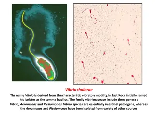
The Vibrio There are more than 35 species recognized for the genus vibrio, only 12 of them are responsible for human infection (gastrointestinal and extra-intestinal infections). Vibrio cholerae: is the most medically important species which is responsible for the cholera disease They are Gram-negative curved rods aerobic with a wide distribution in the nature (Found in marine and surface waters) Actively motile using polar flagellum. The organisms produce an enterotoxin that lead to cause cholera ( which is a watery diarrhea that can rapidly led to dehydration and even death if it is not treated) Only two serogroups (O1 and O139) cause cholera in human Other serogroups (more than 200 ) may cause enteritis and sepsis Other species that can cause infection to human : V. parahaemolyticus, V. vulnificus, V. minicus and V. alginolyticus
Vibrio cholerae Morphology and Identification A. Typical Organisms V cholerae is a curved, comma-shaped, gram-negative bacterium Actively motile using polar flagellum. The organism can revert to straight rods due to subculturing or prolonged cultivation They are non-capsulated and non-spore forming B. Culture V. cholerae on agar plates forms moist, smooth, translucent round colonies They grow at temperature range from 16 -40 0C , but their optimum growth at 37 0C Grow well on alkaline pH (between 7.4 and 9.6) (optimum pH 8.2). Sensitive to acidic pH (below 6), Not halophilic
Gram-stained Vibrio cholera in a specimen Transmission electron microscope image of Vibrio cholerae Source of the picture CDC / Public domain https://upload.wikimedia.org/wikipedia/commons/b/b0/Vibrio_cholerae_gram_stai n_CDC.jpg Source of the picture: Tom Kirn, Ron Taylor, Louisa Howard - Dartmouth Electron Microscope Facility / Public domain https://commons.wikimedia.org/wiki/File:Vibrio_cholerae.jpg
B. Culture B. Culture The bacteria grow well on a wide range of media including Ordinary media such as (nutrient agar, MacCkonkey agar and blood agar). Selective media : such as thiosulfate-citrate- bile-sucrose (TCBS) agar : the bacteria form Yellow colonies as they ferment sucrose, while other vibrios that do not ferment sucrose produce green colonies. The yellow colonies can be easily recognised on the media which has dark-green background Enrichment media : Alkaline peptone water (pH 8.6) Useful for preliminary culturing of the bacteria from stool or other specimens yellow colonies of Vibrio cholerae on TCBS agar Source of picture: Microrao, JJMMC, Davangere, Karnataka, India / Public domain https://commons.wikimedia.org/wiki/File:Vibrio_cholerae_on_TCBS_agar.jpg
String test: A single colony or A loopful of vibrio liquid growth is mixed with a drop of 0.5 % sodium deoxycholate or sodium taurocholate on a slide or petri dish. If the test is positive, the sodium deoxycholate or sodium taurocholate will lysis the bacteria cells, leading to release the DNA from the sells making the suspension mucoid and form a string when taken by loop. This test differentiate V. cholera (string +ve) from Aeromonas spp (string ve) (Both are isolated from diarrheal stool, show similar biochemical properties) and other vibrio species (string ve). String test of Vibrio cholerae Source of image : CDC, Courtesy: Public Health Image Library http://www.publicdomainfiles.com/show_file.php?id=13539910812826
Antigenic Structure and Biological Classification Flagellar (H)antigen: Many Vibrios share a single (H)antigen , which is heat labile and it is not immunogenic (antibodies against this antigen is not protect the host from vibrio cholera infections) Somatic (O) antigen: Depending on variability in O antigen , Vibrio cholerae were divided into 206 different O serogroups These serogroups can be devided to three main groups: O group 1 (O1) O group 139 (O139) non-O1/non-O139 groups Only V. cholera strains of (O1) and (O139) cause classic cholera Other non-O1/non-O139 strains may cause cholera-like disease.
Strains of O1 serogroup of V. cholera are also divided into two different biotypes : classic and El Tor. These two biotypes also divided into different serotypes depending on some determinant in the O antigen These serotypes include : Ogawa, Inaba, and Hikojima. The biotypes of V. cholerae O1 group differ in some tests as in the following table: Tests Vibrio biotypes Voges- Proskauer hemolysin resistant to polymyxin B Group IV phage susceptibility Classical -ve -ve +ve +ve El Tor +ve +ve -ve -ve
Gardner and Venkatraman Classification. Vibrio Vibrios with common flagellar (H) antigen, share similar biochemical characteristic A heterogeneous collection of vibrios Group B (V. cholerae) Group A Non O1 group (involves O139) O1 group Somatic (O) antigen: Classical El Tor Biotypes Serotypes, depending on minor antigenic variation Inaba Inaba Ogawa Ogawa Hikojima Hikojima
Vibrio cholerae Enterotoxin It is heat-labile enterotoxin Multimric, composed of (1 A subunit + 5 B subunits ) B subunits bind to the GM1 ganglioside receptors on the intestinal epithelial cells. This will promotes the entry of subunit A in to the epithelial cells The subunit A then will be cleaved into A1 and A2, A1 is the active portion of the A subunit that enter the cell and activate of adenylate cyclase (which is an enzyme that responsible for production of cyclic adenosine monophosphate (cAMP) in the cell ) leading to overproduction of cAMP. This consequently lead to inhibition of the absorption of the sodium and chloride by the cells lining the microvilli, along with increased secretion of sodium-dependent chloride. This results in blocking the absorption of water, Increased outflow of water and electrolytes into the gut lumen, with consequent diarrhea.
Pathogenesis This bacteria is restricted pathogen for human, but it also growth in brackish water and marine water The infection can happen by ingestion the organism via contaminated water or food In human with normal gastric acidity, the infection dose should be about 1010 Cells or more ( as the bacteria is sensitive to the acidity of the stomach) , while persons with achlorhydria or hypochlorhydria , as few as 102 104 bacterial cells is enough to cause the infection. Reducing the acidity of the stomach due to taking any medication leading to increase the sensitivity to infection with V. cholerae Vibrio cholerae is not invasive and do not reach the bloodstream, it attach to the microvilli of the brush border of intestinal epithelial cells. At this site, the bacteria multiply and produce cholera toxin which responsible for developing the clinical manifestation of the cholera
Clinical Findings The incubation period is 12 hours 3 days (this depends mainly on the number of ingested organisms) The symptoms involve a sudden onset of nausea, vomiting and profuse watery diarrhea with abdominal cramps. The colourless watery stools rice water stool is a characteristic feature for the Cholera. The stool contains flecks of mucus, epithelial cells, and large numbers of vibrios. in sever cholera, the volum of fluid loos is about 1 L/h , and such rapid loss of fluid and electrolytes leads to profound dehydration, circulatory collapse, and anuria as well as renal failar. The infection without treatment is correlated with a high rate of mortality ( between 25% and 50%), but with treatment it can be decreased to 1% or less. Infection with El Tor biotype tends to be milder than the infection caused by classic biotype.
Diagnostic Laboratory Tests A. Specimens Watery stool: should be collected as early as possible in the course of the diarrihea and inculcated within 2-4 hrs after collection into the suitable medium If that will take time, the stool specimens can be mixed with transport medium under cooling conditions (refrigeration) B. Smears Direct microscopic examination as well as Gram staining film for smears made from stool samples is not recommended as the results is difficult to be interpreted . The rapid diagnosis can be achieved by using dark-field or phase contrast microscopy to monitor rapid motility of vibrios (shooting star) This also can be achieved by inhibition the motility of the vibrios by adding the anti- cholera diagnostic antisera
C. Culture Specimens can be plated on different media including peptone agar, blood agar ( pH near 9.0), and TCBS agar After 18 hrs of incubation , V. cholerae typical colonies can be seen on the agar. D. Serological tests V. cholerae organisms can be also identified by slide agglutination tests using anti-O group 1 or group 139 antisera. Further serological tests can also be used for differentiation between the different serotypes (Inaba, Ogawa and Hikojima) E. Biochemical tests : The organisms can be further identified by a variety of biochemical reactions such as IMViC, TSI and others
Treatment The treatment is mainly depends on the water and electrolyte replacement to correct the severe dehydration and salt depletion. This can be achieve by Oral rehydration therapy (ORT) or intravenous rehydration (in sever cases) The antimicrobial thereby is of a secondary importance, but it can be affective to reduce the duration and the amount of the shedding vibrios in the stool. Oral tetracycline is vary affective antibiotic therapy Erythromycin and / or azithromycin are effective therapy in children and in pregnant women Other antibiotics : fluoroquinolones and doxycycline. Resistance of V cholera to antibiotics such as resistance to tetracycline has been noticed and it emerged, especially in area where cholera is endemic or epidemic. Genes responsible for such feature carried on transmissible plasmids
Epidemiology, Prevention, and Control Cholera is considered as both endemic and epidemic disease There were six cholera pandemics occurred between 1817 and 1923 The causative agents for these pandemics were V. cholerae O1 of the classic biotype They were mainly originated in Asia, particularly in Indian subcontinent. A seven pandemic (1961) in Indonesia, and spread to Asia, the Middle East, and Africa. The causative V cholerae was El Tor biotype. The eight pandemic (1992 1993), a spread of the serotype O139 strains in the Indian subcontinent and to other Asian countries was considered to be the eighth pandemic of cholera.
Epidemiology, Prevention, and Control The disease is endemic in India and Southeast Asia. It spreads to other geographical sites in the world by the means of shipping, trade, and pilgrim migration. It spread by water, food, flies as well as contact with individuals that have mild or early infection. In majority, only 1 5% of susceptible people who exposed to V. cholerae can develop disease. People can rarely carry the bacteria asymptomatically ( carriers state) more than 3 4 weeks, and there is no long term carriage state in humans . There is no another animal host is known for V. cholerae
Epidemiology, Prevention, and Control The bacteria can survive in different aquatic habitats for a given periods (up to 3 weeks) Such aquatic habitats are considered as the natural reservoir for V. cholera, as the bacteria can attach to algae, copepods, and crustacean shells and then can survive for years if the environment conditions is suitable. Control measures can be: Improvement of sanitation such as sewage treatment , water purification and prevent of food contamination Isolation of patients, disinfection and disposal of their excretes. Chemoprophylaxis with antibiotics (to household contacts) can limit the spread of the bacteria
Vaccination Many different vaccines have been developed against V. cholerae include: Single dose , living cholera oral vaccine. Killed oral cholera vaccines. Whole cell V. cholerae O1 with recombinant B subunit of cholera toxin vaccine. One whole-cell V. cholerae O1 and O139 used in Vietnam Another one whole-cell V. cholerae O1 and O139 for global use Injectable phenol-inactivated strains of V. cholerae still used in some countries (not recommende by WHO, lower effectivity with short period of protection)
Other vibrios Vibrio parahaemolyticus It is halophilic and marine microorganism Gram ve , curved shape , motile Causes acute gastroenteritis The infection is acquired by ingestion of contaminated seafood such as raw fish or shellfish The incubation period is 12 24 hours The main clinical manifestations are : nausea, fever, and watery diarrhea The infection is self-limited (subside spontaneously in 1 4 days), and only water and electrolyte replacement is required . The bacteria grow well on TCBS and produce green colonies as it does not ferment sucrose.
V. vulnificus V. vulnificus It is a medically important species of vibrio Causing the second serious infection caused by Vibrios after V. cholerae It is halophilic marine microorganism, specifically apt to be found in oysters Does not ferment the sucrose and form green colonies on TCBS agar. Lactose fermentation +ve ( it is the key differentiation feature of this species , as most other vibrio species are ve for lactose fermentation). Diseases caused by this species : Severe wound infections, Bacteremia, Gastroenteritis
References 1. Reidle, S., Morse, S. A., Meitzner, T., and Miller, S. 2019. Jawetz, Melnick & Adelberg s Medical Microbiology , Twenty-Eight Edition. The McGraw-Hill education, Inc. USA 2. Kumar, S. 2012. Textbook of microbiology. Jaypee Brother Medical Publishers (P) Ltd. New Delhi, India. 3. https://commons.wikimedia.org 4. http://www.publicdomainfiles.com 5. Public Health Image Library