Understanding the Mediastinum: Boundaries, Divisions, and Contents
The mediastinum is a critical anatomical region in the thoracic cavity that houses various structures like the heart, thoracic vertebrae, diaphragm, and sternum. Divided into superior and inferior sections, it contains essential organs, vessels, nerves, and lymph nodes. Understanding its boundaries, divisions, and contents is crucial for medical students and professionals for comprehensive anatomical knowledge.
- Mediastinum Anatomy
- Thoracic Cavity Structures
- Superior and Inferior Divisions
- Organ Systems
- Medical Education

Download Presentation

Please find below an Image/Link to download the presentation.
The content on the website is provided AS IS for your information and personal use only. It may not be sold, licensed, or shared on other websites without obtaining consent from the author. Download presentation by click this link. If you encounter any issues during the download, it is possible that the publisher has removed the file from their server.
E N D
Presentation Transcript
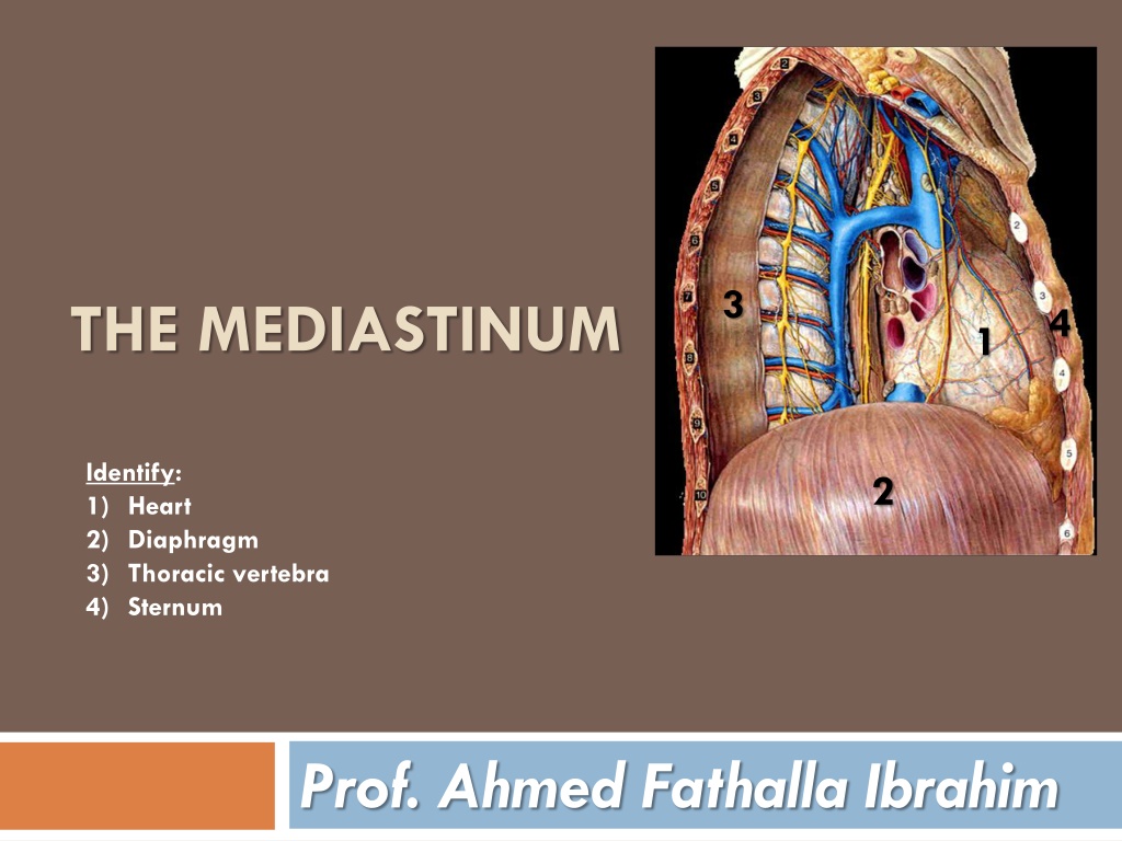
3 THE MEDIASTINUM 4 1 Identify: 1) Heart 2) Diaphragm 3) Thoracic vertebra 4) Sternum 2 Prof. Ahmed Fathalla Ibrahim
OBJECTIVES At the end of the lecture, students should be able to: Define the Mediastinum . Differentiate between the divisions of the mediastinum. List the boundaries and contents of each division. Describe the relations between the important structures in each division.
THE MEDIASTINUM It is a partition between right & left pleural sacs & lungs. It includes all the structures which lie in the intermediate compartments of the thoracic cavity.
BOUNDARIES OF MEDIASTINUM Superior: Thoracic outlet Inferior: Diaphragm Anterior: Sternum Posterior: Thoracic vertebrae Lateral: Lungs & pleurae
DIVISIONS OF THE MEDIASTINUM It is divided by a horizontal plane S extending from sternal angle to A lower border of 4th thoracic vertebra into: M P 1. Superior mediastinum (S): above the plane 2. Inferior mediastinum: below the plane, Inferior mediastinum is subdivided into: Middle mediastinum (M): contains heart Anterior mediastinum (A): in front of heart Posterior mediastinum (P): behind heart
BOUNDARIES OF SUPERIOR MEDIASTINUM Superior: Thoracic outlet Inferior: Horizontal plane Anterior: Manubrium of sternum Posterior: Upper 4 thoracic vertebrae Lateral: lungs & pleurae
CONTENTS OF SUPERIOR MEDIASTINUM Thoracic duct Left vagus nerve Trachea Right vagus nerve Left common carotid artery Left subclavian artery Left recurrent laryngeal nerve Brachiocephalic artery Left brachiocephalic vein Right phrenic nerve Thymus Right brachiocephalic vein Left phrenic nerve S V C Esophagus
CONTENTS OF SUPERIOR MEDIASTINUM c b a A A B FROM SUPERFICIAL TO DEEP: 1. Thymus gland 2. Veins: -Right & left brachiocephalic -Superior vena cava 3. Arteries: -Arch of aorta (A) & its branches a-Brachiocephalic artery b-Left common carotid c-Left subclavian 4. Nerves: - Right & left phrenic -Right & left vagus 5. Trachea 6. Esophagus 7. Thoracic duct 8. Lymph nodes C
CONTENTS OF SUPERIOR MEDIASTINUM 4 ARTERIES: arch of aorta, brachiocephalic, left common carotid, left subclavian 4 NERVES: right & left vagus, right & left phrenic 3 VEINS: right & left brachiocephalic, SVC 2 TUBES: trachea & esophagus 1 GLAND: thymus 1 DUCT: thoracic duct
BOUNDARIES OF POSTERIOR MEDIASTINUM Superior: Horizontal plane Inferior: Diaphragm Anterior: Heart Posterior: Thoracic vertebrae from T5 to T12 Lateral: Lungs & pleurae
CONTENTS OF POSTERIOR MEDIASTINUM 1. Esophagus 2. Vagus nerves: around esophagus 3. Thoracic duct: posterior to esophagus 4. Azygos vein: posterior & to the right of esophagus 5. Descending aorta: posterior & to the left of esophagus 6. Right & left sympathetic trunks 7. Lymph nodes
CONTENTS OF POSTERIOR MEDIASTINUM Right & left Vagus nerves (esophageal plexus) Left Sympathetic Trunk Right Sympathetic Trunk
MIDDLE MEDIASTINUM SITE: Between anterior & posterior mediastinum CONTENTS: Heart & pericardium Ascending Aorta Pulmonary trunk Superior & inferior vena cava Right & left pulmonary veins Right & left phrenic nerves Lymph nodes Left Pulmonary veins Right Pulmonary veins Left Phrenic nerve Right Phrenic nerve Heart Inferior vena cava
ANTERIOR MEDIASTINUM BOUNDARIES: Superior: Horizontal plane Inferior: Diaphragm Anterior: Body & xiphoid process of sternum Posterior: Heart Lateral: Lungs & pleurae CONTENTS: Thymus gland Lymph nodes
LEVEL OF T4 Level of: Sternal angle Second costal cartilage Level of: Bifurcation of trachea Bifurcation of pulmonary trunk Beginning & termination of arch of aorta
VAGUS NERVE Left common carotid It is the 10th cranial nerve. Left subclavian The right vagus descends to the right side of trachea, forms the posterior esophageal plexus & continues in abdomen as posterior gastric nerve. The left vagus descends between left common carotid & left subclavian arteries, forms the anterior esophageal plexus & continues in abdomen as anterior gastric nerve.
PHRENIC NERVE ROOT VALUE: C3,4,5 COURSE IN THORAX: The right phrenic descends on the right side of SVC & heart. The left phrenic descends on the left side of heart. Both nerves terminate in the diaphragm. SUPPLY: 1) Motor & sensory fibers to diaphragm. 2) Sensory fibers to pleurae (mediastinal and central part of diaphragmatic pleura) & pericardium (fibrous and parietal layer of serous pericardium).
LYMPHATIC VESSELS IN THORAX Lymph form right side of head & neck Lymph from left side of head & neck Lymph from right upper limb Lymph from left upper limb end end Lymph from right side of thorax Lymph from left side of thorax Superior Mediastinum Posterior Mediastinum Cysterna chyli (contains lymph from lower half of body)
THORACIC DUCT BEGINNING: It is the continuation of cysterna chyli. COURSE: It passes through aortic opening of diaphragm. It ascends in posterior mediastinum (posterior to esophagus). It ascends in superior mediastinum (to the left of esophagus). TRIBUTARIES: It receives: Lymphatics from all body EXCEPT: right side of thorax, right upper limb and right side of head & neck. END: It ends in the left brachiocephalic vein. N.B.: The right lymphatic duct receives lymphatic from right side of thorax, right upper limb and right side of head & neck. It ends in the right brachiocephalic vein.
AORTA ASCENDING AORTA: Beginning: at aortic orifice of left ventricle. Course: in middle mediastinum End: continues as arch of aorta (at level of T4) ARCH OF AORTA: Course: in superior mediastinum End: continues as descending thoracic aorta (at level of T4) DESCENDING AORTA: Course: in posterior mediastinum End: continues as abdominal aorta through diaphragm Arch Asc. Ao. Descending aorta
QUESTIONS QUESTIONS
QUESTION 1 Which one of the following structures is present in the superior mediastinum? 1) Ascending aorta 2) Arch of aorta 3) Descending aorta 4) Pulmonary trunk
QUESTION 2 Which one of the following structures is present in both superior & posterior mediastinum? 1) Superior vena cava 2) Pulmonary trunk 3) Trachea 4) Esophagus
QUESTION 3 Which one of the following structures lies on the left side of esophagus in the posterior mediastinum? 1) Superior vena cava 2) Descending aorta 3) Azygos vein 4) Pulmonary trunk
THANK YOU THANK YOU