Rectal Prolapse and Rectal Tears in Animals: Causes and Treatments
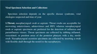
Rectal prolapse and rectal tears are common conditions observed in cattle, buffaloes, and small ruminants. Prolonged tenesmus, increased intraabdominal pressure, rectal inflammation, and other factors contribute to the development of these issues. Early diagnosis and treatment are crucial to prevent necrosis and further complications. Treatment involves reduction, lavage, application of astringent solutions, and appropriate suturing techniques. Rectal tears, although rare, require prompt medical attention based on the extent of damage.

Download Presentation

Please find below an Image/Link to download the presentation.
The content on the website is provided AS IS for your information and personal use only. It may not be sold, licensed, or shared on other websites without obtaining consent from the author. Download presentation by click this link. If you encounter any issues during the download, it is possible that the publisher has removed the file from their server.
E N D
Presentation Transcript
UNIT-5 (REGIONAL SURGERY-II) Rectal prolapse, Rectal tear. Atresia ani et recti, Atresia coli, Recto-vaginal fistula. Dr. Mithilesh Kumar Asstt. Cum Jr. Scientist Deptt. Of Surgery and Radiology BVC, Patna
RECTAL PROLAPSE Observed in cattle, buffaloes and small ruminants. Result of prolonged tenesmus, increased intraabdominal pressure due to bloat, rectal inflammation and irritation, diarrhea and act of parturition. Also due to intestinal neoplasia, foreign body, perineal hernia, constipation and congenital defects. Incomplete only prolapse of mucosa. Complete rectal wall prolapse. Contamination and trauma of prolapse in contact with air.
Early treatment is necessary to prevent necrosis. DIAGNOSIS:- Visual observation of mass, varied length from anus. Recent case short length and mucosa is bright red. Delayed case red and black mucosa, solid, necrosed. TREATMENT: In fresh case involves short length reduced after lavage a warm astringent solution (Pop In) application and topical anesthetics base. Purse string suture used to narrow the anus opening. Local dressing done with antiseptic solution Local anesthetic could used and epidural anesthetic can be used.
Suture removed in two to three day when straining not visible. Laxative diet is given in few days. Treatment of cause is necessary. Resection of dead tissue and damaged tissue require. Local dressing and antibiotic sprinkled over the prolapse. Rectal stump placed cranial to anal sphincter. Amputation of rectum in chronic prolapse with severe necrosis. A series mattress suture are placed. A second row of continuous sutures. The stump is returned in to normal position.
RECTAL TEARS Following trauma rectal tears occur. Reported rarely in ruminant. Classification of rectal tears. Grade I tear involve mucosa, or mucosa and submucosa Grade II only musculature ruptured Grade III mucosa, submucosa and muscular layer Grade IV All layers teared extend into abdominal cavity. Diagnosis:- Presence of excessive amounts of blood on the groove.
Easily palpable viscera, signs of shock and peritonitis. Epidural anesthesia relaxes the rectum and anal sphincter. Suturing of rectum is relatively tedious because of its peculiar location. Proctorrhaphy by using continuous chromic catgut. For proximal rectal tear Right flank laparotomy.
ATRESIA ANI ET RECTI Atresia ani a congenital is a congenital anomaly in calves, lambs and kids. It is associated with abnormal chromosomes. It is terminal part of G.I. tract located below the root of the tail. Failure of the anal membrane to become perforated, failure of the bowel to become canalized, and interruption of the fetal blood supply to the anus may lead to atresia ani or atresia ani et recti. CLINICAL SIGNS:- Absence of anal opening, tenesmus, bulging of the anal area and manifestation of abdominal discomfort along with
Distended abdomen. In case of atresia recti, blind end does not bulge out on applying abdominal pressure Defect is usually located near the pelvic brim. The general condition of calf is more serious if the malformation is located cranially.
Atresia Ani (No Anus)in calf with Recto- Vaginal Fistula in female calf repaired
2-day old female calf suffered from atresia ani and recto-vaginal fistula.
2-day old male cow calf diagnosed as atresia ani et recti by application of digital abdominal pressure and confirmed later by x ray.
calf suffered from atresia ani et recti. Gas is visualized only in the large intestine in front of the pelvis whereas the caudal part of the intestine had a soft tissue density indicates intestinal segmental aplasia.
Plain radiograph showing the presence of multiple air fluid levels in the abdominal cavity (arrows) and gas in the rectum (Atresia Coli)
TREATMENT:- Animal is controlled in dorsoventral position or lateral position. Anesthesia is attained by local infiltration with xylocaine 2%. A circular incision of 2-3cm diameter in large animal and 1cm in small animal is excised. Patency of opening is maintained by interrupted sutures.. Defecation improved slowly. In case of atresia recti blind end of rectum is freed. In some cases a flank approach to rectum is necessary. The blind end of rectum is dissected free in abdominal cavity moved caudally by traction through pelvic canal processed further same.
Post-operative care (i) Kept on liquid food. (ii) Antibiotic should be given 3 to 4 days. (iii) Suture removed 10-12 days.
ATRESIA COLI Congenital anomaly. Lack of development or closure of colon. Reported in cow calves and lambs. Etiology:-Early pregnancy diagnosis by amniotic palpation before 42 days gestation lead to subsequent damage to amniotic vesicles and fetal blood supply to the intestine cause congenital condition. It disrupts organogenesis. Lack of tail, kidney agenesis, umbilical hernia, cryptorchidism has been also reported.
CLINICAL SIGNS:- Absence of faeces since birth, abdominal distension, dehydration and progressive depression. Acute abdominal pain after colostrum consumption. Depression and abdominal distension. DIAAGNOSIS:- History and clinical signs and confirmed by right flank exploratory laparotomy. Radiographs shows gaseous distention of large intestine with absence of fecal material in distended colon.
Per rectal passage of a well lubricated flexible tube that meets resistance at 10 to 20cm of insertion indicates atresia. The tube should not forcefully advanced as the fragile colon is easily torn. Enema contraindicated. TREATMENT :- Surgery is considered urgent. Distension of large intestine leads to ischaemic necrosis, perforation and peritonitis. Preoperative correction of dehydration, acid base status and abs administered.
Contrast radiograph revealing the presence of stenosis at the junction between the colon and rectum (black arrows) and barium sulphate which was passed through a narrow canal and was then blocked at a blind sac (white arrow)
Right flank laparotomy done under general anaesthesia alongwith local anaesthesia. Proximal distended blind is first exteriorized and dissected free from the mesenteric attachment. Meconium is evacuated through enterotomy of proximal blind end. The distended proximal blind of the spiral loop of the ascending colon is resected to level where a more diameter of proximal loop exists. End to end or side to side anastomosis of the proximal loop of the ascending colon to the descending colon is done. One layer inversion technique has superior for colonic anastomosis.
Second row of continuous chromic catgut sutures can used to prevent leakage. The laparotomy wound closed routinely. Whole milk diet divided into 4 to 6 meals daily.
RECTOVAGINAL FISTULA A fistula is an abnormal connection between two organs. Rectovaginal fistula is abnormal connection between rectum and vagina . SYMTOMS- Bowel contents leak through the fistula allowing gas or stool to pass through vagina. Pain while making bowel movements. Smelly discharge from vulva. Repeated vaginal infection.
Rectovaginal fistula with atresia ani is a congenital condition. Delayed cases showed depression, dehydration and distention of abdomen.
2-day old female calf suffered from atresia ani and recto-vaginal fistula.
A female cross-bred cow calf affected with congenital rectovaginal fistula cum atresia ani and vulvular fusion
Female cow calf after surgical rectification of the anomalies
")


in calf with Recto-")