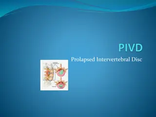
Understanding Prolapsed Intervertebral Disc (PIVD)
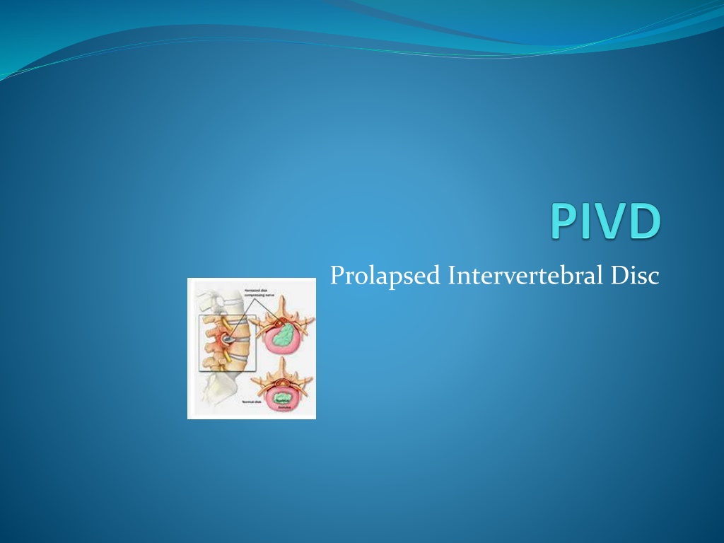
The intervertebral disc, a crucial component of the spine, can be affected by prolapse, leading to various stages of PIVD. The condition involves the nucleus pulposus bulging out due to tears in the annulus fibrosis. The stages of PIVD include bulging disc, protrusion, extrusion, and sequestrization, each signifying different levels of disc displacement and damage. Early detection and appropriate management are essential in addressing PIVD and its impact on spinal health.

Download Presentation

Please find below an Image/Link to download the presentation.
The content on the website is provided AS IS for your information and personal use only. It may not be sold, licensed, or shared on other websites without obtaining consent from the author. Download presentation by click this link. If you encounter any issues during the download, it is possible that the publisher has removed the file from their server.
E N D
Presentation Transcript
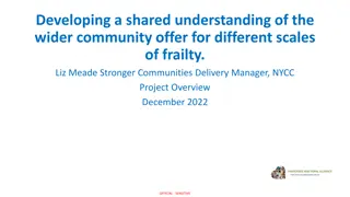
PIVD Introduction The spine consists of many vertebrae they are roughly circle and in between each vertebrae is a disc ( intervertebraedisc ) , the discs are constructed with strong rubber like tissue that allows a spine to become fairly flexible .
It lies between adjacent vertebrae in the vertebral column . A disc includes a stronger fibrous outer part (annulus fibrosis )... a soft jelly like middle part known as nucleus pulposes , disc behave as a shock absorber.
Defination- a spinal disc prolapse is a condition in which tear in the outer fibrous ring that is annulus fibrosis which allows the soft central portion that is nucleus pulposes to buldgeout beyond the damaged outer ring .
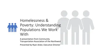
Stages of PIVD There are four stages of PIVD 1) Bulging disc 2) Protrusion 3) extrusion 4) sequestrization
Bulging disc This is a early stage in which the disc is stretched and doesn t return to its normal shape when pressure is relieved , it retains a slight buldgeat one site of the disc in the nucleus pulposes is slipping outwards into the disc fibre but not out of the disc.
Protrusion At this stage the bulge is very prominent and the soft jelly that is nucleus pulposes is slipped out to the inner edge of outer fibre , barely held in by the remaining disc fibre.
Extrussion In this stage the nucleus pulposes has completely spilled out of the disc and now protruding out of the disc fibres.
Sequestrization Here some of the nucleus pulposes is breaking off away from the disc into the surrounding area..causing symptoms or repercussions at an entirely different level of the spine .
PATHOLOGY Tears are almost posterolateral in nature owing to the presence of the posterior longitudinal ligament (PLL) in the spinal canal... Ligaments of the spine are anterior longitudinal ligament ,posterior longitudinal ligament , ligamentum flavum , supraspinal ligament .. This tear in the disc ring may result in the release off inflammatory chemical mediators which may directly results in severe pain .
Disc herniation Disc herniation which can occur to any part of the spine which can irritate the near by nerve ..a condition in which the outermost layer of of the annulus fibrosis but are in still contact but can bulge when the disc is underpressure.
Epidimiology Lumbar disc herniation occurs 15 times more often then cervical disc herniation , cervical disc are affected 8% of the time and thoracic only 1-2% of time . 4.8% males and 2.5% females older than 35 experiences more .
Location in lumbar region it is 95% in L4 and L5 and L5 s1 In cervical region C5 and C6 C7 is the second most common site . In thoracic region accounts for only 0.15% to 4.0% cases
etiology Heavy mannual labour. Repitative lifting and twisting Postural stress Obesity Poor and inadequate strength of trunk Sitting for `long hours Occupational activities Increasing age the disc is more lightly to devlopa weakness with increased age . Participation in sports for example : weight lifting , javenile thr
Pathology There is sequence of changes in the disc which leads to its prolapseand the changes are 1) Nucleus degeneration : softening of the nucleus and its fragmentation , weakening and disintigeration of the posterior part of the annulus . 2) Nucleus displacement : when annulus becomes weak the nucleus tends to bulge through the defect protrusion and then nucleus comes out of the annulus fibrosis and lies under the posterior longitudinal ligament but has not loss contact with parental disc( extrussion )these are the displacement that occur.
PATHOLOGY 3) stage of fibrosis: this is a stage of repair , this begans along side of degeneration than the residual nucleus pulposes becomes fibrosed the extruded nucleus pulposes become flattened fibrosed and finally undergoes calcification .
Sign and symptoms Lumbar Region : 1) severe low back pain 2) pain radiating to the buttocks leg and feet 3) pain may become worse with coughing , sneezing , straining and laughing 4) muscle spasm Tingling and numbness in legs and feet Muscle weakness or atrophy in later stage ( loss of bowel and bladder control in case of caudaequina syndrome .
SIGN AND SYMPTOMS Cervical region : 1) arm muscle weakness Deep pain near or over the shoulder blade on the affected side 2) increased pain when bending neck and turning head to the side ..spasm may also occur 3) burning pain radiating to the shoulder upper arm , forearm and rarely in head and fingers 4) tingling ( a pin and needle sensation ) or numbness in arm .
Investigations :- 1) X-ray of lumbosacral spine findings are a) narrowed disc spaces b) loss of lumbar lordosis c) compensatory sceolosis 2) CT scan of lumbar spine outline of soft tissue , bulging out disc 3) MRI of lumbar spine : intervertebral disc protrussion 2) compression of nerve root.
Examination :- 1) Posture : the patient stands with a rigid flattened lumbar spine the whole trunk is shifted forward . The trunk is tilted to one side . 2) movements : the patient is unable to bend forward muscle spasm in paraspinal muscle ( posterior compartment spine ) 3) tenderness : diffused tenderness in the lumbosacral lies on localized tenderness in the midline or lateral to the spinous process.
TEST 4) Straight leg raising test (SLRT )it is of two types active straight leg raising test (ASLRT ) and passive straight raising test (PSLRT ) this test indicates the nerve root compression . It can be performed unilaterall
TEST 5) lasegue test : modified form of SLRT degree of variation . 6) Slump test : it is a neurological test or orthopaedic test . 7) neurological examination : it would reveal a motar weakness , sensory loss of reflex corresponding to the affected nerve roots . 8) ULTT/ LLTT must be done for proper diagnosis.
Differential diagnosis :- A prolapsed disc is a common cause of low back pain sometimes back pain may be associated with sciatic pain .. Caution must be taken during diagnosis process and should not misguide with other disease like ankylosing spondylities , vascular insufficiency , or spinal tuberculosios ( pott sspine ).
Treatment :- 1) conservative treatment , 2) physiotheraphy treatment , 3) operative treatment .
Conservative treatment :- Rest and anti inflammatory and analgesics . Continue bed rest and traction for first two weeks may lessen the herniation . Rest on hard bed is necessary for not more than 2-4 days .
Physiotheraphy treatment :- Physiotheraphy plays a vital role in the management of PIVD . There are three phase Acute phase Subacutephase Chronic phase
MANAGEMENT Acute phase : rest is recomended as posture and activity modification by avoiding flexed posture, sitting for very longer duration , bending activities Local support :-- as corset that is lumbosacral belt etc.. These measure will enhance and stop reinjury to the herniated disc. Short period of walking at regular intervals .
MANAGEMENT Spinal extension exercises are beneficial in early treatment of disc related sign and symptoms. Soft tissue Manipulation ( massage ) to lessen the local muscle spasm and induces relaxation .
Subacute Phase :- Usually acute symptoms reduction in 6- 10 days . The exercises of acute phase must be performed . Nerve mobility exercises. Isometrics of extensars of spine , Pelvic rocking exercise can be achieved in supine ,sitting position that is cat and camel execise ( bird and dog position ) . Patient should be encouraged to perform aerobic activities walking ,swimming , with patient tolerance .
MANAGEMENT Chronic phase :- hamstring stretching Stretching and adaptability exercising stretching from the lumbar erector spinaeand soft tissues posterior towards the spine that is knee to chest exercise. Core strengtening exercises assist in relieving back pain .. The bridging exercise to strengten the core muscles. Strengtening exercises opposite arm and leg extension exercises on balancing or stabilizing exercises ( knee to chest exercises). All exercises must be performed in gentle way .
SURGICAL MANAGEMENT Operative Treatment are :- 1) Fenestration 2) Laminotomy 3) hemi Laminectomy 4)Laminectomy
Precautions :- Avoid bending , Lifting , Prolonged sitting . Soft mattress should be used, Heavy objects must not be carried . Thank you..