Understanding Hemostasis: Key Concepts in Blood Clotting Mechanisms
Hemostasis, crucial for preventing blood loss, involves platelet formation, clotting factors, and fibrinolysis. This process includes vessel constriction, platelet activation, clot formation, and fibrin breakdown. Platelets, produced in the bone marrow, play a vital role in maintaining vascular integrity. The intricate mechanisms of hemostasis are essential for maintaining homeostasis in response to environmental changes.

Download Presentation

Please find below an Image/Link to download the presentation.
The content on the website is provided AS IS for your information and personal use only. It may not be sold, licensed, or shared on other websites without obtaining consent from the author. Download presentation by click this link. If you encounter any issues during the download, it is possible that the publisher has removed the file from their server.
E N D
Presentation Transcript
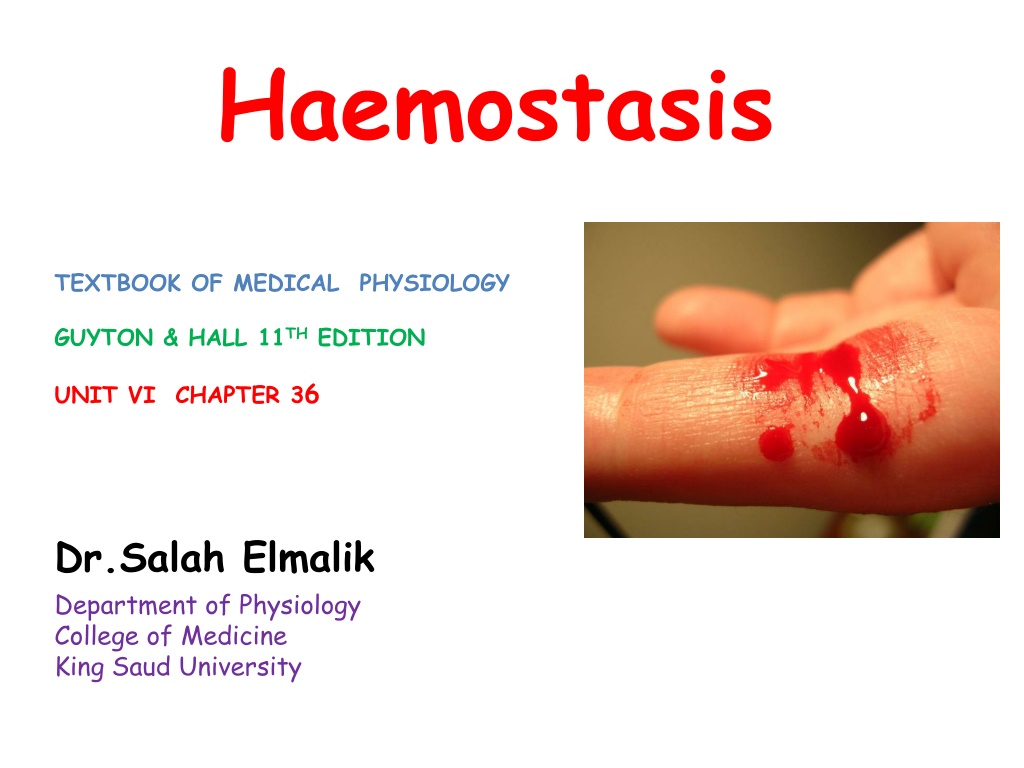
Haemostasis TEXTBOOK OF MEDICAL PHYSIOLOGY GUYTON & HALL 11THEDITION UNIT VI CHAPTER 36 Dr.Salah Elmalik Department of Physiology College of Medicine King Saud University
Haemostasis or Hemostasis NOT Homeostasis The ability to maintain a constant internal environment in response to environmental changes
Objectives At the end of this lecture student should be able to: 1. Describe the formation and development of platelets 2. Recognize different mechanisms of hemostasis 3. Describe the role of platelets in hemostasis. 4. Recognize different clotting factors 5. Describe the cascades of intrinsic and extrinsic pathways for clotting. 6. Recognize process of fibrinolysis and function of plasmin
Megakayocyte and platelets formation
Platelets cont. Site of formation: (Bone marrow) Myeloid stem cell Megakaryoblast Megakaryocyte Platelets
Platelets (Thrombocytes) They are fragments of megakaryocytes formed in the bone marrow. Their production (thrombopoiesis) is regulated by Thrombopoietin, a hormone released from the liver
Platelets - cont Are round/oval disc with diameter about 2-3 m Coated by a glycoprotein layer which prevents their sticking to normal endothelial cells Platelet count = 250,000-500,000/ mm3 life span 8-12 days Active cells contain contractile protein such as actin, myosin, and thrombosthenin Contain high calcium content & rich in ATP
Hemostasis: prevention or stoppage of blood loss. Hemostatic Mechanisms: 1. Vessel wall (Vasoconstriction) 2. Platelets (Production and activation, Platelets Plug formation) 3. Blood coagulation Clot formation (intrinsic & extrinsic pathways) 4. Fibrinolysis
Lumen of blood vessel Platelets Intact endothelium
Memostatic Mechanisms Vessel wall Immediately After injury a localized Vasoconstriction of smooth muscles Mechanism -Humoral factors: local release of thromboxane A2 & serotonin (5HT) from platelets Systemic release of adrenaline - Nervous reflexes (pain nerve impulses)
Platelet Functions Begins with Platelet activation
Platelet Activation Adhesion Shape change Aggregation Release Clot Retraction
Platelet Adhesion Platelets stick to the exposed collagen underlying damaged endothelial cells in vessel wall
Platelet shape change and Aggregation Resting platelet Activated platelet activated platelets
Platelet Aggregation Activated platelets stick together and activate new platelets to form a mass called a platelet plug Plug reinforced by fibrin threads formed during clotting process
Platelet Release Reaction Platelets activated by adhesion Extend projections to make contact with each other Release thromboxane A2, serotonin & ADP activating other platelets Serotonin & thromboxane A2 are vasoconstrictors decreasing blood flow through the injured vessel. ADP causes stickiness
Platelet Plug Aggregation of platelets at the site of injury to stop bleeding Exposed collagen attracts platelets Activated platelets release ADP & Thromboxane A2 (TXA2) the stickiness of platelets Platelets aggregation plugging of the cut vessel Intact endothelium secretes prostacyclin inhibition of aggregation
Activated Platelets Secrete: 1. 5HT vasoconstriction 2. Platelet phospholipid Factor (PF3) clot formation 3. Thromboxane A2 (TXA2) is a prostaglandin formed from arachidonic acid Function: Vasoconstriction Platelet aggregation (TXA2 inhibited by aspirin)
Coagulation: Formation of fibrin meshwork (Threads) to form a CLOT
Clotting Factors Factors Names Fibrinogen Prothrombin Thromboplastin (tissue factor) Calcium Labile factor Stable factor Antihemophilic factor Antihemophilic factor B Stuart-Prower factor Plasma thromboplastin antecedent (PTA) Hageman factor Fibrin stablizing factors I II III IV V VII VIII IX X XI XII XIII
The Coagulation Cascades Intrinsic pathway Extrinsic pathway Activators: Collagen and damaged endothelium Common pathway Activator: Tissue factor (III) (Thromboplastin) XII XII activated XX activated V, Ca, Phospholipids VII VII activated XI XI activated Prothrombin activator Ca III, Ca, Phospholipids IX IX activated Prothrombin Thrombin IX activated, VIII Ca, Phospholipids FibrinogenFibrin XIII Ca Insoluble fibrin
Blood coagulation (clot formation) A series of biochemical reactions leading to the formation of a blood clot within few seconds after injury Prothrombin (inactive thrombin) is activated by a long intrinsic or short extrinsic pathways This reaction leads to the activation of thrombin enzyme from inactive form prothrombin Thrombin will change fibrinogen (plasma protein) into fibrin (insoluble protein) 25
Intrinsic pathway The trigger is the activation of factor XII by contact with foreign surface, injured blood vessel, and glass. Activated factor XII will activate factor XI Activated factor Xl will activate IX Activated factor IX + factor VIII + platelet phospholipid factor (PF3)+ Ca activate factor X Following this step the pathway is common for both intrinsic and extrinsic 26
Extrinsic pathway Triggered by material released from damaged tissues (tissue thromboplastin) Tissue thromboplastin + VII + Ca activate X Common pathway Activated factor X + factor V +PF3 + Ca activate prothrombin activator; a proteolytic enzyme which activates prothrombin. Activated prothrombin activates thrombin Thrombin acts on fibrinogen and change it into insoluble thread like fibrin. Factor XIII + Calcium strong fibrin (strong clot) 27
Activation of Blood Coagulation Intrinsic Pathway: all clotting factors present in the blood Extrinsic Pathway: triggered by tissue factor (thromboplastin) Common Pathway
Thrombin Thrombin changes fibrinogen to fibrin Thrombin is essential in platelet morphological changes to form primary plug Thrombin stimulates platelets to release ADP & thromboxane A2; both stimulate further platelets aggregation Activates factor V 29
Fibrinolysis Formed blood clot can either become fibrous or dissolved. Fibrinolysis (dissolving) = Break down of fibrin by naturally occurring enzyme plasmin therefore prevent intravascular blocking. There is a balance between clotting and fibrinolysis Excess clotting blocking of Blood Vessels Excess fibrinolysis bleeding tendency for 30
Released from injured tissues and vascular endothelium Fibrinolysis Tissue Plasminogen Activator (t-PA) Plasminogen Plasmin (Protein in the blood) Anti-activators Fibrinogen Fibrin Thrombin FDP* FDP*: Fibrin Degradation Products
Plasmin Plasmin is present in the blood in an inactive form plasminogen Plasmin is activated by tissue plasminogen activators (t-PA) in blood. Plasmin digests intra & extra vascular deposit of Fibrin fibrin degradation products (FDP) Unwanted effect of plasmin is the digestion of clotting factors 32
Plasmin Plasmin is controlled by: Tissue Plasminogen Activator Inhibitor (TPAI) Antiplasmin from the liver Uses: Tissue Plasminogen Activator (TPA) used to activate plasminogen to dissolve coronary clots 33