Understanding Coagulation Profile in Hemostasis
Coagulation profile testing is crucial in assessing blood clotting ability. The process of coagulation is essential for hemostasis to prevent bleeding or clotting disorders. This summary explores the steps involved in hemostasis, the clotting cascade, and the triggers for the extrinsic and intrinsic pathways in blood coagulation.
Download Presentation
Please find below an Image/Link to download the presentation.
The content on the website is provided AS IS for your information and personal use only. It may not be sold, licensed, or shared on other websites without obtaining consent from the author. Download presentation by click this link. If you encounter any issues during the download, it is possible that the publisher has removed the file from their server.

Presentation Transcript
Lab# 8 BCH 471 Coagulation Profile
Objectives: 1-To estimate Clotting time, Bleeding time, and Prothrombin time .
The term "coagulation profile, refers to a set of laboratory tests that are used to assess the clotting ability of a person's blood. It provides information about the various factors involved in the coagulation cascade, which is a complex series of chemical reactions that lead to the formation of blood clots.
Coagulation: Coagulation, is a complex process by which blood forms clots. It is an important part of hemostasis, WHICH IS the physiological process that the body employs to stop bleeding and maintain blood within a damaged blood vessel. Disorders of coagulation can lead to an increased risk of bleeding (hemorrhage) or clotting (thrombosis). NOTE: Hemostasis combines the terms hemo (meaning blood ) and stasis (meaning standing still ).
Hemostasis is maintained in the body via three steps 1- Vascular spasm, Damaged blood vessels constrict. 2- Platelet plug formation, Platelets adhere to damaged endothelium to form platelet plug (primary hemostasis) 3- Blood Coagulation, Clots form upon the conversion of fibrinogen to Fibrin (secondary hemostasis).
Clotting Cascade A cascade is a mechanism in which enzymes activate other enzymes sequentially usually leading to an amplification of an initial signal. Pathways Initially independent, then they converge on common pathway leading to the formation of a fibrin clot Extrinsic Intrinsic Each of these pathways leads to the conversion of factor X (inactive) to factor Xa (active)
What triggers extrinsic and intrinsic pathways: Extrinsic Release of biochemicals from broken blood vessels/damaged tissue. Intrinsic No tissue damage, but blood contacts damaged endothelial layer of blood vessel walls.
Clotting time - capillary method Simple test but takes time and rarely done now. Method: Venous blood is taken and placed on glass test tube at 37 C and it observed at time intervals until clotting occurs. Normal blood takes 5-10min to clot. Longer periods Coagulation defects (e.g. Hemophilia, disorder characterized by a deficiency or dysfunction of specific clotting factors.)
Clotting time - capillary method https://www.youtube.com/watch?v=TSlHO CPrKIo
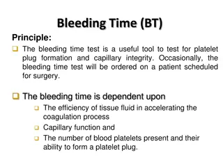
Bleeding Time: Provides assessment of platelet count and function. Method: It is determined by noting time at which blood coming out a small cut, no longer forms a spot on a piece of filter paper placed in contact with cut surface. The normal range from 2-4 min https://www.youtube.com/watch?v= pbQMDlPG0kY
Prothrombin Time (PT) - Test measures how long it takes for a clot to form in a blood sample. - Prothrombin is a protein, which is clotting (coagulation) factors. How fast the blood clots, depends on the amount of clotting factors in your blood. and whether they're working correctly. Method: An excess of tissue factor and Ca2+ ions are added to diluted plasma containing citrate (anticoagulant) and then the time taken for the mixture to clot is measured Normal value 10-15 secs High PT low levels of thrombin. And vice versa. Results from liver disease due to deficiency of prothrombin, fibrinogen, V, VII and X factors
(which contain clotting factors) The image source: https://www.youtube.com/watch?v=q548IZGbt28


")