Understanding Pathological Calcification in Veterinary Pathology
Pathological calcification is the abnormal deposition of calcium salts in soft tissues, with two forms - dystrophic and metastatic calcification. Dystrophic calcification occurs in dying tissues, while metastatic calcification reflects a derangement in calcium metabolism. Dystrophic calcification can be seen in various tissues like necrotic tissue, caseous lymphadenitis, and heart valves, while metastatic calcification is a generalized phenomenon due to elevated extracellular calcium levels.
- Pathological Calcification
- Veterinary Pathology
- Dystrophic Calcification
- Metastatic Calcification
- Calcium Metabolism

Download Presentation

Please find below an Image/Link to download the presentation.
The content on the website is provided AS IS for your information and personal use only. It may not be sold, licensed, or shared on other websites without obtaining consent from the author. Download presentation by click this link. If you encounter any issues during the download, it is possible that the publisher has removed the file from their server.
E N D
Presentation Transcript
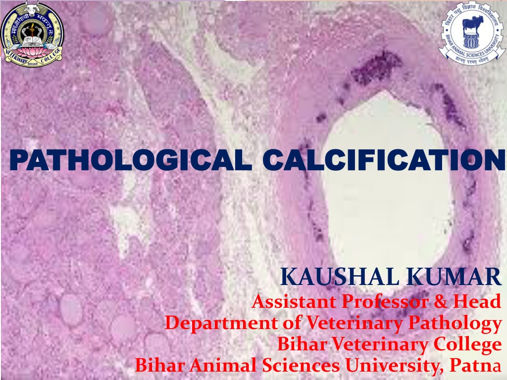
PATHOLOGICAL CALCIFICATION PATHOLOGICAL CALCIFICATION KAUSHAL KUMAR Assistant Professor & Head Department of Veterinary Pathology Bihar Veterinary College Bihar Animal Sciences University, Patna
Pathological Calcification It is a lesion in which calcium salts, usually in the form of calcium phosphate, are deposited abnormally in soft tissues. There are two forms: A. Dystrophic Calcification B. Metastatic Calcification
Types of Pathological Calcification DYSTROPHIC calcification A localized lesion occurs in dying and dead tissues With NORMAL serum calcium level & Ca Metabolism METASTATIC calcification Deposition of Ca salts in NORMAL tissues Always reflects some derangement in Ca Matabolism & hypercalcemia
Types of Pathological Calcification DYSTROPHIC calcification A localized lesion occurs in dying and dead tissues With NORMAL serum calcium level & Ca Metabolism METASTATIC calcification Deposition of Ca salts in NORMAL tissues Always reflects some derangement in Ca Matabolism & hypercalcemia
Dystrophic Calcification Injured and dying cells are not able to maintain normal calcium homeostasis due to increased intracellular calcium levels. Once calcification is initiated deposition of calcium continues. Can occur with any type of tissue necrosis Grossly visible calcification include heart and skeletal muscle and granulomas. Lymph node from a goat with caseous lymphadenitis. The light zones in this photo are areas of dystrophic calcification.
Sites of Dystrophic Calcification Necrotic Tissue Caseous necrosis TB, Caseous Lymphadenitis Fat necrosis Liqifactive necrosis (less Common) Haematoma in vicinity of bone Thrombus-phlabolith Infarcts Cyst-hydatid, cysticercosis Heart valves (Subacute Infective Endocarditis) Tumour-breast cancer- tumoral calcinosis.
Metastatic Calcification When the homeostatic capacity of cells and tissues exceeds Elevated extracellular levels of calcium (hypercalcemia) Typically a generalized phenomenon
Metastatic Calcification Hypercalcemia sufficient to cause metastatic calcification mainly occurs in three situations Hyperparathyroidism Hypervitaminosis D Diseases with extensive destruction of bone.
Site of Metastatic Calcification But there are specific tissues that are prone to become mineralized / calcification Kidneys (nephrocalcinosis) Lungs (alveolar walls) Stomach (acid secreting fundal glands) Blood vessels (internal elastic lamina) Cornea Synovium
Differences Dystrophic Calcification Features Metastatic Calcification Definition Deposition of calcium in dead and degenerated tissue Deposition of calcium salts in normal tissue Calcium Metabolism Normal Deranged Serum Calcium Level Normal Hypercalcemia Reversibility Generally Irreversible Reversible upon correction of metabolic disorder Causes Necrosis, infarcts, thrombi, cysts, tumours, old scars & haematoma e.t.c. Hyperparathyroidism Hypervitaminosis, Prlonged immobilisation Pathogenesis Increased binding of phosphates with necrotic and degenerative tissue; which in turn bind to calcium to form Calcium Phosphate precipitate. Increased precipitates of Cal. phosphate due to hypercalcaemia in lungs, stomach, blood vessels, cornea
thanksthanka Thanks