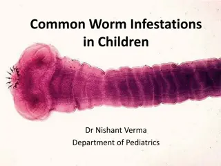
Overview of Ancylostoma duodenale (Hookworm) Infection
Ancylostoma duodenale, a parasitic worm causing Ancylostomiasis, is discussed in this lecture. The infection's distribution, morphology, life cycle, and pathology are detailed, emphasizing the impact on humans. The lifecycle stages, including egg production, larval development, and entry into the human body, are explained with accompanying images. Pathological effects, such as skin lesions and inflammation, due to this hookworm infection are highlighted.

Download Presentation

Please find below an Image/Link to download the presentation.
The content on the website is provided AS IS for your information and personal use only. It may not be sold, licensed, or shared on other websites without obtaining consent from the author. Download presentation by click this link. If you encounter any issues during the download, it is possible that the publisher has removed the file from their server.
E N D
Presentation Transcript
Medical Parasitology Assistant Prof. Dr. Ahmed A. Mohammed Lecture 7 Helminths
Phylum:Nematoda (or Nemathelminths) Class : Phasmidia ex: Ancylostoma duodenale (Hook worm). This Ancylostomiasis and also (old world hookworm). The infection has a world-wide distribution. parasite (worm) causes a disease called The adult worms are found in the small intestine of man, hanging on the mucosa, feeding on the blood and tissue fluids. The living adult worms are pinkish in color, their body is narrow anteriorly with curved head dorsally.
The adult worms are cylindrical, the male copulatory bursa is considerably broader than it is long and is supported by ray. The buccal cavity is wide and ovoid in shape; the cup- shaped mouth capsule is heavily reinforced and provided with many pairs of teeth. Anterior end Posterior end Egg (mouth) (Lateral view right side)
Life cycle: Each female worm lays up to 25.000-30.000 eggs daily. The broadly ovoid eggs have a thin, transparent shell, and are in 2-8 cell stage of cleavage when evacuated. Embryonation to the first rhabditoid larval stage takes place in 24-48hrs on moist sandy loam in a shaded environment at an optimal temperature of about 25 C. The rhabditoid larva is molting to produce the strongyliform larva which is feeding on the bacteria and other substances found in the feces. It grows rapidly, passing through two moltings and become filariformlarva (the infective larva). The filariform larva migrates to the superficial layer of the soil, waiting a chance to be in contact with human skin to penetrate it and reach to the blood or lymph, migrate to the
heart, lungs, pharynx and intestine attached to the villi, then molt once more and attach to the mucosa and growing to adult male and female worms in 5 weeks after their entrance to the body. The infection through foods and water contaminated with eggs is also possible.
Pathology The pathogenicity depends on the number of worms, the infective stage or phase and the feeding of the infected person, as in the following: 1. Cutaneous phase (the skin lesion): At the site of entry on the feet, arms or other surface areas the larvae usually produce a popular eruption. This phase starts soon after the larval penetration to the skin, and causes mechanical injury in the skin layers because it slip through the spaces of the skin or enter the hair follicles which may cause the entry of the pathogenicbacteria with it and leads to inflammation with itch called ground itch, this phase short if the secondary infection with bacteria absent.
2. Pulmonary phase (larval migration through the lungs): It occurs when the juveniles exit from the blood vessels to the follicles then to the trachea and throat. This is lead to bleeding and its dangerous depends on the number of the juveniles. This is occurring in the acute infections and may lead to death. This phase always asymptomatic except the acute infections which are accompanied by dry coughing, sore throat and pneumonitis.
3. Intestinal phase: It is the important phase. When the juveniles reach to the intestine, they stick themselves in the mucosa through the buccal cavity and the teeth, starts with feeding on the blood. This phase accompanied by nausea, slight abdominal pain, anorexia and sometimes diarrhea. But the main symptom is the anemia which results from the continuous blood sucking. The continuity of blood lose leads to lose most the body iron and large amounts of protein which leads to erythrocytopenia. The R.B.Cs. become smallest and the haemoglobin less than that of normal R.B.Cs. The worm sucking about 0.5cm3 of blood daily and about 200cm3 daily in the acute infection.
Oedema around the eye and the lower limbs may be associated. There is also eosinophilia with Charcot-Leyden crystals in the feces appear during the late prepatent period. The infection is very dangerous during pregnancy because the infant requirement for protein and iron will increase. Symptomatology Nausea, headache and irritating cough (during lung migration of larvae). In the middle to late prepatent and early patent periods (29-38 days), there are frequently sever colicky pains, flatulence, diarrhea, loss of weight, dyspnea, dizziness and marked pallor. During this period there is an eosinophilic leukocytosis that may persist at a significantly high level for several months.
In chronic hookworm disease, the signs and symptoms are essentially those of iron-deficiency anemia. Haemoglobin levels may be reduced to 5g/dl or lower, and the heart is greatly enlarged. In children, mental and physical development may be markedly retarded. In addition, local skin manifestations "ground itch" can occur during the penetration of the filariform (L3) larvae. Diagnosis It is based on the detection of eggs in the feces. Examination of the eggs cannot distinguish between Necator americanus and A. duodenale.
Treatment In light to moderate hookworm infections in which the anemia is not severe, specific treatment can usually be undertaken without a preliminary period of supportive treatment. For individuals with low haemoglobin levels, it is desirable to prescribe a diet rich in animal proteins for a week to 10 days before the specific chemotherapy. Iron must also be administered to replace that which is lost during the intestinal haemorrhage caused by the worms. Rarely, a whole blood transfusion may be needed. The most effective drug without significant side effects is probably Mebendazole. Pyrantel pamoate is also effective. One or two weeks after treatment, a follow-up stool examination should be made. If necessary, retreatment may be undertaken in a week to remove the remaining worms.

")


:")