Understanding Neck Anatomy: Triangles, Glands, and Lymph Nodes
Explore the intricate anatomy of the neck, including the various triangles, thyroid and parotid gland structures, as well as cervical lymph nodes. Delve into the essentials of neck mass evaluation, differential diagnosis, and management strategies. Enhance your knowledge of head and neck surgery through detailed visuals and comprehensive information provided by Dr. Manal Bin Manie, Assistant Professor at King Saud University.

Download Presentation

Please find below an Image/Link to download the presentation.
The content on the website is provided AS IS for your information and personal use only. It may not be sold, licensed, or shared on other websites without obtaining consent from the author. Download presentation by click this link. If you encounter any issues during the download, it is possible that the publisher has removed the file from their server.
E N D
Presentation Transcript
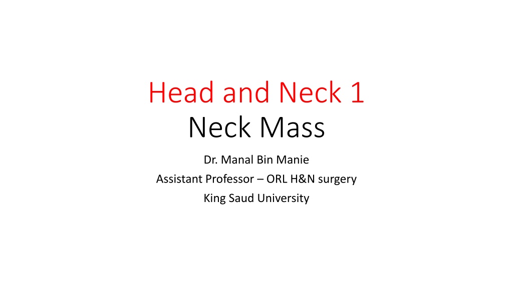
Head and Neck 1 Neck Mass Dr. Manal Bin Manie Assistant Professor ORL H&N surgery King Saud University
Objectives Anatomy of the neck History / physical examination Pathology / differential diagnosis Management/ investigation and treatment
Anterior triangle Posterior triangle
Thyroid gland anatomy Butterfly-shaped gland Isthmus :overlying 2ndto 4th tracheal rings Parathyroids
Parotid gland Lies over the angle of mandible Superficial and deep lobe Facial nerve Stenson s duct : opens in the mouth opposite to maxillary second molar
Submandibular gland Lies inferior and posterior to the mandible Mylohyoid muscle runs through the lobules of the gland and section it into superficial and deep parts Wharton s duct opens at the lingual papilla
Approach to neck mass History Physical exam Age, gender, ethnicity Duration of neck mass Progression Associated symptoms :URTI Voice change Hx of cough, fever, sore throat, night sweats,weight loss Recent travel Insect bite Dental problems Tobacco and alcohol use Exposure to Radiation Family history of malignancy Full head and neck examination including the cranial nerves and nasopharyngolaryngoscopy Location of the mass: midline , lateral Size, consistency, tenderness, mobility Pulsation Skin changes Movement with swallowing or tongue protrusion
Differential diagnosis Pediatric Infective and inflammatory masses Congenital masses Vascular masses Traumatic masses Metabolic, idiopathic and autoimmune conditions Adult Infective and inflammatory masses Neoplastic masses Vascular masses Traumatic masses Metabolic, idiopathic and autoimmune conditions Thyroid gland masses Salivary gland masses Parapharyngeal masses
General approach to a neck mass Patient with a neck mass Diagnosis suggested by history and physical exam Congenital mass Infectious/ inflammatory Neoplastic Course of broad spectrum antibiotics with close FU in 2-4 weeks FNA+/- CT scan CT scan +/- Endoscopy Excisional biopsy Further management based on stage and histology
Cervical lymphadenitis: Most common in children and adolescents Etiology : Bacterial streptococcal and staphylococcal infections , mycobacterial infections, secondary to dental or tonsillitis and rarely cat-scratch disease and actinomyces Viral- EBV, CMV ,herpes simplex virus, others Parasitic toxoplasmosis Fungal- coccidiomycosis Sialadenitis Diagnosis: CBC, CT scan if needed Treatment: antibiotics
Cervical lymphadenitis: Rule out Neck abscess formation if no improvement with antibiotics CT neck with contrast Incision and drainage
Tuberculous cervical lymphadenitis: Scrofula Most common manifestation of extrapulmonary TB Non tender If untreated , spontaneous discharge and sinus formation CT scan may show necrotic/cystic nodal matting FNA/ excisional biopsy Treatment: antimycobacterial medications
Mumps ( viral parotitis): Viral infection caused by paramyxovirus Droplet infection and fomites Children are more affected Fever, malaise, parotid swelling Orchitis , ophritis, aseptic meningitis , unilateral SNHL Treatment is supportive , hydration and analgesics
Acute suppurative parotitis/sialadenitis : Commonly seen in elderly , diabetic , debilitated and dehydrated patients Staph aureus is the usual causative organism Fever, swelling , pus from stenson s duct Antibiotics and hydration
Neoplastic masses Benign : Lipoma , fibroma, neuroma and schwannoma Malignant: Primary neck tumors sarcoma, salivary gland tumors, thyroid gland tumors, parathyroid gland tumors Lymphoma Metastasis
Thyroid gland nodules: benign thyroid nodules are very common 5-10 % are malignant Hot vs cold
Thyroid Nodule - Evaluation U/S FNA Thyroid Function Tests CT
Thyroid gland nodules: Risk factors for malignancy: History : External radiation during childhood Age <20 or >60 years Male gender Family history of thyroid cancer Hoarseness, dysphagia Rapid growth Physical exam : Firm or hard Fixed to soft tissue Lymphadenopathy
Ultrasound characteristics of thyroid nodules that increase suspicious of malignancy : Taller than wide shape Speculated margin Microcalcifications Marked hypo echogenicity Increased vascularity
Fine needle aspiration FNA: Safe and minimally invasive Indicated for nodules > 1 cm or nodules with suspicious features of malignancy US guided FNA
CT scan indications: Recurrent disease Lymph node metastasis Vocal cord paralysis Fixation of tumor to adjacent structures or skin Huge goiter , retrosternal extension
Malignant Thyroid Lesions 1. Well Differentiated (85%) Papillary Thyroid Carcinoma (PTC) Follicular Thyroid Carcinoma (FTC) Hurthle Cell Carcinoma (HCC) 2. Poor differentiated malignant neoplasms - Medullary thyroid carcinoma (MTC) - Anaplastic thyroid carcinoma (ATC) 3. Other malignant tumors: - Lymphoma - Metastatic tumors
Treatment : Observation FU Thyroidectomy : Malignancy or Suspicious for malignancy Compression symptoms Cosmetic Graves disease Toxic nodule
Post op complications : RLN Injury Hypocalcemia Hematoma
Salivary gland tumors: Pleomorphic adenoma is the most common benign tumor of salivary gland It can arise from parotid, submandibular or minor salivary glands Slow growing , usually seen in the third or fourth decade , with propensity to females Encapsulated, Pseudopods
Salivary gland tumors: Mucoepidermoid carcinoma is the most common malignant tumor of salivary gland Can invade the facial nerve Slow growing Low grade and high grade Surgical excision
Thyroglossal duct cyst: Cystic midline swelling Affecting young children but can occur at any age Increases in size with URTI +_ sinus
Thyroglossal duct cyst: Moves with tongue protrusion because of its attachment to foramen cecum It may contain the only functioning thyroid tissue Rarely malignant <1% Investigations :Ultrasound
Thyroglossal duct cyst: Treatment : Surgical excision including the body of hyoid bone and core of tongue tissue to prevent recurrence ( Sistrunk s procedure)
Dermoid/epidermoid cyst : Cystic mass resulting from congenital epithelial inclusion or rest Epidermoid : epithelial elements only, fluid content Dermoid : epithelial elements plus dermal substructure ( hair, sebaceous glands) Typically seen in the midline of the neck , usually in the submental region Treatment is complete surgical excision
Branchial cyst : Common in the second decade of life Swelling in the upper part of the neck anterior to SCM Anomalies of second branchial arch are the most common May be associated with a sinus or a fistula
Branchial cyst : A second arch branchial sinus has an external opening at the junction of the lower and middle third of the anterior border of SCM and may excrete mucoid discharge , it may have internal opening in the tonsillar fossa Treatment is surgical excision along with its tract , if present
Cystic hygroma : It occurs most commonly in the posterior triangle of the neck It arises from obstruction or sequestration of jugular lymph sac Seen in neonate, early infancy or childhood May cause difficulty in labor Soft, cystic and partially compressible Treatment is surgical excision


:")