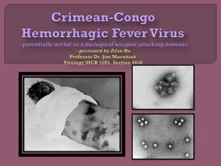
Virus-Cell Interactions and Infections

 
Page
  
|
 
1
 
cellular
 
tropism
 
of
 
viruses
 
is
 
determined
 
by the
  
presence
  
of
  
appropriate
  
cellular
receptors
  
and
  
frequently,
  
cell-type
  
specific
  
transmission factor.
 
Viruses
 
typically
 
encode
genes
 
that
 
modulate
 
host-cell
 
function
 
for
 
their
 
own
 
benefit
 
and of
  
course
  
the
  
host
  
has
elaborate
  
innate
  
defense
  
to
  
restrict
  
viral
  
functions.
  
Thus
  
the
  
viral
  
and cellular
  
factor
that
  
influence
  
the
  
outcome
  
of
  
infection
 
are
  
often
  
in
 
delicate
  
balance
  
and
  
easily
shifted
 
one
 
way
 
or
 
the
 
other.
 
Virus
  
infection
  
causes
  
a
  
wide
  
variety
 
of
  
potentiality
 
victorious
  
changes
  
in
  
the
  
many
different kinds
 
of
 
cells
 
that
 
occur
 
in
 
the
 
animal
 
host.
 
The
 
disruption
 
of
 
cellular
 
function,
 
the
induction
 
of cell
  
death
  
or
  
transformation
  
or
  
the
  
activation
  
of
  
an
  
inappropriate
immune
  
response
  
are
  
all potentiality
 
manifested
  
as
 
disease
 
by
 
the
  
infected
  
host.
 Many
 
virus-cell
 
interactions
 
are
 
transient
 
and
 
asynchronous,
 
involving
 
intermediate
 
steps
on
 
the pathway,
  
each
 
featuring
 
dynamic
  
interactions
 
between
 
different
  
viral
  
and
  
cellular
components.
 
V
I
R
U
S
-
C
E
L
L
 
I
N
T
E
R
A
C
T
I
O
N
 
 
Page
  
|
 
2
T
Y
P
E
S
 
O
F
 
V
I
R
U
S
-
C
E
L
L
 
I
N
T
E
R
A
C
T
I
O
N
Viral
 
infection
 
may
 
be
 
Cytocidal
 
or
 
non
 
cytocydal
 
and
 
productive
 
or
 
non-productive
 
that
is,
 
not all
 
infection
 
leads
 
to
 
cell
 
death
 
or
 
the
 
production
 
and
 
release
 
of
 
new
 
versions.
However
 
criticalchanges
 
can
 
occur
 
in
 
virus
 
infected
 
cells
 
regardless
 
of
 
whether
 
the
infection
 
is
 
productive
 
or
 
not.
Some
 
of
 
the
 
most
 
important
 
of
 
all
 
nonproductive
 
virus
 
cell
 
interactions
 
are
 
those
associated
 
with 
persistent
  
infections
  
or
  
latent
  
infections.
  
The
  
term
  
persistent
infection
  
simply
  
describes
  
an infection
 
that
 
lasts
 
a
 
long
 
time.
 
The
 
term
 
latent
 
infection
describes
 
an
 
infection
 
that
 
"exists
 
but
 
is not
 
exhibited,"
 
i.e.,
 
an
 
infection
 
in
 
which
infectious
 
virions
 
is
 
not
 
formed.
 
In
 
either
 
case,
 
the
 
virus or
 
its
 
genome
 
is
 
maintained
indefinitely
 
in
 
the
 
cell,
 
either
 
by
 
the
 
integration
 
of
 
the
 
viral
 
nucleic acid
 
into
 
the
 
host
 
cell
DNA
 
or
 
by
 
carriage
 
of
 
the
 
viral
 
nucleic
 
acid
 
in
 
the
 
form
 
of
 
an
 
episome.
 
In these
  
instances,
the
  
cell
  
survives,
  
indeed
  
may
 
divide
  
repeatedly.
  
In
  
some
  
instances
  
such
  
cells
never
  
release
  
virions,
  
in
  
others
  
the
  
infection
  
may
  
become
  
productive
  
when
induced
  
by
  
an appropriate
   
stimulus.
    
Persistent
    
or
    
latent
    
infections
    
may
   
also
be
    
associated
    
with
   
cell 
transformation.
  
The
  
various
  
types
  
of
  
interaction
  
that
  
can
occur
  
between
  
virus
  
and
  
cell
  
are summarized.
 
FIGURE:
  
VIRUS-CELLINTERACTION
 
Page
  
|
 
3
 
T
y
p
e
s
 
o
f
 
V
i
r
u
s
-
C
e
l
l
 
I
n
t
e
r
a
c
t
i
o
n
 
Page
  
|
 
4
 
 
Figure:
 
Unstained
 
confluent
 
monolayers
 
of
 
the
 
three
 
main
 
types
 
of
 
cultured
 
cells
 
as
 
they
 
appear
 
by
 
low
 
power
 
light
 
microscopy
 
through
 
the
 
wall
 
of
 
the
 
glass
 
or
 
plastic
 
flask
 
in
 
which
 
they
 
form
 
a
 
monolayer.
 
(A)
 
Primary
 
monkey
 
kidney
 
epithelial
 
cells
 
obtained
 
directly
 
by
 
the
 
dissociation
 
of
 
cells
 
from
 
a
 
kidney;
 
this
 
produces
 
a
 
mixed
 
population
 
of
 
mainly
 
epithelial
 
cells.
  
(B)
  
Diploid
 
cell
 
line
  
of
 
fetal
 
fibroblasts.
  
(C)
 
Continuous
 
line
 
of
 
malignant
 
epithelial
 
cells.


C
Y
T
O
C
I
D
A
L
 
C
H
A
N
G
E
S
 
I
N
 
V
I
R
U
S
-
I
N
F
E
C
T
E
D
 
C
E
L
L
S


Cytopathic
  
viruses
 
kill
 
the
 
cells
 
in
 
which
 
they
 
replicate.
 
When
 
a
 
monolayer
 
of
 
cultured
cells
 
is inoculated
 
with
 
a
 
cytopathic
 
virus,
 
the
 
first
 
round
 
of
 
infection
 
yields
 
progeny
virus
 
that
 
spreads through
 
the
 
medium
 
to
 
infect
 
adjacent
 
as
 
well
 
as
 
distant
 
cells
eventually
 
all
 
cells
 
in
 
the
 
culture may
  
become
  
infected.
   
The
  
resulting
  
cell
  
damage
is
  
known
  
as
  
a
   
cytopathic
  
effect
  
(CPE).
Cytopathic
  
effect
  
can
  
usually
  
be
  
observed
  
by
  
low-power
  
light
  
microscopy
  
of
unstained
  
cell cultures.
 
 
M
e
c
h
a
n
i
s
m
s
 
o
f
 
C
e
l
l
 
D
a
m
a
g
e
So
 
many
 
pathophysiologic
 
changes
 
occur
 
in
 
cells
 
infected
 
with
 
cytopathic
 
viruses
 
that
the
 
death of
 
the
 
cell
 
usually
 
cannot
 
be
 
attributed
 
to
 
any
 
particular
 
event;
 
rather,
 
cell
death
 
may
 
be
 
the
 
final result
  
of
  
the
  
cumulative
  
action
  
of
  
many
  
insults.
 
I
n
h
i
b
i
t
i
o
n
 
o
f
 
H
o
s
t
 
C
e
l
l
 
N
u
c
l
e
i
c
 
A
c
i
d
 
S
y
n
t
h
e
s
i
s
 
Inhibition
  
of
   
host
   
cell
  
DNA
   
synthesis
   
is
   
common
   
in
   
viral
   
infections.
   
It
is
   
an
   
inevitable consequence
 
of
 
viral
 
inhibition
 
of
 
host
 
cell
 
protein
 
synthesis
 
and
its
 
effect
 
on
 
the
 
machinery
 
of DNA
 
replication,
 
but
 
some
 
viruses
 
employ
 
more
specific
 
mechanisms.
 
For
 
example,
 
poxviruses produce
  
a
  
DNAse
  
that
  
degrades
cellular
  
DNA,
  
and
  
herpes
  
viruses
  
specifically
  
displace
  
the synthesis
 
of
 
host
 
cell
DNA
 
with
 
their
 
own
 
synthetic
 
processes.


I
n
h
i
b
i
t
i
o
n
 
o
f
 
H
o
s
t
 
C
e
l
l
 
R
N
A
 
T
r
a
n
s
c
r
i
p
t
i
o
n
 
Many different classes of viruses including poxviruses, rhabdoviruses, reoviruses,
paramyxoviruses,
 
and
 
picornaviruses,
 
inhibit
 
host
 
cell
 
RNA
 
transcription.
 
In
some
 
instances,
 
this inhibition
 
may
 
be
 
the
 
indirect
 
consequence
 
of
 
viral
 
effects
on
 
host
 
cell
 
protein
 
synthesis,
 
which decreases
 
the
 
availability
 
of
 
transcription
factors
 
required
 
for
 
RNA
 
polymerase
 
activity.
 
In
 
other instances,
 
viruses
 
encode
specific
 
transcription
 
factors
 
for
 
the
 
purpose
 
of
 
regulating
 
the
 
expression
of
 
their
 
own
 
genes
 
and,
 
in
 
some
 
instances;
 
these
 
factors
 
modulate
 
the
 
expression
of
 
cellular
 
genes as
  
well.
  
For
  
example,
  
herpesviruses
  
encode
  
proteins
  
that
bind
  
directly
  
to
  
specific
  
viral
  
DNA sequences,
 
thereby
 
regulating
 
the
transcription
 
of
 
viral
 
genes.
 
Figure:
 
Cytopathic
 
effects
 
produced
 
by
 
different
 
viruses.
 
The
 
cell
 
monolayers
 
are
 
shown
 
as
 
they
 
would
normally
  
be
  
viewed
  
in
  
the
  
laboratory,
  
unfixed
  
and
  
unstained.
  
(A)
  
Typical
  
cytopathology
  
of
  
an
enterovirus:
 
rapid
 
rounding
 
of
 
cells,
 
progressing
 
to
 
complete
 
cell
 
lysis.
 
(B)
 
Typical
 
cytopathology
 
of
 
a
herpesvirus:
 
focal
 
areas
 
of
 
swollen
 
rounded
 
cells.
 
(C)
 
Typical
 
cytopathology
 
of
 
a
 
paramyxovirus:
 
focal
clusters
 
of
 
cells
 
are
 
fused
 
to
 
form
 
syncytia.
 
(D)
 
Hemadsorption:
 
erythrocytes
 
adsorb
 
to
 
infected
 
cells
that
 
have
 
incorporated
 
hemagglutinin
 
into
 
the
 
plasma
 
membrane
.
 
I
n
h
i
b
i
t
i
o
n
 
o
f
 
P
r
o
c
e
s
s
i
n
g
 
o
f
 
H
o
s
t
 
C
e
l
l
 
m
R
N
A
s
 
Many
   
viruses,
   
including
   
vesicular
   
stomatitis
   
viruses,
   
influenza
   
viruses,
   
and
herpesviruses, interfere
 
with
 
the
 
splicing
 
of
 
cellular
 
primary
 
mRNA
 
transcripts
 
that
 
are
needed
 
to
 
form
 
mature mRNAs.
 
In
 
some
 
instances,
 
spliceosomes
 
are
 
formed,
 
but
subsequent
 
catalytic
 
steps
 
are
 
inhibited.
For
 
example,
 
a
 
protein
 
synthesized
  
in
 
herpesvirus-
 
infected
 
cells
 
suppresses
 
RNA
splicing
 
and leads
 
to
 
reduced
 
amounts
 
of
 
cellular
 
mRNAs
 
and
 
the
 
accumulation
 
of
primarymRNA
 
transcripts.
 
I
n
h
i
b
i
t
i
o
n
 
o
f
 
H
o
s
t
 
C
e
l
l
 
P
r
o
t
e
i
n
 
S
y
n
t
h
e
s
i
s
 
The
  
shutdown
  
of
  
host
  
cell
  
protein
  
synthesis,
  
while
   
viral
  
protein
  
synthesis
  
continues,
is
  
a characteristic
  
of
  
many
  
virus
  
infections.
  
This
  
shutdown
  
is
  
particularly
  
rapid
  
and
profound
  
in picornavirus
  
infections,
  
but
  
it
  
is
  
also
  
pronounced
  
in
  
togavirus,
influenzavirus,
  
rhabdovirus, poxvirus,
 
and
 
herpesvirus
 
infections.
 
With
 
some
  
other
viruses,
 
the
 
shutdown
 
occurs
 
late
 
in
 
the course
 
of
 
infection
 
and
 
is
 
more
 
gradual,
 
whereas
with
 
noncytocidal
 
viruses,
 
such
 
as
 
pestiviruses, arenaviruses,
   
and
   
retroviruses,
   
there
is
   
no
   
shutdown
  
and
   
no
   
cell
  
death.
   
The
   
mechanisms underlying
 
the
 
shutdown
 
of
host
 
cell
 
protein
 
synthesis
 
are
 
varied:
 
some
 
are
 
as
 
mentioned
 
earlier, whereas
 
others
include
 
(1)
 
the
 
production
 
of
 
viral
 
enzymes
 
that
 
degrade
 
cellular
 
mRNAs,
 
(2)
 
the
production
 
of
 
factors
 
that
 
bind
 
to
 
ribosomes
 
and
 
inhibit
 
cellular
 
mRNA
 
translation,
 
and
 
(3)
the alteration
 
of
  
the
  
intracellular
  
ionic
  
environment
  
favoring
  
the
  
translation
  
of
  
viral
mRNAs
 
over cellular
 
mRNAs.
 
Most
 
importantly,
 
some
 
viral
 
mRNAs
 
simply
 
out
 
compete
cellular
 
mRNAs
 
for cellular
 
translation
 
machinery
 
by
 
mass
 
action;
 
i.e.,
 
the
 
large
 
excess
 
of
viral
 
mRNA
 
outcompetes cellular
 
mRNA
 
for
 
host
 
ribosomes.
 
Viral
 
proteins
 
may
 
also
inhibit
 
the
 
processing
 
and
 
transport
 
of cellular
   
proteins
   
from
   
the
   
endoplasmic
reticulum,
   
and
   
this
   
inhibition
   
may
   
lead
   
to
   
their degradation.
 
This
 
effect
 
is
 
seen
 
in
lentivirus
 
and
 
adenovirus
 
infections.
 
C
y
t
o
p
a
t
h
i
c
 
E
f
f
e
c
t
s
 
o
f
 
T
o
x
i
c
 
V
i
r
a
l
 
P
r
o
t
e
i
n
s
 
Large
 
amounts
 
of
 
various
 
viral
 
components
 
may
 
accumulate
 
in
 
the
 
cell
 
late
 
in
 
infection.
Most
 
cell
 
damage
 
has
 
been
 
recognized
 
as
 
the
 
supervening
 
of viral
 
replication
 
events
 
on
cellular
 
events.
 Some exceptions 
for
 
example,
 
the
 
toxicity
 
of
 
adenovirus
 
penton
 
and
 
fiber
proteins
 
seems
 
direct and
 
independent
 
of
 
adenovirus
 
replication.
 
C
y
t
o
p
a
t
h
i
c
 
C
h
a
n
g
e
s
 
I
n
v
o
l
v
i
n
g
 
C
e
l
l
 
M
e
m
b
r
a
n
e
s
 
Cellular
  
membranes
 
participate
  
in
 
many
 
phases
  
of
 
viral
 
replication,
  
from
 
viral
attachment
  
and entry,
 
to
  
the
  
formation
 
of
 
replication
 
complexes,
  
to
  
virion
 
assembly.
Viruses
 
may
 
alter
 
plasma
 
membrane
 
permeability,
 
affect
 
ion
 
exchange
 
and
 
membrane
 
potential,
 
induce
 
the
 
synthesis
of
 
new intracellular
  
membranes,
  
and
  
induce
  
the
  
rearrangement
  
of
  
previously
  
existing
membranes.
  
A generalized
 
increase
 
in
 
membrane
 
permeability,
 
detected
 
by
 
entry
 
into
cells
 
of
 
normally
 
excluded macromolecules
   
or
   
escapes
   
of
   
intracellular
   
molecules,
occurs
   
early
   
during
   
picornavirus, alphavirus,
 
reovirus,
 
rhabdovirus,
 
and
 
adenovirus
infections.
 
Most
 
importantly,
 
enveloped
 
viruses also
 
direct
  
the
  
insertion
 
of
 
their
  
surface
glycoproteins,
  
including
 
fusion
 
proteins,
  
into
  
host
  
cell membranes
 
as
 
part
 
of
 
their
budding
 
process,
 
often
 
leading
 
to
  
membrane
 
fusion
 
and
  
syncytium formation.
 
C
e
l
l
 
M
e
m
b
r
a
n
e
 
F
u
s
i
o
n
 
a
n
d
 
S
y
n
c
y
t
i
u
m
 
F
o
r
m
a
t
i
o
n
 
A
   
conspicuous
   
feature
   
of
   
infection
   
of
   
cell
   
monolayers
   
by
   
lentiviruses,
paramyxoviruses, morbilliviruses,
 
pneumoviruses,
 
some
 
herpesviruses,
 
and
 
some
 
other
viruses
 
is
 
the
 
production
 
of 
syncytia
   
which
  
result
  
from
   
the
  
fusion
  
of
  
an
   
infected
cell
  
with neighboring
 
infected
  
or
 
uninfected
  
cells.
 
Such
 
syncytia
 
may
 
represent
 
an
important
 
mechanism
 
of
 
viral
 
spread
 
in
 
tissues. Cell membrane
 
fusion
 
is
 
mediated
 
by
 
viral
fusion
 
proteins
 
or
 
fusion
 
domains.
 
N
O
N
-
C
Y
T
O
C
I
D
A
L
 
C
H
A
N
G
E
S
 
I
N
 
V
I
R
U
S
-
I
N
F
E
C
T
E
D
 
C
E
L
L
S
 
Non-Cytocidal
 
viruses
 
usually
 
don’t
 
kill
 
cell
 
in
 
which
 
they
 
will
 
replicate.
 
On
 
the
contrary,
 
they often
  
cause
  
persistent
  
infection,
  
in
 
which
  
infected
  
cell
  
produce
  
and
release
  
virions
 
but
  
overall cellular
 
metabolism
 
is
 
little
 
affected.
 
In
 
many
 
instances
 
the
infected
 
cells
 
even
 
continue
 
grow
 
and divided.
 
E
f
f
e
c
t
 
o
f
 
N
o
n
-
C
y
t
o
c
i
d
a
l
 
V
i
r
a
l
 
I
n
f
e
c
t
i
o
n
 
o
n
 
F
u
n
c
t
i
o
n
 
o
f
 
S
p
e
c
i
a
l
i
z
e
d
 
C
e
l
l
 
Some
 
viruses
 
such
 
as
 
pestiviruss,
 
arenaviruses
 
and
 
retroviruses
 
that
 
do
 
not
 
shut
 
down
 
host
cell DNA,
   
RNA
   
or
   
protein
   
synthesis
   
or
   
do
    
not
   
killed
   
their
   
host
   
cell
   
produce
important pathophysiologic
 
changes
 
in
 
their
 
host
 
by
 
affecting
 
crucial
 
functions
 
that
 
are
neither
 
associated with
 
the
 
integrity
 
of
 
the
 
cells
 
or
 
their
 
basic
 
housekeeping
 
functions.
 
I
n
c
l
u
s
i
o
n
 
B
o
d
i
e
s
 
A
  
characteristic
  
morphological
  
change
  
in
  
cells
  
infected
  
by
  
certain
  
virus
  
is
  
the
formation
  
of Inclusion
 
Bodies,
 
which
 
may
 
be
 
recognized
 
by
 
light
 
microscopy
 
following
fixation
 
and
 
staining.
Depending
  
on
  
the
  
virus
  
Inclusion
  
Bodies
  
may
  
be
  
intranuclear
  
or
  
intracytoplasmic,
single
  
or multiple,
 
large
 
or
 
small,
 
round
 
or
 
irregular
 
shape
 
and
 
acidophilic
 
or
 
basophilic.
 
P
o
l
a
r
i
t
y
 
o
f
 
V
i
r
a
l
 
I
n
f
e
c
t
i
o
n
 
In
 
the
 
course
 
of
 
their
 
replication
 
viruses
 
belonging
 
to
 
several
 
families
 
of
 
enveloped
 
virus
insert their
 
glycoprotein
 
into
 
plasma
 
membrane
 
of
 
their
 
host
 
cell,this
 
insertion
 
is
 
not
 
a
random
 
matter.
For
 
example
 
in
 
reservoir
 
host
 
species
 
rabies
 
virus
 
is
 
shed
 
from
 
the
 
apical
 
surface
 
of
salivary
 
gland epithelial
 
cells
 
where
  
it
  
enters
 
the
 
saliva
 
and
  
is
 
transmitted
  
by
 
bite.
However,
  
lentiviruses
 
bud from
 
basolateral
 
plasma
 
membranes
 
and
 
pass
 
directly
 
from
 
cell
to
 
cell
 
or
 
become
 
disseminated through
 
tissue
 
spaces
 
and
 
the
 
bloodstream.
 
U
l
t
r
a
s
t
r
u
c
t
u
r
a
l
 
C
h
a
n
g
e
s
 
i
n
 
V
i
r
u
s
-
I
n
f
e
c
t
e
d
 
C
e
l
l
s
 
Early
 
changes
 
in
 
cell
 
structure often
 
are
 
dominated
 
by
 
proliferation
 
of
 
various
 
cell
membranes:
 
for
 
example,
 
herpesviruses
 
cause increased
 
synthesis,
 
even
 
reduplication,
 
of
nuclear
 
membranes;
 
flaviviruses
 
cause
 
proliferation
 
of the
  
endoplasmic
  
reticulum;
picornaviruses
  
and
  
caliciviruses
  
cause
  
a
 
distinctive
  
proliferation
 
of vesicles
   
in
   
the
cytoplasm;
   
and
   
many
   
retroviruses
   
cause
   
peculiar
   
fusions
   
of
   
cytoplasmic
membranes.
  
Other
  
ultrastructural
  
changes
  
that
  
are
  
prominent
  
in
  
many
  
viral
  
infections
include disruption
  
of
  
cytoskeletal
  
elements,
  
mitochondrial
  
damage,
  
and
  
changes
  
in
the
  
density
 
of
  
the cytosol.
  
Late
  
in
  
the
  
course
  
of
  
infection,
  
many
  
cytolytic
  
viruses
cause
  
nuclear,
  
organelle,
  
and cytoplasmic
 
rarefaction
 
and/or
 
condensation,
 
with
 
terminal
loss
 
of
 
host
 
cell
 
membrane
 
integrity.
Slide Note
Embed
Share

Viral infections exhibit diverse cellular tropism based on receptor interactions, impacting cell function through modulation and potential host defense responses. Infections can lead to various outcomes, from cell death to persistent or latent states, with nuanced effects on cellular behavior. Different virus-cell interactions highlight the complexity and consequences of viral infections on host cells.

  • Virus-Cell Interaction
  • Cellular Tropism
  • Viral Infections
  • Host Defense
  • Cell Function

Uploaded on Jul 22, 2024 | 0 Views


Download Presentation

Please find below an Image/Link to download the presentation.

The content on the website is provided AS IS for your information and personal use only. It may not be sold, licensed, or shared on other websites without obtaining consent from the author. Download presentation by click this link. If you encounter any issues during the download, it is possible that the publisher has removed the file from their server.

E N D

Presentation Transcript


  1. Page|1 VIRUS VIRUS- -CELL CELLINTERACTION INTERACTION cellular tropism of viruses is determined by the presence of appropriate cellular receptors and frequently, cell-type specific transmission factor. Viruses typically encode genes that modulate host-cell function for their own benefit and of course the host has elaborate innate defense to restrict viral functions. Thus the viral and cellular factor that influence the outcome of infection are often in delicate balance and easily shifted one way or the other. Virus infection causes a wide variety of potentiality victorious changes in the many different kinds of cells that occur in the animal host. The disruption of cellular function, the induction of cell death or transformation or the activation of an inappropriate immune response are all potentiality manifested as disease by the infected host. Many virus-cell interactions are transient and asynchronous, involving intermediate steps on the pathway, each featuring dynamic interactions between different viral and cellular components.

  2. Page|2 TYPES TYPESOF Viral infection may be Cytocidal or non cytocydal and productive or non-productive that is, not all infection leads to cell death or the production and release of new versions. However criticalchanges can occur in virus infected cells regardless of whether the infection is productive or not. Some of the most important of all nonproductive virus cell interactions are those associated with persistentinfections or latentinfections. The term persistent infection simply describes an infection that lasts a long time. The term latent infection describes an infection that "exists but is not exhibited," i.e., an infection in which infectious virions is not formed. In either case, the virus or its genome is maintained indefinitely in the cell, either by the integration of the viral nucleic acid into the host cell DNA or by carriage of the viral nucleic acid in the form of an episome. In these instances, the cell survives, indeed may divide repeatedly. In some instances such cells never release virions, in others the infection may become productive when induced by an appropriate stimulus. Persistent or latent infections may also be associated with cell transformation. The various types of interaction that can occur between virus and cell are summarized. OFVIRUS VIRUS- -CELL CELLINTERACTION INTERACTION FIGURE: VIRUS-CELLINTERACTION

  3. Page|3 Types Typesof ofVirus Virus- -Cell CellInteraction Interaction TYPE OF INFECTION PRODUCTION OF INFECTIOUS VIRIONS EFFECTS ON CELL EXAMPLES Cytocidal Morphologic changes in Cells (cytopathic effects); Inhibition of protein, RNA and DNAsynthesis; Cell death Alphaherpesviruses, enteroviruses, Reoviruses Yes No cytopathic effect; Little metabolic disturbance; cells Continue to divide; may Be loss of the special Functions of some Differentiated cells Pestiviruses, Persistent, productive Arenaviruses, rabies virus, most Retroviruses Yes Persistent, nonproductive Canine distemper Virus in brain Polyomavirus, Adenoviruses No,but virus may Be Induced Usually nil Alteration in cell morphology; cells can be Passaged indefinitely; May produce tumors When transplanted to Experimental animals No, oncogenic Murine, avian leukosis, and DNA viruses Transformation sarcoma Viruses Yes, oncogenic retroviruses

  4. Page|4 Figure: Unstained confluent monolayers of the three main types of cultured cells as they appear by low power light microscopy through the wall of the glass or plastic flask in which they form a monolayer. (A) Primary monkey kidney epithelial cells obtained directly by the dissociation of cells from a kidney; this produces a mixed population of mainly epithelial cells. (B) Diploid cell line of fetal fibroblasts. (C) Continuous line of malignant epithelial cells. CYTOCIDAL CYTOCIDALCHANGES CHANGESIN INVIRUS VIRUS- -INFECTED INFECTEDCELLS CELLS Cytopathic viruses kill the cells in which they replicate. When a monolayer of cultured cells is inoculated with a cytopathic virus, the first round of infection yields progeny virus that spreads through the medium to infect adjacent as well as distant cells eventually all cells in the culture may become infected. The resulting cell damage is known as a cytopathiceffect (CPE). Cytopathic effect can usually be observed by low-power light microscopy of unstained cell cultures. Mechanisms Mechanismsof So many pathophysiologic changes occur in cells infected with cytopathic viruses that the death of the cell usually cannot be attributed to any particular event; rather, cell death may be the final result of the cumulative action of many insults. ofCell CellDamage Damage

  5. Inhibition Inhibitionof ofHost HostCell CellNucleic NucleicAcid AcidSynthesis Synthesis Inhibition of host cell DNA synthesis is common in viral infections. It is an inevitable consequence of viral inhibition of host cell protein synthesis and its effect on the machinery of DNA replication, but some viruses employ more specific mechanisms. For example, poxviruses produce a DNAse that degrades cellular DNA, and herpes viruses specifically displace the synthesis of host cell DNA with their own synthetic processes. Inhibition Inhibitionof ofHost HostCell CellRNA RNATranscription Transcription Many different classes of viruses including poxviruses, rhabdoviruses, reoviruses, paramyxoviruses, and picornaviruses, inhibit host cell RNA transcription. In some instances, this inhibition may be the indirect consequence of viral effects on host cell protein synthesis, which decreases the availability of transcription factors required for RNA polymerase activity. In other instances, viruses encode specific transcription factors for the purpose of regulating the expression of their own genes and, in some instances; these factors modulate the expression of cellular genes as well. For example, herpesviruses encode proteins that bind directly to specific viral DNA sequences, thereby regulating the transcription of viral genes. Figure: Cytopathic effects produced by different viruses. The cell monolayers are shown as they would normally be viewed in the laboratory, unfixed and unstained. (A) Typical cytopathology of an enterovirus: rapid rounding of cells, progressing to complete cell lysis. (B) Typical cytopathology of a herpesvirus: focal areas of swollen rounded cells. (C) Typical cytopathology of a paramyxovirus: focal clusters of cells are fused to form syncytia. (D) Hemadsorption: erythrocytes adsorb to infected cells that have incorporated hemagglutinin into the plasma membrane.

  6. Inhibition Inhibitionof Many viruses, including vesicular stomatitis viruses, influenza viruses, and herpesviruses, interfere with the splicing of cellular primary mRNA transcripts that are needed to form mature mRNAs. In some instances, spliceosomes are formed, but subsequent catalytic steps are inhibited. For example, a protein synthesized in herpesvirus- infected cells suppresses RNA splicing and leads to reduced amounts of cellular mRNAs and the accumulation of primarymRNA transcripts. ofProcessing Processingof ofHost HostCell CellmRNAs mRNAs Inhibition Inhibitionof ofHost HostCell CellProtein ProteinSynthesis Synthesis The shutdown of host cell protein synthesis, while viral protein synthesis continues, is a characteristic of many virus infections. This shutdown is particularly rapid and profound in picornavirus infections, but it is also pronounced in togavirus, influenzavirus, rhabdovirus, poxvirus, and herpesvirus infections. With some other viruses, the shutdown occurs late in the course of infection and is more gradual, whereas with noncytocidal viruses, such as pestiviruses, arenaviruses, and retroviruses, there is no shutdown and no cell death. The mechanisms underlying the shutdown of host cell protein synthesis are varied: some are as mentioned earlier, whereas others include (1) the production of viral enzymes that degrade cellular mRNAs, (2) the production of factors that bind to ribosomes and inhibit cellular mRNA translation, and (3) the alteration of the intracellular ionic environment favoring the translation of viral mRNAs over cellular mRNAs. Most importantly, some viral mRNAs simply out compete cellular mRNAs for cellular translation machinery by mass action; i.e., the large excess of viral mRNA outcompetes cellular mRNA for host ribosomes. Viral proteins may also inhibit the processing and transport of cellular proteins from the endoplasmic reticulum, and this inhibition may lead to their degradation. This effect is seen in lentivirus and adenovirus infections. Cytopathic CytopathicEffects Effectsof ofToxic ToxicViral ViralProteins Proteins Large amounts of various viral components may accumulate in the cell late in infection. Most cell damage has been recognized as the supervening of viral replication events on cellular events. Some exceptions for example, the toxicity of adenovirus penton and fiber proteins seems direct and independent of adenovirus replication. Cytopathic CytopathicChanges ChangesInvolving InvolvingCell CellMembranes Membranes Cellular membranes participate in many phases of viral replication, from viral attachment and entry, to the formation of replication complexes, to virion assembly. Viruses may alter plasma

  7. membrane permeability, affect ion exchange and membrane potential, induce the synthesis of new intracellular membranes, and induce the rearrangement of previously existing membranes. A generalized increase in membrane permeability, detected by entry into cells of normally excluded macromolecules or escapes of intracellular molecules, occurs early during picornavirus, alphavirus, reovirus, rhabdovirus, and adenovirus infections. Most importantly, enveloped viruses also direct the insertion of their surface glycoproteins, including fusion proteins, into host cell membranes as part of their budding process, often leading to membrane fusion and syncytium formation. Cell CellMembrane MembraneFusion Fusionand andSyncytium SyncytiumFormation Formation A conspicuous feature of infection of cell monolayers by lentiviruses, paramyxoviruses, morbilliviruses, pneumoviruses, some herpesviruses, and some other viruses is the production of syncytia which result from the fusion of an infected cell with neighboring infected or uninfected cells. Such syncytia may represent an important mechanism of viral spread in tissues. Cell membrane fusion is mediated by viral fusion proteins or fusion domains. NON NON- -CYTOCIDAL CYTOCIDALCHANGES CHANGESIN INVIRUS VIRUS- -INFECTED INFECTEDCELLS CELLS Non-Cytocidal viruses usually don t kill cell in which they will replicate. On the contrary, they often cause persistent infection, in which infected cell produce and release virions but overall cellular metabolism is little affected. In many instances the infected cells even continue grow and divided. Effect Effectof ofNon Non- -Cytocidal CytocidalViral ViralInfection Infectionon onFunction Functionof ofSpecialized SpecializedCell Cell Some viruses such as pestiviruss, arenaviruses and retroviruses that do not shut down host cell DNA, RNA or protein synthesis or do not killed their host cell produce important pathophysiologic changes in their host by affecting crucial functions that are neither associated with the integrity of the cells or their basic housekeeping functions.

  8. Inclusion InclusionBodies Bodies A characteristic morphological change in cells infected by certain virus is the formation of Inclusion Bodies, which may be recognized by light microscopy following fixation and staining. Depending on the virus Inclusion Bodies may be intranuclear or intracytoplasmic, single or multiple, large or small, round or irregular shape and acidophilic or basophilic. Polarity Polarityof ofViral ViralInfection Infection In the course of their replication viruses belonging to several families of enveloped virus insert their glycoprotein into plasma membrane of their host cell,this insertion is not a random matter. For example in reservoir host species rabies virus is shed from the apical surface of salivary gland epithelial cells where it enters the saliva and is transmitted by bite. However, lentiviruses bud from basolateral plasma membranes and pass directly from cell to cell or become disseminated through tissue spaces and the bloodstream. Ultrastructural UltrastructuralChanges Changesin inVirus Virus- -Infected InfectedCells Cells Early changes in cell structure often are dominated by proliferation of various cell membranes: for example, herpesviruses cause increased synthesis, even reduplication, of nuclear membranes; flaviviruses cause proliferation of the endoplasmic reticulum; picornaviruses and caliciviruses cause a distinctive proliferation of vesicles in the cytoplasm; and many retroviruses cause peculiar fusions of cytoplasmic membranes. Other ultrastructural changes that are prominent in many viral infections include disruption of cytoskeletal elements, mitochondrial damage, and changes in the density of the cytosol. Late in the course of infection, many cytolytic viruses cause nuclear, organelle, and cytoplasmic rarefaction and/or condensation, with terminal loss of host cell membrane integrity.

More Related Content

giItT1WQy@!-/#giItT1WQy@!-/#giItT1WQy@!-/#giItT1WQy@!-/#giItT1WQy@!-/#giItT1WQy@!-/#