Overview of Middle Ear Infections: Microbiology, Classification, and Risk Factors
Middle ear infections, known as otitis media (OM), are common, especially in infants. The microbiology, anatomy, classification, epidemiology, pathogenesis, and risk factors of middle ear infections are discussed in detail. Factors like URTI, allergies, and exposure to pathogens contribute to the development of OM, affecting individuals of different ages due to various anatomical and medical conditions.

Download Presentation

Please find below an Image/Link to download the presentation.
The content on the website is provided AS IS for your information and personal use only. It may not be sold, licensed, or shared on other websites without obtaining consent from the author.If you encounter any issues during the download, it is possible that the publisher has removed the file from their server.
You are allowed to download the files provided on this website for personal or commercial use, subject to the condition that they are used lawfully. All files are the property of their respective owners.
The content on the website is provided AS IS for your information and personal use only. It may not be sold, licensed, or shared on other websites without obtaining consent from the author.
E N D
Presentation Transcript
MICROBIOLOGY OF MIDDLE EAR INFECTIONS
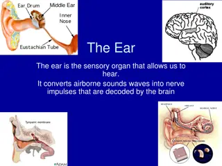
Definitions Middle ear is the area between the tympanic membrane and the inner ear including the Eustachian tube. Otitismedia (OM) is inflammation of the middle ear.
OM-Classification Acute OM http://t2.gstatic.com/images?q=tbn:1tH2Q7X7lqkCwM:http://www.middleearimplants.com/images/fully-implantable-middle-ear-device.jpg Secretory( Serous) OM Chronic OM
http://t0.gstatic.com/images?q=tbn:F1j8DXYrYIv89M:http://www.alleganyhealthdept.com/images/infant.pnghttp://t0.gstatic.com/images?q=tbn:F1j8DXYrYIv89M:http://www.alleganyhealthdept.com/images/infant.png OM- Epidemiology Most common in infants 6 to 18 months of age (2/3 of cases). Improves with age, why ? The Eustachian Tube which vents the middle ear to the nasopharynx , is horizontal in infants, difficult to drain naturally, its surface is cartilage ,and lymphatic tissue lining is an extension of adenoidal tissue from back of the nose. Accompanied with viral URTI http://t3.gstatic.com/images?q=tbn:R9V79ruGIs2kaM:http://content.onestepahead.com/assets/images/product/detail/13735_1.jpg
OM-Pathogenesis and Risk Factors URTI or allergic condition cause edema or inflammation of the tube. Functions of the tube ( ventilation, protection and clearance ) disturbed. Oxygen lost leading to negative pressure Pathogens enter from nasopharynx into middle ear. Colonization and infection result. http://t0.gstatic.com/images?q=tbn:by3MmKmS-3rwXM:http://faculty.ksu.edu.sa/75719/Pictures%2520Library/Respiratory%2520system/Upper%2520respiratory%2520tract.jpg
OM- Other risk factors Anatomic abnormalities Medical conditions such as Cleft palate ,obstruction due to adenoid or NG tube or malignancy, immune dysfunction. Exposure to pathogens from day care. Exposure to smoking. http://t1.gstatic.com/images?q=tbn:J3ptxBEM_0ul-M:http://www.biomedcentral.com/content/figures/1471-2350-5-15-1.jpg http://t2.gstatic.com/images?q=tbn:xqzWfJzPWrDXrM:http://buckheadent.net/images/tons.gif
Images of acute OM http://t1.gstatic.com/images?q=tbn:ewRNPlXlpEjGlM:http://de.academic.ru/pictures/dewiki/79/Otitis_media_schollig.jpg http://t3.gstatic.com/images?q=tbn:f0Vfl7IkhnkVjM:http://upload.wikimedia.org/wikipedia/commons/3/39/Otitis_media_incipient.jpg http://t1.gstatic.com/images?q=tbn:qDwZNRe8K2SM0M:http://www.meddean.luc.edu/lumen/MedEd/medicine/pulmonar/pdself/Serous_ottitis_media.jpg
Images of chronic OM http://t1.gstatic.com/images?q=tbn:Xmx1Gilw3kPdRM:http://www.evmsent.org/images/tmperf.jpg http://t2.gstatic.com/images?q=tbn:1SDGBm5dQug-kM:http://www.uthsc.edu/otolaryngology/images/567.jpg
Images of serous OM http://t3.gstatic.com/images?q=tbn:1QsfEh0rhFpSaM:http://www.rcsullivan.com/www/toml0911.jpg http://t2.gstatic.com/images?q=tbn:VXNqbOkLsY1mmM:http://www.fpnotebook.com/_media/EntSerousOtitisMedia.jpg http://t2.gstatic.com/images?q=tbn:-obOj53sdnbekM:http://www.texasent.com/userfiles/image//Otitis%2520Media%2520Fig3.JPG
Microbiology of OM http://t1.gstatic.com/images?q=tbn:avYNveKQwqRGfM:http://www.msevans.com/cnsinfections/h-influenzae.jpg http://t0.gstatic.com/images?q=tbn:MGJUENYImbkqwM:http://textbookofbacteriology.net/themicrobialworld/S.pneumoniae1.jpg http://t2.gstatic.com/images?q=tbn:Na9KOCmMV9oAwM:http://www.gslabs.com/images/saureus2.jpg http://t2.gstatic.com/images?q=tbn:mlz6bFjW6h_T-M:http://www.vetbact.org/vetbact/include/getvetbaktimage.php%3Fimgid%3D238%26imgtable%3Dvetbact_images%26images%3D0 http://t3.gstatic.com/images?q=tbn:I37L4EcCa2rF5M:http://farm3.static.flickr.com/2240/2402321868_539a568ec7_o.jpg http://t2.gstatic.com/images?q=tbn:MkzqdnjQrCYNzM:http://www.mc.maricopa.edu/~johnson/labtools/Dbiochem/opto4b.jpg
Microbiology of OM-continue Go to fullsize image Go to fullsize image Go to fullsize image Go to fullsize image Go to fullsize image Go to fullsize image Go to fullsize image
OM-Microbiology-Bacterial Causes Acute OM < 3months of age S.pneumoniae,(40%) group B Streptococcus, H.influenzae (non typable) ,Gram negative bacteria and P.aeruginosa > 3 months of age S.pneumoniae, H.influenzae ,others eg, S.pyogenes, Moraxella catarrhalis, S.aureus
OM-Microbiology-cont. Chronic OM Serous OM Mixed flora in 40% of cases P.aeruginosa, H.influenzae, S.aureus, Proteus species, K.pneumoniae, Moraxella catarrhalis, anaerobic bacteria. Same as chronic OM, but Most of the effusions are sterile Few acute inflammatory cells
OM-Viral causes RSV -74% of viral isolates Rhinovirus Parainfluenza virus Influenza virus http://t3.gstatic.com/images?q=tbn:gIH_nUuEjo67FM:http://template.bio.warwick.ac.uk/staff/easton/IMAGES/Diagrams/3dvirus.jpg
Clinical presentation Acute OM Mostly Bacterial ,often a complication of viral URTI First 1-2 days: Fever (39 C), irritability, earache , muffled nose. Bulging tympanic membrane ,poor mobility and obstruction by fluid or inflammatory cells on otoscopicexamination.
3-8 days: Pus and ear exudatedischarge spontaneously and pain and fever begin to decrease. 2-4 weeks : Healing phase, discharge dies up and hearing becomes normal.
Serous OM Collection of fluid within the middle ear as a result of negative pressure produced by altered eustachian tube function. Represent a form of chronic OM or allergy- related inflammation Tends to be chronic , with non purulent secretions. Cause hearing deficit.
Chronic OM Usually result from unresolved acute infection due to in adequate treatment or host factors that perpetuate the inflammatory process. Result in destruction of middle ear structures and significant risk of permanent hearing loss.
Diagnostic approaches of OM Clinical examination Tympanometry( detect presence of fluid) Gram stain and culture of aspirated fluid to determine the etiologic agents. http://t1.gstatic.com/images?q=tbn:9V291FPaYcZ7lM:http://www.tchain.com/otoneurology/testing/images/audio5.gif http://t1.gstatic.com/images?q=tbn:qDwZNRe8K2SM0M:http://www.meddean.luc.edu/lumen/MedEd/medicine/pulmonar/pdself/Serous_ottitis_media.jpg
Management of OM Acute OM requires antimicrobial therapy & careful follow up. Antimicrobial usually empirical depending on the most likely bacterial pathogens, usually to cove S.pneumonia and H.influenzae. Drainage of exudate may be required. Chronic or serous OM need complex management, possibly surgical.
Complications Intracranial Intracranial Hearing loss Tympanic membrane perforation Mastoiditis Cholestatoma Labyrinthitis others Meningitis Extraduralabscess Suduralempyema Brain abscess others http://t1.gstatic.com/images?q=tbn:UKwRtXWPyE792M:http://top-10-list.org/wp-content/uploads/2009/09/Meningitis.jpg http://t3.gstatic.com/images?q=tbn:nwKnADJQFFwfDM:http://www.ferne.org/Lectures/acep_2005_peds/perron_pic_11.jpg http://t3.gstatic.com/images?q=tbn:33OTr-ilCLStzM:http://de.academic.ru/pictures/dewiki/77/Mastoiditis1.jpg