Overview of Jejunum and Ilium Histology and Vascular Supply
The jejunum and ileum are segments of the small intestine with distinct features in terms of position, diameter, wall thickness, and lymphoid aggregates. They receive arterial blood supply from branches of the superior mesenteric artery and drain into various venous tributaries. Histologically, the GI tract is composed of mucosa, submucosa, muscularis externa, and serosa layers, each serving specific functions. The esophagus and stomach also exhibit unique histological features related to mucosa, glands, and muscle layers.
Download Presentation

Please find below an Image/Link to download the presentation.
The content on the website is provided AS IS for your information and personal use only. It may not be sold, licensed, or shared on other websites without obtaining consent from the author.If you encounter any issues during the download, it is possible that the publisher has removed the file from their server.
You are allowed to download the files provided on this website for personal or commercial use, subject to the condition that they are used lawfully. All files are the property of their respective owners.
The content on the website is provided AS IS for your information and personal use only. It may not be sold, licensed, or shared on other websites without obtaining consent from the author.
E N D
Presentation Transcript
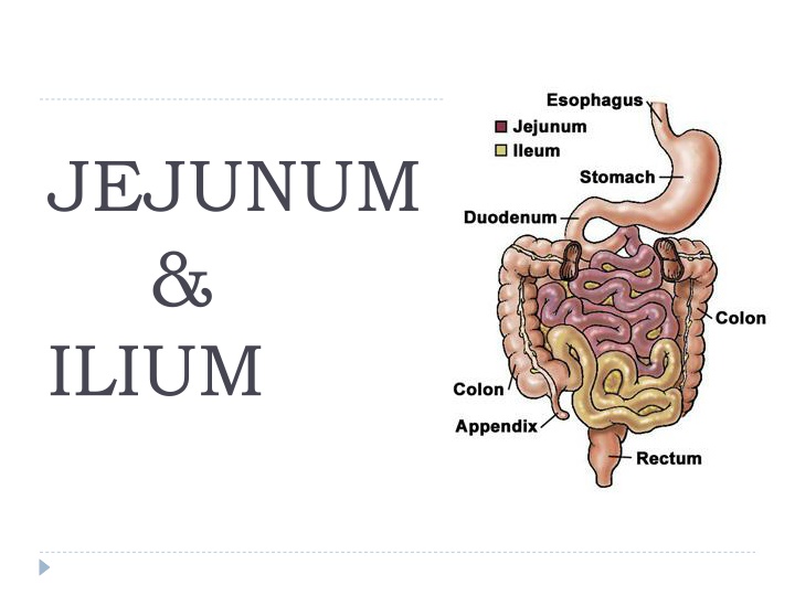
JEJUNUM & ILIUM
Features Jejunum Ilium Position Left infracolic compartment upto umbilical region Pelvic region ends in rt.iliac fossa by opening into ilioceacal valve. Median external diameter Median internal diameter Walls 4cm 3.5cm 2.5cm 2cm Thicker Thinner Plica circularis More pronounced Less (flat in terminal part) Lymphoid aggregates Less More
Vascular Supply Arterial Jejunal & ilial branches of Sup.mesenteric artery. Venous drainage- jejunal, ilial, iliocolic, rt.colic, middle colic, rt. Gastroepiploic, pancreaticoduodenal tributeries into sup. Mesentric vein. Lymphatic drainage- mesenteric lymph nodes . Innervation:- Sympathetic- mid thoracic spinal segment via splanchnic nerve and sup. mesentric ganglions Parasympathetic:-ant. and right vagus nerve .
Histology of GI Tract Entire GI tract is formed of 4 layers 1.Mucosa- a) lining epithilium b) lamina propria- made up of connective tissue containing glands, blood and lymph vessels c)Muscularis mucosa- made up of 2 layers inner circular and outer longitudinal. 2. Submucosa- dense irregular connective tissue containing blood vessels, lymphatic vessels, nerve plexus and occasional glands. 3. Muscularis externa- inner circular and outer longitudinal layer containing myenteric nerve plexus. 4.Serosa simple squamous epithilium(peritoneum)
Histology of oesophagus 1.Mucosa- non-keratinized stratified squamous epithilium. Lamina propria oesophageal cardiac glands (lower part) Muscularis mucosa- single longitudinal layer of smooth muscle. 2. submucosa- oesophageal glands 3.Muscularis externa- upper1/3 skeletal muscle middle 1/3- skeletal & smooth Lower1/3-smooth Arranged in 2 layers inner circular& outer longitudinal 4.Adventitia loose connective tissue without peritoneum
Histology of stomach Mucosa- simple columnar epithilium having surface mucous cells, invaginations are called gastric pits. Lamina propria- long tubular fundic glands which contain 1.Mucous neck cells- low columnar secrets soluble mucus. 2.Chief/zymogenic/ peptic cells- low cuboidal secrets pepsinogen,lipase, amylase. 3.Parietal/oxyntic cells- large pyramidal secrets hcl and gastric intrinsic factor 4. Enteroendocrine cells- unicellular secrets gastrin, somatostatin and histamine. Submucosa irregular dense connective tissue containing b.v. ,l.v.& n.p. Muscularis externa inner oblique, outer longitudinal and middle circular. Serosa- loose connective tissue covered with peritoneum.
Histology of small intestine Special features- Plica circularis/ valves of kerckring- mucosa & submucosa is thrown into permanent folds which are numerous in distal duodenum and jejunum and less in ilium. Villi- a finger like projection lined by surface epithilium with lamina propria projecting into it. Villus contain core of loose connective tissue, fenestrated blood capillaries and a central blind ended lymphatic vessel called lacteal. these are large and numerous in duodenum and smaller in ilium. Intestinal glands/ crypts of lieberkuhn- invaginations of epithilium into lamina propria which open on luminal surface of intestine at base of intervillous space. It contains :
Conti.. enterocyte columnar absortive cells their surface bear up to 3000 microvilli. Microvilli posses a specialized glycoprotein called glycocalyx. Goblet cells- produce mucus which protect against microorganisms and toxins. Paneth cells- found in deeper parts of crypts(duodenum) secretion protects against bacterial flora. Stem cells- source of most of cell type of intestinal epithilium. Enteroendocrine cells- secretes local harmones Cholecytokinin (inc.activity of g.b. & pancreas) Gastric inhibitory polypeptide (inhibit gastric secretion and motility) Secretin (stimulate release of insulin in pancreas) Motilin (stimulate gastric and intestinal motility).
Conti. Microfold (M) cells-epithilial cells overlying lymphoid aggregates in intestinal wall. They taken up antigen from int.lumen and come in contact with cells of immune system and produce antibodies (IgA). Peyer s patches- aggregate lymphoid follicles present in lamina propria of small intestine especially ilium which protect gut against invasion of microorganisms.
Duodenum Mucosa- simple columnar with microvilli & few goblet cells r seen lamina propria contains crypts of lieberkuhn which contain columnar, goblet, paneth and enteroendocrine cells. Muscularis mucosa is made up of inner circular and outer longitudinal layer.Plica circularis r seen. Submucosa contain brunner s gland which secrets alkaline mucus to neutralize acid chyme. Muscularis externa is made up of inner circular and outer longitudinal layer containing myenteric plexus. Serosa contain only 1stpart of duodenum.
Jejunum & ilium Jejunum is same as duodenum with absence of brunner s and payer patches. Mucosa is lined by simple columnar epithilium with microvilli. Prominent intestinal villi and plica circularis. Ilium is same as jejunum mucosa is lined by simple columnar epithilium with microvilli. Villi are short and few. Lamina propria contains peyer s patches. M cells r found overlying lymphoid follicles. Muscularis mucosa is thin than jejunum.
