Microscopic Structure and Histology of Kidney and Urinary Tract
Explore the intricate details of kidney anatomy, nephron structure, glomerular filtration barrier, and the histology of renal cortex and medulla, renal corpuscle, proximal and distal tubules, juxtaglomerular apparatus, as well as the urinary bladder and urethra in both male and female. Delve into the functional units of the kidney and understand the composition of the uriniferous tubule, nephron types, and the crucial role of the proximal tubule in kidney function.

Download Presentation

Please find below an Image/Link to download the presentation.
The content on the website is provided AS IS for your information and personal use only. It may not be sold, licensed, or shared on other websites without obtaining consent from the author.If you encounter any issues during the download, it is possible that the publisher has removed the file from their server.
You are allowed to download the files provided on this website for personal or commercial use, subject to the condition that they are used lawfully. All files are the property of their respective owners.
The content on the website is provided AS IS for your information and personal use only. It may not be sold, licensed, or shared on other websites without obtaining consent from the author.
E N D
Presentation Transcript
Kidney, Ureter, Urinary bladder & Urethra Red: important. Black: in male|female slides. Gray: notes|extra. Editing file Editing file Editing file
OBJECTIVES The microscopic structure of the renal cortex and medulla. The histology of renal corpuscle, proximal and distal tubules, loop of Henle, and collecting tubules & ducts. The histological structure of juxtaglomerular apparatus. The functional structures of the different parts of the kidney. The microscopic structure of the Renal pelvis and ureter. The microscopic structure of the urinary bladder and male and female urethra Histology team 437 | Renal block | All lectures
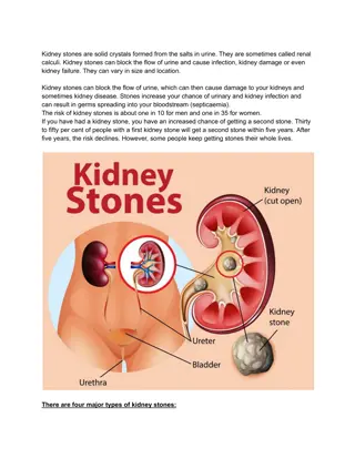
KIDNEY Cortex: Dark brown and granular. Content of cortex (renal corpuscle, PCT, loop of Henle, DCT, part of collecting tubule) Medulla: 6-12 pyramid-shape regions (renal pyramids) content of medulla ( collecting duct, loop of Henle, collecting tubule) The base of pyramid is toward the cortex (cortico-medullary border) The apex (renal papilla) toward the hilum, it is perforated by 12 openings of the ducts of Bellini (Papillary collecting ducts) in region called area cribrosa. The apex is surrounded by a minor calyx. 3 or 4 minor calyces join to form 3 or 4 major calyces that form renal pelvis. Pyramids are separated by cortical columns of Bertin (renal column) o o o o o o o URINIFEROUS TUBULE It is the functional unit of the kidney. Is formed of: 1- Nephron. 2-Collecting tubule. The tubules are densely packed. The tubules are separated by thin stroma and basal lamina. o o o o Histology team 437 | Renal block | All lectures
NEPHRON There are 2 types of nephrons: a- Cortical nephrons. b- Juxtamedullary nephrons. (Juxta = near) It is formed of : 1- Renal corpuscle. 2- Proximal tubule. 3- Thin limbs of Henle s loop. 4- Distal tubule o o 1. Renal Corpuscle Glomerulus; (tuft of fenestrated capillaries "without diaphragm ) Bowman s capsule; (Parietal layer, urinary space and visceral layer or podocytes) In front of bowman s capsule known as vascular pole ,Back of bowman s capsule known as Urinary pole. Mesangial cells; (intra-glomerular cells). o o o Histology team 437 | Renal block | All lectures
GLOMERULAR FILTRATION BARRIER Called glomerular endothelium. Endothelial wall of the glomerular capillaries. The glomerular basal lamina (inner and outer laminae rarae and middle lamina densa). Visceral layer of Bowman s capsule (podocytes) Cell come contact with glomerulus known as podocyte (podocyte = viscera layer) Podocytes have primary (major) processes and secondary (minor) processes pedicles . Between pedicles (on the surface of capillaries) there are filtration slits that have filtration slit diaphragms o o o o o o Histology team 437 | Renal block | All lectures
2. Proximal tubule: o It is composed of simple cuboidal epithelium with acidophilic cytoplasm. o The cells have striated or brushborder and lateral inter-digitations. o They have well-defined basal lamina. o It's is more than tubule distal in the cortex. Most of tubular sections in cortex related to PCT PCT cells have brush border (microvilli) to help in reabsorptions 3. Thin limbs of Henle s loop: o It has 3 regions: 1-Descending thin limb. 2-Crest of Henle s loop. 3-Ascending thin limb. o It is longer in juxta-medullary nephron than in cortical nephron. o It is composed of simple squamous epithelium. o Loop of Henle s and tubules extend to medulla 4. Distal tubule: o It starts at the macula densa. o macula densa (tall columnar & narrow cells) o The Distal convoluted tubule is formed of low cuboidal epithelium. o Because distal convoluted tubules are much shorter than proximal convoluted tubules, any section of renal cortex presents many more sections of proximal convoluted tubules. o Distal tubules drain into collecting tubules. o N.B. Cells in distal tubule come in contact with afferent & efferent arterioles is macula densa o N.B. More reabsorption occurs in the proximal tubules. Histology team 437 | Renal block | All lectures
COLLECTING TUBULES Endothelial wall of the glomerular capillaries. Are composed of simple cuboidal epithelium. They aren t part of nephron. They have 3 regions: 1-Cortical: Simple Cuboidal Epithelium. 2-Medullary: Simple Cuboidal Epithelium. 3-Papillaryducts (ducts of Bellini): Simple Columnar Epithelium. They open in area cribrosa. They are impermeable to water except in presence of ADH. Collecting tubule lined with cuboidal E.P once it change to columnar E.P called collecting duct o o o o o o o o o o RENAL INTERSTITIUM It is a very flimsy, scant amount of loose connective tissue that contains: 1-Fibroblasts. 2-Macrophages. 3-Interstitial cells: They secrete medullipin I, which is converted in the liver into medullipin II, that lowers blood pressure. o Histology team 437 | Renal block | All lectures
From 436 team Explanation from Dr.Raeesa The renal corpuscle has two poles 1- vascular pole 2- urinary pole for the passage of what will give me urine later. The vascular pole is for the blood entry and excitation of arterioles: (Afferent + Efferent) The blood enters the afferent arteriole and goes out when its filtered through efferent arteriole. In the space between the two arterioles there is distal tubule, part of it is adherent to the arterioles. So the triangle of Juxtaglomerular apparatus is formed of 3 things: part of afferent, part of efferent, part of distal tubule and in between there is extra mesangial cells. There are changes only in the cells of distal tubule adherent to the afferent arterioles: The changes: 1- Columnar epithelium with oval nuclei 2- The number of cells increase 3- There is macula dense There are changes in the arterioles in the part adherent to the distal tubule. the changes: 1- Modification in the tunica media (smooth muscle), so instead of spindle shaped cell it will be cuboidal cell. After modification it called Juxtaglomerular cells which is responsible for renin secretion. Histology team 437 | Renal block | All lectures
Juxtaglomerular Apparatus When DCT come in contact with afferent & efferent arterioles some modification happened to some cell: 1) Cells in distal tubule come in contact with afferent & efferent arterioles change from cuboidal to columnar cells called macula dense 2) smooth muscle cell in wall of afferent & efferent arterioles come in contact with DCT change to cuboidal cells called juxtaglomerular cell It has 3 components: A-The macula densa of distal tubule: Tall cells with centrally-placed nuclei B-Juxtaglomerular cells: of afferent glomerular arteriole (modified smooth muscle of tunica media), Nuclei are round with granular cytoplasm. They secrete renin. C-The extraglomerular mesangial cells. o o Renal Calyces Each calyx accepts urine from the renal papilla of a renal pyramid. They are lined with transitional epithelium, lamina propria & smooth muscle. Minor calyces merge to form major calyces (with same lining tissue as minor calyces). Major calyces open into renal pelvis. Urinary track epithelium start from minor calyces until part of urethra o o o o o Histology team 437 | Renal block | All lectures
Ureter Mucosa: Is formed of transitional epithelium and lamina propria. Muscularis (muscular coat): Is formed of 2 layers of smooth muscle in the upper 2/3: 1) Inner longitudinal. 2) Outer circular. Is formed of 3 layers of smooth muscle in the lower 1/3: 1) Inner longitudinal. 2) Middle circular. 3) Outer longitudinal. Adventitia: fibrous C.T. covering (Without serosa). Serosa -> adventitia + visceral layer of peritoneum (mesothelium) o o o Urinary bladder It has the same structure as the lower third of ureter. (All same lower third of ureter except urinary bladder with serosa while ureter without serosa) Superficial layer of transitional epithelium has dome-shaped cells (in empty bladder). It has 3 layers of smooth muscle: 1) Inner longitudinal. 2) Middle circular. 3) Outer longitudinal. Its outer covering is adventitia or serosa. Part of urinary bladder cover with Adventitia and other part cover with Serosa o o o o o Histology team 437 | Renal block | All lectures
Urethra Male urethra (divided into 3 regions) Female urethra is lined with transitional epithelium 1-Transitional epithelium near the bladder 2-Pseudostratified columnar epithelium 3 Stratified squamous non-keratinized epithelium Prostatic urethra (3 cm) Epithelium Sub- that contains glands of Littre (mucus- secreting glands) Membranous urethra (1 cm) Its longest part is lined with Stratified columnar epithelium with patches of pseudostratified columnar epithelium epithelial fibroelastic C.T. Penile (spongy) urethra (16 cm) inner longitudinal and outer circular layers Smooth muscle N.B. In navicular fossa (enlarged terminal portion): Stratified squamous non- keratinized epithelium. N.B. The lamina propria contains mucus secreting glands of Littre. o o N.B. The female urethra is shorter than the male urethra Histology team 437 | Renal block | All lectures
QUESTIONS: Q1: Which of them structure dark brown and granular? A) Cortex B) Medulla C) Major calyces D) Minor calyces Q2: The base of pyramid is? A) Away from the cortex B) Toward the cortex C) Helium D) minor calyx 5- A 4- C Q3: Pyramids are separated by? A) Renal papilla B) Renal vein C) Thin stroma & basal lamina D) Cortical column of Bertin 3- D 2- B 1- A Q4: The uriniferous tubules are separated by? A) Renal papilla B) Renal vein C) Thin stroma & basal lamina D) Cortical column of Bertin Q5: What is the function of medullipin II ? A) Decrease blood pressure B) Increase temperature C) Increase blood pressure D) Decrease temperature Histology team 437 | Renal block | All lectures
Q6: The cytoplasm of Proximal convoluted tubule is? A) Basophilic B) Acidophilic C) Both A&B D) Non of them Q7: Describe the position of filtration slits? A) Between pedicles B) Podocytes C) Both A&B D) Non of them Q8: Describe the Glomerulus? A) Tuft of fenestrated capillaries B) Intra-glomerular cells C) Parietal layer D) Visceral layer 10- A 9- B 8- A Q9: Proximal convoluted tubule is composed of? A) Simple squamous epithelium B) Simple cuboidal epithelium C) Simple columnar epithelium D) Low cuboidal epithelium 7- A 6- B Q10: Thin limb of Henle's loop is composed of? A) Simple squamous epithelium B) Simple cuboidal epithelium C) Simple columnar epithelium D) Low cuboidal epithelium Histology team 437 | Renal block | All lectures
Q11: Distal convoluted tubule is composed of? A) Simple squamous epithelium B) Simple cuboidal epithelium C) Simple columnar epithelium D) Low cuboidal epithelium Q12: Papillary ducts of collecting tubule is composed of? A) Simple squamous epithelium B) Simple cuboidal epithelium C) Simple columnar epithelium D) Low cuboidal epithelium 15- A 14- C Q13: Which of them cells secrete medullipin I ? A) Fibroblasts B) Fibroclasts C) Macrophages D) Interstitial cells 13- D 12- C 11- D Q14: Which of them is type of nephron? A) Cortical B) Juxtamedullary C) Both A&B D) Non of them Q15: Which of them tubules isn't part of nephron? A) Collecting tubules B) Proximal tubules C) Thin limb of Henle's loop D) All of them Histology team 437 | Renal block | All lectures
Q16: Which of them structure secrete renin? A) mesangial cells B) Juxtaglomerular cells C) Membranous urethra D) serosa muscle Q17: How many layers does the upper 2/3 of ureter have? A) 5 layers B) 4 layers C) 3 layers D) 2 layers Q18: How many layers does the urinary bladder have? A) 5 layers B) 4 layers C) 3 layers D) 2 layers 20- A 19- D 18- C Q19: Epithelium of female urethra is? A) Pseudostratified columnar epithelium B) Transitional epithelium C) Stratified squamous non-keratinized epithelium D) All of them 17- D 16- B Q20: Describe the nuclei & cytoplasm of Juxtaglomerular cells? A) Round with granular cytoplasm B) Round with basophilic cytoplasm C) Oval with granular cytoplasm Histology team 437 | Renal block | All lectures
Q21: Mucosa of ureter is formed of? A) Transitional epithelium B) Lamina propria C) smooth muscle D) A & B Q22: Prostatic urethra is lined with? A) Transitional epithelium B) Simple squamous epithelium C) A & B D) Non of them Q23: Urinary bladder has the same structure as the? A) Kidney B) Upper ureter C) Lower ureter D) Urethra 25- B 24- D Q24: Renal Calyces are lines with? A) Transitional epithelium B) Lamina propria C) smooth muscle D) All of them 23- C 22- A 21- D Q25: Penile (spongy) urethra is lined with? A) Simple columnar epithelium with patches of pseudostratified columnar epithelium B) Stratified columnar epithelium with patches of pseudostratified columnar epithelium C) Transitional epithelium with patches of pseudostratified columnar epithelium D) Stratified squamous non-keratinized epithelium with patches of pseudostratified columnar epithelium Histology team 437 | Renal block | All lectures
Team leaders : Khalid Fayez Alshehri Rawan Mohammad Alharbi Special thank for: Marwah Jassim Alkhalil Twitter.com/Histology437 HistologyTeam437@gmail.com Histology team 437 | Renal block | All lectures