Neuroglia: The Connective Tissue of the Nervous System
Neuroglia, a vital component of the nervous system, play a crucial role in providing support, protection, and defense to neural tissue. Recent research has highlighted their involvement in various disorders, including autism spectrum disorders (ASD). This article explores the functions and contributions of different types of glial cells, emphasizing their significance in the regulation of cerebral environment and synaptic development.

Download Presentation

Please find below an Image/Link to download the presentation.
The content on the website is provided AS IS for your information and personal use only. It may not be sold, licensed, or shared on other websites without obtaining consent from the author.If you encounter any issues during the download, it is possible that the publisher has removed the file from their server.
You are allowed to download the files provided on this website for personal or commercial use, subject to the condition that they are used lawfully. All files are the property of their respective owners.
The content on the website is provided AS IS for your information and personal use only. It may not be sold, licensed, or shared on other websites without obtaining consent from the author.
E N D
Presentation Transcript
NEUROGLIA: The connective tissue of nervous system COMPILED BY Prof. Sudhir Kumar Awasthi Dept. Of Life Sciences CSJMU
Neuroglia are a large class of neural cells of ectodermal (astroglia, oligodendroglia, and peripheral glial cells) and mesodermal (microglia) origin. Neuroglial cells provide homeostatic support, protection, and defense to the nervous tissue. Pathological potential of neuroglia has been acknowledged since their discovery. Research of the recent decade has shown the key role of all classes of glial cells in autism spectrum disorders (ASD), although molecular mechanisms defining glial contribution to ASD are yet to be fully characterized. This narrative conceptualizes recent findings of the broader roles of glial cells, including their active participation in the control of cerebral environment and regulation of synaptic development and scaling, highlighting their putative involvement in the etiopathogenesis of ASD.
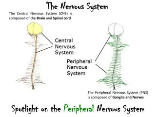
Neuroglia cells represent the most numerous cell family in the central nervous system with 5 10 glial cells per neuron or 350 billion cells per brain. Glial cells retain their ability to divide; provide metabolic and structural support for neurons, and maintain conditions that allow adequate functioning of neurons. They are less electrically excitable than neurons and do not form chemical synapses Based upon their appearance, function, and origin, four types of glial cells have been identified in the central nervous system: astrocytes, oligodendroglia, ependymal cells, and microglia.
Astrocytes Astrocytes are the most numerous type of glia and account for about one half of all cells in the brain. They are divided into two categories of glia, the protoplasmic astrocytes and the fibrous astrocytes. Protoplasmic astrocytes are present predominantly in gray matter and are characterized by many, often short, branched processes. Sometimes, these processes form sheets that can separate different neuronal cell processes or neurites and form close associations with synapses. Protoplasmic astrocytes are involved in many regulatory processes they buffer K+and Cl ions, they regulate extracellular neurotransmitter concentrations through specific uptake mechanisms, and they play an important role in the formation of synapses by, for example, secreting substances that stimulate the development of synapses. In addition, some astrocytes release neurotrophic and gliotrophic factors, such as glial-derived neurotrophic factor, which promotes survival of select neurons.
Fibrous astrocytes Fibrous astrocytes are present predominantly in white matter (ie, myelinated fiber tracts). These cells have long, thin processes that do not form sheets. Both types of astrocytes form expansions at the end of their processes, the astrocytic end feet. These structures completely cover the basement membrane of capillaries and small arterioles as well as the outer surface of the brain forming an insulating layer, the glia limitans. Thus, astrocytes form a physical barrier which is important for the control of movement of small molecules into the brain.
Oligodendrocytes They are relatively small glial cells that develop from precursor cells in the ventral ventricular zone during the late prenatal and early postnatal period. The main function of oligodendrocytes is to provide an insulating cover that surrounds individual axons and thereby allows an increase in the speed of conductance of electrical signals. Oligodendrocytes have several processes that can flatten into sheet-like structures and wrap many times around portions of individual axons.
Each process of an oligodendrocyte covers a small segment of an axon, and a narrow gap or node of Ranvier of uncovered axon remains between adjacent segments. These nodes contain a large number of Na ion channels and are critical for the saltatory conduction of action potentials. Each oligodendrocyte can form several myelin sheets covering different segments of one axon, or they can provide myelin to several segments of different axons. Usually, axons are covered by myelin sheets from the initial segment to the terminal.
Ependymal cells Ependymal cells form a simple, cuboidal epithelium that lines the inner surfaces of the brain, including the ventricles and the central canal in the spinal cord. Ependymal cells are linked to each other by desmosomes while the base of the cells is in contact with astrocytes. In most regions of the ventricular system ependymal cells have kinocilia at their apical surface which are thought to be important for moving the cerebrospinal fluid throughout the ventricles and central canal of the spinal cord.
In certain regions of the lateral, third and fourth ventricles, the ependymal epithelium is continuous with the choroid plexus which consists of highly specialized and polar cells that form cellular sheets and float in the cerebrospinal fluid. The cells of the choroid plexus rest on a basement membrane, the cells are connected to each other by tight junctions and the lateral plasma membranes form extensive interlocking folds with adjacent choroid plexus cells. Underneath the choroid plexus cells lies a dense network of fenestrated capillaries that also lack tight junctions. This allows plasma filtrate to freely move from the blood into the extravascular region, however, due to the junctional complexes between adjacent choroid plexus cells, movement of solutes toward the apical ventricular surface is prevented.
Instead, transport and exchange mechanisms move ions and micronutrients through the choroid plexus cells, which sets up an osmotic gradient that is neutralized by water that follows passively The cerebrospinal fluid consists mostly of water and ions, some glucose and very small amounts of protein. The cerebrospinal fluid leaves the ventricular system at the foramen magnum and the aperture of Luschka of the fourth ventricle as well as at the terminal cisterna of the spinal cord to reach the subarachnoid space Thus, both internal and external surfaces of the brain are bathed in cerebrospinal fluid.
Microglia Microglia are small cells with short processes that are found throughout the central nervous system. In contrast to the other families of glial cells, microglia are not derived from a neuroectodermal lineage. Instead, they originate from the bone marrow mesoderm and are derived from monocytes. Microglia enter the central nervous system early in development and remain inconspicuous for a long period of time during which they migrate through the brain, screening for immunologically relevant events. Thus, microglia participate in the management of inflammatory and immune responses in the brain. Microglia can become highly phagocytic after insults to the brain, such as stroke, when they remove cellular debris or other pathological substances, such as beta- amyloid. In addition, they are antigen-presenting cells and they release cytokines that attract other cells, including neutrophils and lymphocytes to move across the blood brain barrier into the central nervous system.
REFERENCES Fundamentals of Neuroscience edited by Larry Squire & Darwin Berg (2010) Principles of Neurobiology by Princeton Math(2012) Principles of Neurobiology by Liqun Luo (2020)