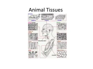
Connective Tissues: Characteristics, Classification, and Types
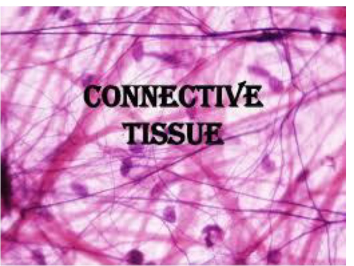
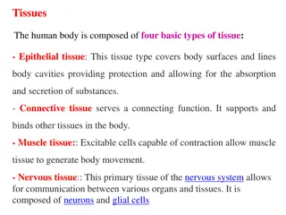
Connective tissues play a crucial role in supporting and connecting various tissues in the body. They are highly vascularized and develop only from mesoderm during embryonic origin. Consisting of cells and intercellular matter secreted by the cells, these tissues are categorized based on cell types and intercellular matter present. Areolar (Loose) Connective Tissue, a common type, includes fibroblasts, macrophages, and mast cells. They contain protein gel with collagen and elastin fibers, providing strength and elasticity. Local inflammation processes involve the dilation of blood vessels, increased permeability, and pain stimulation.

Download Presentation

Please find below an Image/Link to download the presentation.
The content on the website is provided AS IS for your information and personal use only. It may not be sold, licensed, or shared on other websites without obtaining consent from the author.If you encounter any issues during the download, it is possible that the publisher has removed the file from their server.
You are allowed to download the files provided on this website for personal or commercial use, subject to the condition that they are used lawfully. All files are the property of their respective owners.
The content on the website is provided AS IS for your information and personal use only. It may not be sold, licensed, or shared on other websites without obtaining consent from the author.
E N D
Presentation Transcript
General Characteristics 1. Embryologic origin develop only from mesoderm 2. the connective tissues consist of cells and intercellular matter secreted by the cells 3. connective tissues are highly vascularized 4. generally function to connect and support the other tissues of the body
Classification of Connective Tissues 1. based on the type of cell or cells 2. based on the type of intercellular matter found between the cells
Types of Connective Tissue 1. Areolar (Loose) Connective Tissue a. cells present (1) fibroblasts - principal type of cell - secrete proteins into the spaces between the cells (2) macrophages big eater, swallow bacteria, viruses engulf foreign agents
(3) mast cells secrete histamine & other chemical mediators of inflammation
Local Inflammation (1) local dilation of blood vessels = erythema (in the injured area) (2) local increased permeability of blood vessels = swelling (edema) - more leaky (3) local stimulation of pain fibers = pain
b. intercellular matter (1) protein gel (much like Jello ) containing protein fibers (a) collagen protein fibers confer strength to the tissue (b) elastin protein fibers confer elasticity to the tissue (2) All proteins are synthesized and secreted by the fibroblasts
c. Areolar connective tissue is the most widely distributed tissue in the body serving to support and nourish (via the blood vessels) the other tissues of the body
Adipose (Fat Tissue) a. Cells present - closely packed adipose (fat) cells, each containing a large fat-filled vacuole b. Intercellular Matter: - small amount of protein fibers, secreted by the fat cells c. Adipose tissue provides a reservoir of food (for energy), insulates against heat loss, supports and protects the organs it encloses
Fat Tissue (Adipose) Location (1) under the skin (subcutaneous fat) (2) around the kidneys and eyeballs (3) buttocks and breasts Fat cells secrete a little bit of collagen
3. Dense Fibrous Connective Tissue a) cells present - fibroblasts b) intercellular matter - principally collagen protein fibers, secreted by the fibroblasts, which confers strength to the tissue c) there are 2 sub-types of Dense Fibrous Connective tissue based upon how the protein fibers are arranged
Dense Fibrous Connective Tissue Is simpler than areolar connective tissue Contains fibroblasts and collagen
2 types of Dense Fibrous Connective Tissue 1. Dense Regularly-Arranged Fibrous Connective Tissue collagen is arranged in one direction so it means it is really strong in one direction 2. Dense Irregularly-Arranged Fibrous Connective Tissue the collagen is running all around so it is relatively strong in all directions
Dense Regularly-Arranged Fibrous Connective Tissue
Dense Irregularly-Arranged Fibrous Connective Tissue
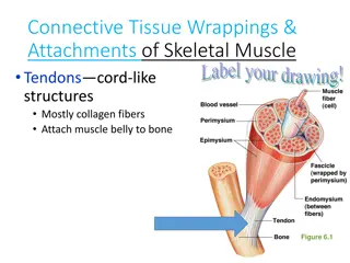
(1) Regularly Arranged Dense Fibrous Connective Tissue (a) the protein fibers are arranged in parallel bundles (b) locations: (1) tendons cords that connect muscles to bones (2) ligaments cords that connect bones together * it is the collagen that makes them strong
Other locations for regularly arranged dense fibrous connective tissue (3) dura mater fibrous connective tissue that encloses the brain and spinal cord - tough mother - it is one of the layers around the brain and spinal cord (4) fascia fibrous connective tissue that encloses muscles (5) perichondrium and periosteum fibrous connective tissue that encloses cartilage and bones
Irregularly Arranged Dense Fibrous Connective Tissue (a) the protein fibers are interwoven, running in all directions (b) location: (1) dermis of the skin (2) scar tissue
Stratified Squamous Non Keratinized Epithelium
4. Cartilage unusual connective tissue - no blood vessels, that is why they do not heal very well a. cells present: - chondrocytes, usually arranged in groupings of 2 to 4 cells, surrounded by a pocket of fluid called a lacuna b. intercellular matter: - protein gel (much like Jello ) containing protein fibers
c. cartilage contains no blood vessels (non- vascular), and is thus an exception to the general pattern of connective tissue d. cartilage tissue is capable of growing in both length and width (thickness) e. there are 3 sub-types of cartilage (based upon the nature of the intercellular matter):
(1) Hyaline Cartilage (a) the intercellular matter is made up of a gel membrane with collagen protein fibers (secreted by the chondrocytes) (b) locations (1) the embryonic lung bones of the body (2) front of the nose (3) the trachea & larynx
(2) Fibrocartilage (a) the intercellular matter is made-up of a gel containing large bundles of collagen protein fibers (b) locations: - the intervertebral disks
(3) Elastic Cartilage (a) the intercellular matter is made up of a gel containing elastin protein fibers (b) locations the external ear * Hardest is bone followed by fibrocartilage
Bone Tissue a. cells present - osteocytes, each surrounded by a pocket of fluid called a lacuna b. intercellular matter: (1) calcium salts (calcium phosphate & calcium carbonate) or (Ca(PO4)2 & CaCO3) (2) collagen protein fibers interwoven in the calcium salts
c. bone tissues are highly vascularized d. bone tissue is only capable of growing in width (thickness) e. there are 3 subtypes of Bone Tissue (based upon its organization):
a. Compact Bone need to recognize under a microscope - the osteocytes are arranged in concentric circles, called Haversian Systems b. Spongy (Cancellous) Bone Tissue - the osteocytes are arranged in a spongy network


 mast cells – secrete histamine & other")
 mast cells – secrete histamine & other")
 mast cells – secrete histamine & other")
 mast cells – secrete histamine & other")
 mast cells – secrete histamine & other")


")
")
")


 Regularly Arranged Dense Fibrous")


")


 Hyaline Cartilage")
 Hyaline Cartilage")
 Fibrocartilage")
 Fibrocartilage")
 Elastic Cartilage")
 Elastic Cartilage")