Exploring the World of Cells and Microscopy
Delve into the fascinating world of cells by discovering their sizes, from a giraffe's nerve cell to a human egg, and learn about different types of cells like cheek cells and onion cells. Explore the use of electron microscopes for detailed imaging, compare light and electron microscopes, and understand the workings of a scanning electron microscope. Discover the magnification capabilities, resolution differences, and types of radiation used in light and electron microscopes. Uncover the preparation processes and costs associated with each type of microscope.

Download Presentation

Please find below an Image/Link to download the presentation.
The content on the website is provided AS IS for your information and personal use only. It may not be sold, licensed, or shared on other websites without obtaining consent from the author. If you encounter any issues during the download, it is possible that the publisher has removed the file from their server.
You are allowed to download the files provided on this website for personal or commercial use, subject to the condition that they are used lawfully. All files are the property of their respective owners.
The content on the website is provided AS IS for your information and personal use only. It may not be sold, licensed, or shared on other websites without obtaining consent from the author.
E N D
Presentation Transcript
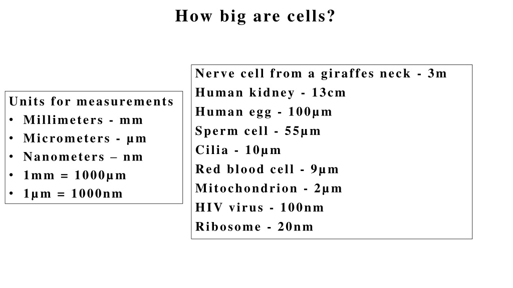
How big are cells? Nerve cell from a giraffes neck - 3m Human kidney - 13cm Human egg - 100 m Sperm cell - 55 m Cilia - 10 m Red blood cell - 9 m Mitochondrion - 2 m HIV virus - 100nm Ribosome - 20nm Units for measurements Millimeters - mm Micrometers - m Nanometers nm 1mm = 1000 m 1 m = 1000nm
How big are cells? plasma membrane A p p r o x i m a t e l y 6 0 m i n d i a m e t e r Cheek cell cytoplasm nucleus
How big are cells? Onion cells A p p r o x i m a t e l y 2 0 0 m i n d i a m e t e r
To see more detail an electron microscope can be used Electron microscopes use an electron beam instead of light, which is focussed using electromagnets. The specimen has to be specially prepared and held inside a vacuum chamber from which the air has been pumped out (because electrons do not travel very far in air). The image is formed as a photograph (called an electron micrograph) or as an image on a TV screen.
Electron microscope high voltage electron gun Specimen is dead and dehydrated. Black and white image [or false colour]. Objects can be magnified up to x500000! anode condenser lens specimen objective aperture lens Intermediate lens projector lens fluorescent screen
Comparing light and electron microscope Light microscope Electron microscope Magnification x1500 X500,000 Resolution 250nm 0.25nm Type of radiation used Light Electrons Focussed by Glass lenses Electromagnets Type of material that can be viewed Living/moving/dead/abiotic Dead/abiotic Size Small and portable Large and static Preparation and cost of material Cheap and easy Difficult and expensive
Scanning Electron Microscope (SEM) Work in a similar way like Transmission Electron Microscope (TEM)but are designed to make images of the surfaces of objects.


")