Clinical Anatomy and Muscles of the Face Explained
Detailed information on the clinical anatomy of the face, covering the skin's criteria, skin tension lines, muscles of facial expression, muscles acting on the forehead, muscles of the eyelids, and muscles of the mouth. Understand the structure, function, and importance of each component in facial anatomy.

Download Presentation

Please find below an Image/Link to download the presentation.
The content on the website is provided AS IS for your information and personal use only. It may not be sold, licensed, or shared on other websites without obtaining consent from the author.If you encounter any issues during the download, it is possible that the publisher has removed the file from their server.
You are allowed to download the files provided on this website for personal or commercial use, subject to the condition that they are used lawfully. All files are the property of their respective owners.
The content on the website is provided AS IS for your information and personal use only. It may not be sold, licensed, or shared on other websites without obtaining consent from the author.
E N D
Presentation Transcript
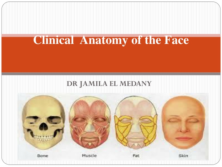
Clinical Anatomy of the Face DR JAMILA EL MEDANY
Criteria of Skin of the Face Numerous Sweat and Sebaceous glands. Connected to the underlying bone by Loose connective tissue. No Deep Fascia in the face, because of this, facial lacerations tend to gap. The looseness of the subcutaneous tissue enables fluid and blood to accumulate in the loose connective tissue following bruising of the face. Similarly , facial inflammation causes considerable swelling.
SKIN TENSION LINES (STLs) They are the result of a complex interaction between internal and external factors involving the skin. The intrinsic framework of the skin, which consists of elastin and collagen, progressively loosens with age. Its interaction with the muscles of facial expression leads to the development of (STLs ). Generally, STLs are perpendicular to the underlying muscles of the face. Aging, particularly, tends to accentuate the appearance of STL Incisions along these wrinkle lines heal with minimal scaring.
Muscles acting on the Fore head Frontalis muscle: It creates the horizontal wrinkles on the forehead and assists with eyebrow elevation giving the face a surprised looking. The corrugators and procerus muscles: Are the antagonistic muscles on the forehead. The dominant muscle of the Nose is the Nasalis muscle, which consists of Nasal and Alar components. Its function is to compress and dilate the nares.
MUSCLES OF THE EYE LIDS The Orbicularis Oculi muscles are a complex of muscles surrounding the eyes; these assist with closing the eye tightly. This complex lies superficially in the eyelid skin and is encountered with even a shallow incision. It has orbital part & palpebral part. Palpebral part : closes the eye lightly (as in plinking and sleeping). Orbital part : Closes the eye firmly (as in exposure to strong light).
MUSCLES OF THE MOUTH The mouth has the most extensive network of facial musculature and accounts for much of an individual's capability of facial expression. The Orbicularis Oris encircles the mouth and is the major component of the lips. The major functions of the orbicularis oris muscle are to pull the lips against the teeth, to draw the lips together, to pull the corners of the mouth together, and to pucker the mouth. This muscle is also extremely important for the phonation of sounds that rely on the lips, such as the pronunciation of the letters M,V,F, and P.
Quadratus Labii Superioris Muscles A group of 6 muscles, controls the upper mouth. The 6 muscles are as follows: Zygomaticus major muscle It helps in forming the lower nasolabial fold and is primarily responsible for smiling Zygomaticus minor muscle -Arises just medially to the zygomaticus major and assists with its functions. Levator labii superioris muscle - helps to elevate the medial part of the upper lip and assists the zygomatic muscles with open smiling ( Levator anguli oris muscle - The most deeply positioned of the lip elevators; it assist with lip elevation Risorius muscle - assists with smiling; the risorius is not always present.
MUSCLE OF THE CHEEK (BUCCINATOR The buccinator muscle is neither an elevator nor a depressor of the lip; it arises just posterior and medial to the last molar tooth and extends forward to become continuous with the orbicularis oris muscle The buccinator muscle is the major component of the cheek musculature and prevents overdistension of the cheek (eg, in playing a wind instrument). This muscle assists the orbicularis oris muscle in whistling.
MUSCLES OF MASTICATION (A) Temporalis, Masseter, Lateral & Medial pterygoids. Supplied by the mandibular nerve
ACTION OF MUSCLES OF MASTICATION SIDE TO SIDE MOVEMENT
Sensory Nerve Supply The Trigeminal nerve (the fifth cranial nerve, or simply CN V) is the nerve responsible for sensation in the face and motor functions such as biting and chewing. It has three major branches: the ophthalmic nerve (V1), the maxillary nerve (V2), and the mandibular nerve (V3). The ophthalmic and maxillary nerves are purely sensory, and the mandibular nerve has sensory (or "cutaneous") and motor functions.[1]
OPHTHALMIC NERVE Supplies : Forehead. Upper Eye Lid & Conjunctiva. Side of the Nose down to and including the tip. MAXILLARY NERVE Supplies : Posterior part of the side of the nose. Lower eye lid. Cheek & Upper lip . Lateral side of the orbital opening. Mandibular Nerve Supplies: Lower lip& chin. Lower part of the face &cheek. Temporal region. Part of the auricle, external auditory meatus and outer surface of tympanic membrane
Causes Treatmentdepends mostly on the use of & Antiepileptic (AED) & Antidepressant drugs (ADD) to relieve pain. In some cases Section of the sensory root is necessary.
Motor Nerve Supply (Facial Nerve) (7th cranial nerve). It gives motor supply to all muscles of facial expression. It does not supply the Skin. It enters the inner ear through the internal auditory meatus It passes through the middle ear (tympanic cavity) and leaves it to enter the Parotid gland. Within the parotid gland: It passes forward in a Horizontal direction At the anterior border of the gland, it divides into its Five terminal branches.
Bells Palsy It is a condition that causes the facial muscles to weaken or become paralyzed. It's caused by trauma to the 7th cranial nerve, and is not permanent. The most common cause is idiopathic but it can results from exposure to cold , tumor of parotid gland, lacerations of the face.
Manifestations Food accumulates during chewing and often must be continually removed by a finger. Patients frequently dab their eyes and mouth with a handkerchief to wipe the fluid (tears & saliva)which runs from the dropping lid and mouth. The fluid and constant wiping result in localised skin irritation.
Arterial Supply of Face The face has a rich blood supply so that it is rare for skin flaps in the face to necrose in plastic surgery. Facial wounds bleed freely but heal rapidly.
Facial Artery It arches upward Deep to the Submandibular salivary gland. It curves around the inferior margin of the body of the mandible at the anterior border of the masseter muscle. It runs upward in a tortuous course to the angle of the mouthdeep to the muscles. This is to accommodate itself to neck movements such as those of the pharynx in deglutition; and facial movements such as those of the mandible,lips, and cheeks. It ascends along the side of the nose to the medial angle of the eye and terminates as the Angular artery.
Branches of Facial artery MENTAL INFERIOR LABIAL SUPERIOR LABIAL LATERAL NASAL
Where to feel Facial Pulsation? As the artery crosses the inferior margin of the body of the mandible at the anterior border of the masseter muscle.
Compression of the Facial Artery The facial artery can be occluded by pressure against the mandible as the vessel crosses it. Because of the numerous anastomoses of the arteries of the face, compression of the facial artery on one side does not stop all bleeding from a lacerated facial artery or one of its branches. In lacerations of the lip, pressure must be applied on both sides of the cut to stop bleeding
Venous Drainage of the face It is drained mainly by Anterior & Posterior Facial (Retromandibular) veins. Anterior Fcial vein: Formed close to medial angle of the eye by union of: Supratrochlear & Supraorbital veins Descends in the face behind the facial artery. In the neck it joins the anterior branch of retromandibular to form Common Facial that ends in Internal jugular vein (IJV
Dangerous Area of the Face It is a triangular area bounded by: the Side of nose, Medial angle of the eye and Upper lip.
Thrombophlebitis of the Cavernous sinus This may result from lacerations of the nose or by squeezing pustules on the side of the nose and upper lip (dangerous area) In patients with inflammation of the facial vein with secondary thrombus, pieces of the infected clot may extend into the intracranial venous system and produce thrombophlebitis of the cavernous sinuses.
How it Happens? Blood from the nose, medial angle of the eye and lips usually drains inferiorly through the facial vein especially when the person is erect. Because the facial vein has no valves, blood may pass through it in the opposite direction to the inferior ophthalmic vein and enters the Cavernous Sinus
Lymph Drainage of Face Lymphatic nodes are categorized into several groups: Parotid lymph nodes: Receive lymph from the side of the face and scalp Submandibular lymph nodes: Get lymph from the upper lip and part of the lower lip as well as most of the oral cavity Submental lymph nodes: Get lymph from the chin, tip of the tongue and center of the lower lip Lymph from these nodes eventually drains into the Deep Cervical lymph nodes. The deep cervical lymph nodes drain into the Jugular Lymphatic Trunk, which joins the internal jugular vein or brachiocephalic vein on the right side and Thoracic Duct on the left side.


")


")
")