Pelvic Anatomy: Bones, Joints, and Muscles
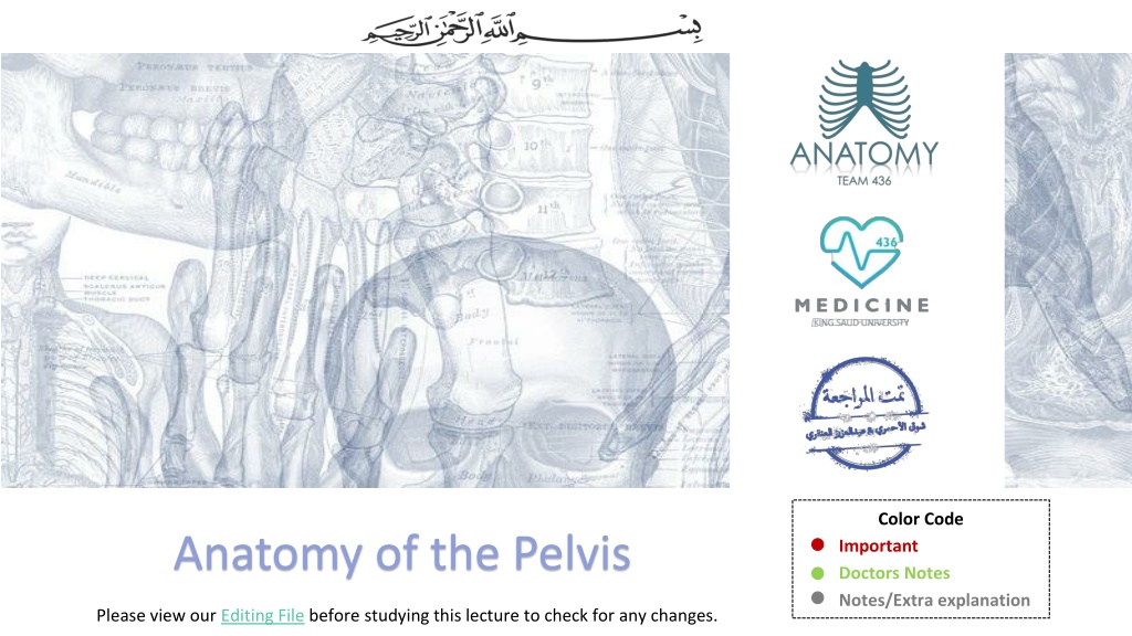
Anatomy of the Pelvis
Anatomy of the Pelvis
Please view our
Editing File
before studying this lecture to check for any changes.
Objectives
At the end of the lecture, students should be able to:
Describe the anatomy of the
pelvic wall
,
bones
,
joints
&
muscles
.
Describe the
boundaries
and
subdivisions
of the pelvis.
Differentiate the different
types
of the
female pelvis
.
Describe the
pelvic walls
&
floor
.
Describe the
components
&
function
of the
pelvic diaphragm
.
List the
arterial
&
nerve
supply
List the
lymph
&
venous
drainage of the pelvis.
م
لا
ح
ظ
ة
:
ل
م
ا
ن
ك
ت
ب
ا
و
ن
ق
ص
د
م
و
ج
و
د
ف
ق
ط
في
ج
س
م
ا
ل
ن
س
ا
ء
ا
و
ا
ل
ر
ج
ا
ل
ب
ي
ن
م
ا
ا
ذ
ا
ك
ت
ب
ن
ا
ا
و
ن
ق
ص
د
ا
خ
ت
ل
ا
ف
ف
ي
س
ل
ا
ي
د
ز
ا
ل
م
ح
ا
ض
ر
ة
ب
ين
ا
ل
د
ك
ت
و
ر
و
ا
ل
د
ك
ت
و
ر
ة
In females
In males
Girls
’ slides
Boys
’ slides
o
The
bony pelvis
is composed of
four
bones:
•
Two hip bones, which form the
anterior
and
lateral
walls.
•
Sacrum and coccyx, which form the
posterior
wall.
o
These
4
bones
are connected by
4
joints
and
lined by
4
muscles
.
o
The bony pelvis with its joints and muscles
form a strong
basin-shaped
(
حوض
)
structure
(with multiple foramina),
o
The pelvis
contains
and
protects
the:
(1) lower parts of the alimentary &
(2) urinary tracts &
(3) internal organs of reproduction.
I
n
t
r
o
d
u
c
t
i
o
n
The four joints are:
1- Symphysis pubis (2ry Cartilaginous joint)
Anteriorly
2-
Two
Sacroiliac joints. (Synovial joints)
Posteriolateraly
3- Sacrococcygeal joint
(
2ry
Cartilaginous joint)
Posteriorly
Between sacrum & coccyx
The pelvis is divided into two parts by the
pelvic brim
or inlet
:
P
e
l
v
i
s
*recall these are the same boundaries as the perineum
Extra
False pelvis
Inlet
True pelvis
Outlet
Perineum
ب
ا
ل
ت
ر
ت
ي
ب
ي
ك
و
ن
Pelvis
F
e
m
a
l
e
v
s
.
M
a
l
e
What is the main differences between male and female
pelvis?
1.
In female the
Sacrum
is usually
wider
and
shorter,
while in male is long and curved
.
2.
Also, the
Angle of the pubic
arch is
wider
.
3.
The
promontory
and the
ischial spines
are
less
projecting,
while the male is inverted.
Types of Female Bony Pelvis
o
Information of the shape and dimensions of the
female pelvis is of
great importance for obstetrics.
o
Why?
because it is the bony canal through which
the
child passes during birth
.
o
There are 4 types:
A.
Android
(resembles male pelvis)
B.
Gynecoid
(typical female type)
C.
Anthropoid
(has both male and female characteristics)
D.
Platypelloid
(least common)
**Gynecoid
is the only type that can give normal birth , the
other three types can’t.
Oval shaped
Heart shaped
P
e
l
v
i
c
W
a
l
l
s
The walls are formed by bones
and ligaments that are lined with
muscles covered with fascia and
parietal peritoneum.
10:26
P
e
l
v
i
c
W
a
l
l
s
P
e
l
v
i
c
W
a
l
l
s
o
It is formed by the
levator ani
and the
coccygeus
muscles
and their covering fasciae.
o
It is incomplete
anteriorly
to allow passage of:
•
the urethra in
males
and
•
urethra and vagina in
females
.
o
Posteriorly
the muscles of each side meet together.
P
e
l
v
i
c
D
i
a
p
h
r
a
g
m
•
It is a wide thin sheet-like muscle that has a linear origin .
•
ORIGIN
:
(look at the dotted yellow line)
1.
Back of the body of the pubis
2.
Tendinous arch of the obturator fascia
3.
Spine of the ischium
(ischial spine)
.
•
Its fibers are divided into 3 parts:
1.
Pubococcygeus
2.
Puborectalis
3.
Iliococcygeus
(Ischiococcygeus)
02:54
Pelvic Diaphragm
L
e
v
a
t
o
r
e
s
A
n
i
M
u
s
c
l
e
s
1.
Pubococcygeus
o
Origin:
originates from the posterior surface of
the body of the pubis and passes downward &
medially
o
Insertion:
inserted into the perineal body,
anococcygeal body and coccyx.
o
Action:
•
forms a sling around the prostate or the
vagina*:
Levatores Ani Muscles
A
n
t
e
r
i
o
r
F
i
b
e
r
s
In males
Levator prostate:
1.
Supports prostate
2.
Stabilizes perineal body
*While the muscle goes from origin to insertion it surrounds
these 2 organs (prostate or vagina) and supports them.
In females
Sphincter vaginae:
1.
Supports vagina
2.
Stabilizes perineal body
4. Ischiococcygeus*
(only on the
girls
’ slides)
o
Origin:
Arises from the ischial spine
o
Insertion:
inserted into the anococcygeal body &
coccyx.
3. Iliococcygeus
o
Origin:
Inserted into
the anococcygeal body and
the coccyx
Levatores Ani Muscles
I
n
t
e
r
m
e
d
i
a
t
e
F
i
b
e
r
s
P
o
s
t
e
r
i
o
r
F
i
b
e
r
s
2. Puborectalis
(
(شكلها زي اللجام بتاع الخيل
•
forms a sling around the recto-anal Junction.
•
It has a very important role in maintaining
fecal
continence
.
•
Its tone is important to maintain the angle
between rectum & anal canal. When it relaxes,
the angle is gone and defecation can occur
*
ا
ل
د
ك
ا
ت
ر
ه
ا
خ
ت
ل
ف
و
ا
في
ه
ذ
ه
ا
ل
ع
ض
ل
ة
د
.
ا
ب
و
ا
ل
م
ك
ا
ر
م
ق
ا
ل
ا
ن
ه
ا
ف
ي
ا
س
م
ث
ا
ن
ي
ل
ـ
م
و
ج
و
د
ه
ف
ي
ب
ع
ض
ا
ل
ك
ت
ب
.
و
د
.
ج
م
ي
ل
ة
ت
ق
و
ل
في
ا
خ
ت
لا
ف
ف
ي
ا
ل
ا
ر
ا
ء
ب
ح
ي
ث
ا
ل
ب
ع
ض
ي
ع
ت
بر
ه
ا
ج
ز
ء
م
ن
.
ر
ي
ح
و
ا
ب
ا
ل
ك
م
و
ا
ن
س
و
ا
ع
ن
ه
ا
.
ا
ه
م
ش
يئ
ر
ك
ز
و
ا
ع
لى
ا
ل
ث
لا
ث
ه
ا
لا
و
لى
لا
ن
ه
ا
هي
ا
لا
س
ا
س
ي
ة
iliococcygeus
coccygeus
Actions of levator ani:
1.
The muscles of the two sides form an efficient muscular sling that
supports
and
maintains
the
pelvic viscera
in position.
(so if it is injured rectal or vaginal prolapse may occur)
2.
They
resist the rise in intra pelvic pressure
during the straining and expulsive efforts of the abdominal
muscles (as in coughing).
3.
They have a very important role in
maintaining fecal continence
(puborectalis)
by acting as a sphincter at
the anorectal junction .
4.
They serve as a
vaginal sphincter
in the female.
(
The most important muscle in labour is levator ani: should be
relaxed so the vaginal sphincter isn
’
t closed)
Nerve supply to levator ani:
1. perineal branch of the
fourth sacral nerve (S4)
upper surface
.
2. perineal branch of the
pudendal nerve
lower surface
.
Levatores Ani Muscles
S
u
p
p
l
y
&
A
c
t
i
o
n
The importance of the different supplies is if one of these nerves is affected, the other is still intact
This table is a summary of the slides before.
Pelvic Diaphragm
C
o
c
c
y
g
e
u
s
M
u
s
c
l
e
This muscle is bigger in animals because they have tails.
I) Internal iliac artery (IIA)
o
One of the 2 terminal branch of the
Common
iliac artery
.
o
Course:
1.
Arises in front of the sacroiliac joint
2.
It descends downward & backwards over the
pelvic inlet.
3.
It
divides
at the upper border of the
greater sciatic foramen
into:
Anterior & Posterior divisions
.
A
r
t
e
r
i
e
s
o
f
t
h
e
P
e
l
v
i
s
Extra
Extra
Extra
The abdominal aorta divides into 2 common iliac arteries. Then at the sacroiliac joint the common iliac divides into external and internal iliac arteries. The
external iliac continues as the femoral artery, while the internal iliac supplies the pelvis and perineum.
Then at the greater sciatic foramen the internal iliac
divides into anterior and posterior divisions.
P
A
II) Ovarian artery (in female):
o
Arises from the
abdominal aorta.
08:29
A
r
t
e
r
i
e
s
o
f
t
h
e
P
e
l
v
i
s
I
n
t
e
r
n
a
l
I
l
i
a
c
A
r
t
e
r
y
•
Posterior division Supplies:
(supplies only walls)
1.
Posterior abdominal wall.
2.
Posterior pelvic wall.
3.
Gluteal region.
•
Anterior division supplies
:
(supplies wall & viscera)
1.
Gluteal region.
2.
Perineum.
3.
Pelvic viscera.
4.
Medial (adductor) region of thigh
(by obturator
artery).
5.
The fetus (through the umbilical arteries).
Extra
A
r
t
e
r
i
e
s
o
f
t
h
e
P
e
l
v
i
s
I
n
t
e
r
n
a
l
I
l
i
a
c
A
r
t
e
r
y
* Supplies urinary bladder:
In male (superior and inferior vesical arteries)
In female (superior vesical & vaginal artery)
A
r
t
e
r
i
e
s
o
f
t
h
e
P
e
l
v
i
s
I
n
t
e
r
n
a
l
I
l
i
a
c
A
r
t
e
r
y
Summary of the Branches
(only on the
girls
’ slides)
:
*recall the arteries that
have tortuous course:
1.
Facial artery
2.
Splenic artery
3.
Uterine artery
S
u
p
p
l
y
o
f
t
h
e
P
e
l
v
i
s
V
e
n
o
u
s
D
r
a
i
n
a
g
e
1. Internal iliac veins:
•
It collect tributaries corresponding to the
branches of the internal iliac artery.
•
joins the external iliac vein in front of the
sacroiliac joint
to form the
common iliac
vein
(the common iliac veins join at the level of L5
to give the inferior vena cava)
.
2. Ovarian vein:
•
Right
vein drains into IVC.
•
Left
vein drains into left renal Vein.
The veins correspond to the arteries.
Clinical point:
There are venous plexuses around uterus, bladder, rectum…
Their importance is that they can drain to vertebral venous plexuses
So venous blood can shift to vertebral venous plexuses ascend through foramen magnum then to occipital sinus in cranium
That means if there is cancer in either uterus, bladder, rectum the metastasis reaches the brain quickly (faster than other places)
S
u
p
p
l
y
o
f
t
h
e
P
e
l
v
i
s
L
y
m
p
h
a
t
i
c
D
r
a
i
n
a
g
e
o
The lymph nodes and vessels are
arranged in a chain along the main
blood vessels.
o
Thus, there are
external iliac nodes
,
internal iliac nodes
, and
common iliac
nodes
.
o
Lymph from Common iliac nodes & the
(Ovaries, uterine tubes & fundus of
uterus) passes to
Lateral aortic
(paraortic) nodes
which then passes to
cisterna chyli (thoracic duct)
S
u
p
p
l
y
o
f
t
h
e
P
e
l
v
i
s
N
e
r
v
e
S
u
p
p
l
y
M
C
Q
s
1-The bony pelvis is composed of four bones :
A-are connected by 3 joints and lined by 4 muscles.
B-are connected by 4 joints and lined by 3 muscles.
C-are connected by 4 joints and lined by 4 muscles.
D-are connected by 2joints and lined by 8 muscles.
2-The Sacroiliac joints is :
A- Anteriolateral cartilaginous join
B-posteriomedial cartilaginous joint
C- posteriolateral Synovial joint
D- Anteriomedial Synovial joint
3- The False pelvis is bounded by :
A- T11
B-C6
C-L1
D-S2
4- Which of the following is true about the PELVIC INLET:
A- Bounded Posteriorly by the Coccyx
B- Bounded Anteriorly by the Ileopectineal (arcuate) lines.
C- Bounded lateraly by the Promontory of sacrum
D-Bounded posteriorly by the ala of sacrum.
5-Which of the following is a female of the pubic arch angle
:
A- 45
B- 65
C-75
D- 85
6-
A 28 year old women ,school teacher from
Dublin
had a positive pregnancy test. Her obstetrician informed her that it is
impossible for her to deliver normally because of the type of her bony pelvis ,
which of the following types of pelvis can have normal vaginal delivery?
A- Gynecoid Pelvis
B- Android Pelvis
C- Anthropoid Pelvis
D- Platypelloid Pelvis
Answers: 1-C, 2-C, 3-C , 4-D, 5-D, 6-A, 7-A, 8-D, 9-B.
7-No muscles are found in :
A- The anterior pelvic wall
B- The posterior pelvic wall
C- The lateral pelvic wall
D- The medial pelvic wall
8-Which of the following is true about the piriformis muscle :
A- Origin from the Inner surface of the obturator membrane and the hip bone.
B- inserted in the pelvic surface of the middle 3 sacral vertebrae.
C- Supplied by the lumber plexus.
D- leaves the pelvis through the greater sciatic foramen.
9-Which of the following is true about the PELVIC DIAPHRAGM :
A- It is incomplete posteriorly to allow
passage of the anal canal.
B- it forms the INFERIOR PELVIC WALL.
C- Main (true) pelvic cavity below it.
D- formed by the levator ani and the coccygeus muscles without their covering
fasciae.
17. Woman is undergoing hysterectomy and during the ligation of uterine
artery which of the following will be endanger:-
A.
Ureter
B.
Urinary bladder
C.
Ovarian artery
Answers: 10:C, 11:A, 12:B, 13:C, 14:C, 15:B, 16:B,
17
:A
10. Which one the following fibers of Levatores Ani muscles considered
as the intermediate part?
A.
Pubococcygeus
B.
Ischiococcygeus
C.
Iliococcygeus
11. The Nerve supply to the Coccygeus muscle
:
A.
Branches of 4
th
and 5
th
sacral nerves
B.
Branches of pudendal nerve
C.
both
12: The
medial
umbilical ligament results from?
A.
Fibrosed umbilical vein
B.
Fibrosed umbilical artery
C.
Superior vesical artery
13. Which of the following arteries supply the urinary bladder in
female
:
A.
Uterine artery
B.
Inferior vesical artery
C.
Vaginal artery
M
C
Q
s
14. The right ovarian vein drains into:
A.
Renal vein
B.
Common iliac vein
C.
Inferior vena cava
15. The relaxation of which of the following muscle fibers lead to defecation?
A.
Pubococcygeus
B.
Puborectalis
C.
Coccygeus
16
.
The ovarian artery originate from
:
A.
Uterine artery
B.
Abdominal aorta
C.
Internal iliac artery
Q1: A 14 years old patient was diagnosed with
osteosarcoma in the distal femur 3 months after
starting the treatment the doctor found that the
tumor have metastasized to the right
hip bone.
A-What forms the LATERAL PELVIC WALLLATERAL :
It is formed by:
1- Part of the hip bone below the pelvic inlet,
2- Obturator internus and its covering fascia & the
obturator fascia.
3- Sacrotuberous ligament.
4- Sacrospinous ligament.
B- What is the action of
the Obturator internus
muscle
:
Action: Lateral rotator of the femur at the hip joint.
Q2: What is the nerve supply of levator ani muscles?
1. perineal branch of the fourth sacral nerve.
2. perineal branch of the pudendal nerve.
Q3: The arterial supply of urinary bladder in male and female are:
In male : superior vesical artery + inferior vesical artery
In female : superior vesical artery + vaginal artery
Q4: Define the anterior divisions of Internal Iliac Artery :-
Obturator Artery - Inferior Gluteal Artery - Vaginal Artery -
Middle Rectal Artery - Internal Pudendal Artery - Uterine
Artery - Inferior Vesical Artery - Umbilical Artery Gives:
superior vesical artery and Artery of vas deferens.
Q5: Describe nerve supply of the pelvis :-
Somatic > Sacral plexus
Autonomic >
-
Sympathetic: Pelvic part of sympathetic trunk
-
Parasympathetic: Pelvic splanchnic nerves From S 2 , 3 & 4.
S
A
Q
s
Cartilaginous
synovial
S
U
M
M
A
R
Y
Leaders:
Nawaf AlKhudairy
Jawaher Abanumy
References:
1- Girls’ & Boys’ Slides
2- Greys Anatomy for Students
3- TeachMeAnatomy.com
anatomyteam436@gmail.com
@anatomy436
Feedback
Members:
Ashwaq Almajed
Ameera Niazi
Dania Alkelabi
Do'aa abdulfattah
Muneerah Alzayed
Nourah Al Hogail
Nouf Aloqaili
Sarah Alshamrani
Explore the comprehensive overview of the anatomy of the pelvis, including its walls, bones, joints, muscles, and differentiations between male and female pelves. Learn about the boundaries, subdivisions, and pelvic floor components, as well as the arterial, nerve, lymph, and venous supplies. Delve into the distinctions between the false and true pelvis, types of female pelves, and their significance in obstetrics. Unravel the intricacies of the bony pelvis structure and function through detailed doctors' notes and illustrations.

Download Presentation

Please find below an Image/Link to download the presentation.
The content on the website is provided AS IS for your information and personal use only. It may not be sold, licensed, or shared on other websites without obtaining consent from the author.If you encounter any issues during the download, it is possible that the publisher has removed the file from their server.
You are allowed to download the files provided on this website for personal or commercial use, subject to the condition that they are used lawfully. All files are the property of their respective owners.
The content on the website is provided AS IS for your information and personal use only. It may not be sold, licensed, or shared on other websites without obtaining consent from the author.
E N D
Presentation Transcript
Color Code Important Doctors Notes Notes/Extra explanation Anatomy of the Pelvis Please view our Editing File before studying this lecture to check for any changes.
Objectives At the end of the lecture, students should be able to: Describe the anatomy of the pelvic wall, bones, joints & muscles. Describe the boundaries and subdivisions of the pelvis. Differentiate the different types of the female pelvis. Describe the pelvic walls & floor. Describe the components & function of the pelvic diaphragm. List the arterial & nerve supply List the lymph & venous drainage of the pelvis. Boys slides Girls slides : In males In females
Introduction Introduction o The bony pelvis is composed of four bones: Two hip bones, which form the anterior and lateral walls. Sacrum and coccyx, which form the posterior wall. o These 4 bones are connected by 4 joints and lined by 4 muscles. o The bony pelvis with its joints and muscles form a strong basin-shaped ( ) structure (with multiple foramina), o The pelvis contains and protects the: (1) lower parts of the alimentary & (2) urinary tracts & (3) internal organs of reproduction. The four joints are: 2- Two Sacroiliac joints. (Synovial joints) Posteriolateraly 3- Sacrococcygeal joint (2ry Cartilaginous joint) Posteriorly Between sacrum & coccyx 1- Symphysis pubis (2ry Cartilaginous joint) Anteriorly
False pelvis Inlet True pelvis Outlet Perineum Pelvis Pelvis The pelvis is divided into two parts by the pelvic brim or inlet: False or greater pelvis Above the brim (part of the abdominal cavity) True or lesser pelvis Below the brim. Has 3 parts: Pelvic brim Bounded by: 1. A Cavity: The cavity is a short, curved canal, with a shallow anterior wall and a deeper posterior wall. It lies between the inlet and the outlet. Posteriorly: Lumbar vertebrae. Laterally: Iliac fossae and the iliacus muscle. Anteriorly: Lower part of the anterior abdominal wall. 2. Inlet (oval shape) 3. Outlet* (Diamond shape) Anteriorly: Anteriorly: Symphysis pubis (upper border) Symphysis pubis (lower border) It supports the abdominal contents Posteriorly: Posteriorly : Promontory of sacrum, ala of sacrum. Tip of Coccyx. Laterally: (both sides) Anterolaterally: ischiopubic ramus. Ileopectineal (arcuate) lines. Posterolaterally: Sacrotuberous ligament & ischial tuberosities. (Ileo = illum \ pectineal = pubis) Extra *recall these are the same boundaries as the perineum
Pelvis Female vs. Male Female vs. Male Types of Female Bony Pelvis o Information of the shape and dimensions of the female pelvis is of great importance for obstetrics. o Why? because it is the bony canal through which the child passes during birth. o There are 4 types: A. Android (resembles male pelvis) B. Gynecoid (typical female type) C. Anthropoid (has both male and female characteristics) D. Platypelloid (least common) FEMALE MALE Oval shaped Heart shaped What is the main differences between male and female pelvis? **Gynecoid is the only type that can give normal birth , the other three types can t. 1. In female the Sacrum is usually wider and shorter, while in male is long and curved. 2. Also, the Angle of the pubic arch is wider. 3. The promontory and the ischial spines are less projecting, while the male is inverted.
Image result for youtube Pelvic Walls Pelvic Walls 10:26 Anterior Posterior The walls are formed by bones and ligaments that are lined with muscles covered with fascia and parietal peritoneum. The pelvis has 4 walls: Inferior (floor) lateral 1. Anterior pelvic wall 2. Posterior pelvic wall It is the shallowest wall and is formed by only bones: (1) the posterior surfaces of the bodies of the pubic bones, (2) the 2 pubic rami, and (3) the symphysis pubis. It is large and deeper. Formed by: (1) sacrum, (2) coccyx , (3) piriformis muscles (right and left) and (4) their covering of parietal pelvic fascia.
Pelvic Walls Pelvic Walls 3. Lateral pelvic wall 4. Inferior pelvic wall (floor) It is formed by: 1. Part of the hip bone below the pelvic inlet (the arrow), 2. Obturator internus and its covering fascia, the obturator fascia. 3. Sacrotuberous ligament. 4. Sacrospinous ligament. Basin-like structure which supports the pelvic viscera and is formed by the pelvic diaphragm. It stretches across the lower part of the true pelvis and divides it into: 1. Main (true) pelvic cavity above, which contains the pelvic viscera 2. Perineum below which carries the external genital organs.
Pelvic Walls Pelvic Walls Piriformis muscle (part of posterior pelvic wall) Obturator internus muscle (part of lateral pelvic wall) Origin Pelvic surface (in front of) the middle 3 sacral vertebrae. It leaves the pelvis through the greater sciatic foramen. Origin Inner surface of the obturator membrane and the hip bone. It leaves the pelvis through the lesser sciatic foramen Insertion Greater trochanter of the femur. Insertion Greater trochanter of the femur. Action Lateral rotator of the femur at the hip joint. Action Lateral rotator of the femur at the hip joint. Innervation Sacral plexus (lies in front of the muscles). Innervation Nerve to obturator internus (from sacral plexus).
Pelvic Diaphragm Pelvic Diaphragm o It is formed by the levator ani and the coccygeus muscles and their covering fasciae. o It is incomplete anteriorly to allow passage of: the urethra in males and urethra and vagina in females. o Posteriorly the muscles of each side meet together.
Pelvic Diaphragm Levatores Levatores Ani Muscles Ani Muscles Image result for youtube 02:54 It is a wide thin sheet-like muscle that has a linear origin . ORIGIN: (look at the dotted yellow line) 1. Back of the body of the pubis 2. Tendinous arch of the obturator fascia 3. Spine of the ischium (ischial spine). Its fibers are divided into 3 parts: 1. Pubococcygeus 2. Puborectalis 3. Iliococcygeus (Ischiococcygeus) Extra
Levatores Ani Muscles Anterior Fibers Anterior Fibers 1. Pubococcygeus o Origin: originates from the posterior surface of the body of the pubis and passes downward & medially o Insertion: inserted into the perineal body, anococcygeal body and coccyx. o Action: forms a sling around the prostate or the vagina*: In males In females Sphincter vaginae: 1. Supports vagina 2. Stabilizes perineal body Levator prostate: 1. Supports prostate 2. Stabilizes perineal body Pubococcygeus levator prostatae *While the muscle goes from origin to insertion it surrounds these 2 organs (prostate or vagina) and supports them. sphincter vaginae
Levatores Ani Muscles Intermediate Fibers Intermediate Fibers 2. Puborectalis ( ( It has a very important role in maintaining fecal continence. Its tone is important to maintain the angle between rectum & anal canal. When it relaxes, the angle is gone and defecation can occur forms a sling around the recto-anal Junction. Posterior Posterior Fibers Fibers 4. Ischiococcygeus* (only on the girls slides) 3. Iliococcygeus o Origin: Arises from the ischial spine o Origin: Inserted into the anococcygeal body and the coccyx o Insertion: inserted into the anococcygeal body & coccyx. . . . coccygeus iliococcygeus . * .
Levatores Ani Muscles Supply & Action Supply & Action Nerve supply to levator ani: 1. perineal branch of the fourth sacral nerve (S4) upper surface. 2. perineal branch of the pudendal nerve lower surface. The importance of the different supplies is if one of these nerves is affected, the other is still intact Actions of levator ani: 1. The muscles of the two sides form an efficient muscular sling that supports and maintains the pelvic viscera in position. (so if it is injured rectal or vaginal prolapse may occur) 2. They resist the rise in intra pelvic pressure during the straining and expulsive efforts of the abdominal muscles (as in coughing). 3. They have a very important role in maintaining fecal continence (puborectalis) by acting as a sphincter at the anorectal junction . 4. They serve as a vaginal sphincter in the female. (The most important muscle in labour is levator ani: should be relaxed so the vaginal sphincter isn t closed)
This table is a summary of the slides before. Muscle Origin Insertion Action Innervation 1. 2. 3. perineal body anococcygeal body coccyx Pubococcygeus 1. Support and maintain the pelvic viscera in position. (forms a sling around the prostate or the vagina) forms a sling around the recto- anal Junction. 2. They resist the rise in intra pelvic pressure during the straining and expulsive efforts of the abdominal muscles (as in coughing). Puborectalis 1. Back of the body of the pubis Levator Ani Muscle It has a very important role in maintaining fecal continence. 1. perineal branch of the fourth sacral nerve 2. Tendinous arch of the obturator fascia 1. 2. anococcygeal body coccyx 2. perineal branch of the pudendal nerve. 3. Spine of the ischium. Iliococcygeus 3. Maintaining fecal continence. 4. They serve as a vaginal sphincter in the female. 1. 2. anococcygeal body coccyx Ischiococcygeus
Pelvic Diaphragm Coccygeus Muscle Coccygeus Muscle This muscle is bigger in animals because they have tails. Coccygeus: small triangular muscle. Origin Ischial spine Coccygeus muscle has the same attachment as the sacrospinous ligament! Insertion Lower end of sacrum and coccyx Action Assist the levator ani in supporting the pelvic viscera branches of the 4thand 5thsacral nerves (S4 & S5). Innervation Extra
Image result for youtube IIA Arteries of the Pelvis Arteries of the Pelvis 08:29 I) Internal iliac artery (IIA) o One of the 2 terminal branch of the Common iliac artery. o Course: 1. Arises in front of the sacroiliac joint 2. It descends downward & backwards over the pelvic inlet. 3. It divides at the upper border of the greater sciatic foramen into: Anterior & Posterior divisions. A P Extra II) Ovarian artery (in female): o Arises from the abdominal aorta. Extra Extra The abdominal aorta divides into 2 common iliac arteries. Then at the sacroiliac joint the common iliac divides into external and internal iliac arteries. The external iliac continues as the femoral artery, while the internal iliac supplies the pelvis and perineum. Then at the greater sciatic foramen the internal iliac divides into anterior and posterior divisions.
Arteries of the Pelvis Internal Iliac Artery Internal Iliac Artery Posterior division Supplies: (supplies only walls) 1. Posterior abdominal wall. 2. Posterior pelvic wall. 3. Gluteal region. Anterior division supplies: (supplies wall & viscera) 1. Gluteal region. 2. Perineum. 3. Pelvic viscera. 4. Medial (adductor) region of thigh (by obturator artery). 5. The fetus (through the umbilical arteries). Extra
Arteries of the Pelvis Internal Iliac Artery Internal Iliac Artery Parietal Branches From posterior division From anterior division: 1. Iliolumbar artery. 2. Lateral sacral arteries (2 branches.) 3. Superior gluteal artery. 4. Obturator artery. 5. Inferior Gluteal artery. Visceral Branches (all from anterior division only) 1. Umbilical artery :Gives the superior vesical* artery: The distal part of this artery fibrosed and forms the Medial Umbilical Ligament. 2. Inferior Vesical artery in male or vaginal in female: In the male it supplies, the Prostate and the Seminal Vesicles. It also gives the artery of the Vas Deferens. 3. Middle rectal artery 4. Internal pudendal artery It is the main arterial supply to the perineum. * Supplies urinary bladder: In male (superior and inferior vesical arteries) In female (superior vesical & vaginal artery)
Arteries of the Pelvis Internal Iliac Artery Internal Iliac Artery Summary of the Branches (only on the girls slides): Visceral Branches (In Female) 1. Vaginal artery: Replaces the inferior vesical artery. 2. Uterine artery: has tortuous* course Crosses the Ureter superiorly and supplies the uterus & uterine tubes. N.B. The ureter may be wrongly ligated (cut) in hysterectomy (removal of uterus) 3. Ovarian artery from abdominal aorta *recall the arteries that have tortuous course: 1. Facial artery 2. Splenic artery 3. Uterine artery
Supply of the Pelvis Venous Drainage Venous Drainage The veins correspond to the arteries. 1. Internal iliac veins: It collect tributaries corresponding to the branches of the internal iliac artery. joins the external iliac vein in front of the sacroiliac joint to form the common iliac vein (the common iliac veins join at the level of L5 to give the inferior vena cava). 2. Ovarian vein: Right vein drains into IVC. Left vein drains into left renal Vein. Clinical point: There are venous plexuses around uterus, bladder, rectum Their importance is that they can drain to vertebral venous plexuses So venous blood can shift to vertebral venous plexuses ascend through foramen magnum then to occipital sinus in cranium That means if there is cancer in either uterus, bladder, rectum the metastasis reaches the brain quickly (faster than other places)
Supply of the Pelvis Lymphatic Drainage Lymphatic Drainage o The lymph nodes and vessels are arranged in a chain along the main blood vessels. o Thus, there are external iliac nodes, internal iliac nodes, and common iliac nodes. o Lymph from Common iliac nodes & the (Ovaries, uterine tubes & fundus of uterus) passes to Lateral aortic (paraortic) nodes which then passes to cisterna chyli (thoracic duct)
Supply of the Pelvis Nerve Supply Nerve Supply Nerve Supply Somatic Autonomic Sacral plexus: Sympathetic: Parasympathetic: Pelvic splanchnic nerves (From S 2 , 3 & 4): preganglionic fibers to pelvic viscera & hindgut from ventral (anterior rami) of L4 & L5 (lumbosacral trunk) + S1, S2, S3 and most of S4. It gives pudendal nerve to perineum. Pelvic part of sympathetic trunk: It is the continuation of the abdominal part of sympathetic trunk. It descends in front of the ala of the sacrum. The 2 sympathetic trunks unite inferiorly in front of the coccyx and form a single ganglion (Ganglion Impar). Superior & Inferior Hypogastric plexuses
MCQs MCQs 1-The bony pelvis is composed of four bones : A-are connected by 3 joints and lined by 4 muscles. B-are connected by 4 joints and lined by 3 muscles. C-are connected by 4 joints and lined by 4 muscles. D-are connected by 2joints and lined by 8 muscles. 6- A 28 year old women ,school teacher from Dublin had a positive pregnancy test. Her obstetrician informed her that it is impossible for her to deliver normally because of the type of her bony pelvis , which of the following types of pelvis can have normal vaginal delivery? A- Gynecoid Pelvis B- Android Pelvis C- Anthropoid Pelvis D- Platypelloid Pelvis 2-The Sacroiliac joints is : A- Anteriolateral cartilaginous join B-posteriomedial cartilaginous joint C- posteriolateral Synovial joint D- Anteriomedial Synovial joint 7-No muscles are found in : A- The anterior pelvic wall C- The lateral pelvic wall B- The posterior pelvic wall D- The medial pelvic wall 3- The False pelvis is bounded by : A- T11 B-C6 8-Which of the following is true about the piriformis muscle : A- Origin from the Inner surface of the obturator membrane and the hip bone. B- inserted in the pelvic surface of the middle 3 sacral vertebrae. C- Supplied by the lumber plexus. D- leaves the pelvis through the greater sciatic foramen. C-L1 D-S2 4- Which of the following is true about the PELVIC INLET: A- Bounded Posteriorly by the Coccyx B- Bounded Anteriorly by the Ileopectineal (arcuate) lines. C- Bounded lateraly by the Promontory of sacrum D-Bounded posteriorly by the ala of sacrum. 9-Which of the following is true about the PELVIC DIAPHRAGM : A- It is incomplete posteriorly to allow passage of the anal canal. B- it forms the INFERIOR PELVIC WALL. C- Main (true) pelvic cavity below it. D- formed by the levator ani and the coccygeus muscles without their covering fasciae. 5-Which of the following is a female of the pubic arch angle: A- 45 B- 65 C-75 D- 85 Answers: 1-C, 2-C, 3-C , 4-D, 5-D, 6-A, 7-A, 8-D, 9-B.
MCQs MCQs 10. Which one the following fibers of Levatores Ani muscles considered as the intermediate part? A. Pubococcygeus B. Ischiococcygeus C. Iliococcygeus 14. The right ovarian vein drains into: A. Renal vein B. Common iliac vein C. Inferior vena cava 15. The relaxation of which of the following muscle fibers lead to defecation? A. Pubococcygeus B. Puborectalis C. Coccygeus 11. The Nerve supply to the Coccygeus muscle: A. Branches of 4thand 5thsacral nerves B. Branches of pudendal nerve C. both 16. The ovarian artery originate from: A. Uterine artery B. Abdominal aorta C. Internal iliac artery 12: The medial umbilical ligament results from? A. Fibrosed umbilical vein B. Fibrosed umbilical artery C. Superior vesical artery 17. Woman is undergoing hysterectomy and during the ligation of uterine artery which of the following will be endanger:- A. Ureter B. Urinary bladder C. Ovarian artery 13. Which of the following arteries supply the urinary bladder in female: A. Uterine artery B. Inferior vesical artery C. Vaginal artery Answers: 10:C, 11:A, 12:B, 13:C, 14:C, 15:B, 16:B, 17:A
SAQs SAQs Q1: A 14 years old patient was diagnosed with osteosarcoma in the distal femur 3 months after starting the treatment the doctor found that the tumor have metastasized to the right hip bone. Q2: What is the nerve supply of levator ani muscles? 1. perineal branch of the fourth sacral nerve. 2. perineal branch of the pudendal nerve. Q3: The arterial supply of urinary bladder in male and female are: In male : superior vesical artery + inferior vesical artery In female : superior vesical artery + vaginal artery A-What forms the LATERAL PELVIC WALLLATERAL : It is formed by: 1- Part of the hip bone below the pelvic inlet, 2- Obturator internus and its covering fascia & the obturator fascia. 3- Sacrotuberous ligament. 4- Sacrospinous ligament. Q4: Define the anterior divisions of Internal Iliac Artery :- Obturator Artery - Inferior Gluteal Artery - Vaginal Artery - Middle Rectal Artery - Internal Pudendal Artery - Uterine Artery - Inferior Vesical Artery - Umbilical Artery Gives: superior vesical artery and Artery of vas deferens. B- What is the action of the Obturator internus muscle: Action: Lateral rotator of the femur at the hip joint. Q5: Describe nerve supply of the pelvis :- Somatic > Sacral plexus Autonomic > - Sympathetic: Pelvic part of sympathetic trunk - Parasympathetic: Pelvic splanchnic nerves From S 2 , 3 & 4.
SUMMARY SUMMARY Pelvis composed of 4 bones : internal iliac artery divides to: 1. anterior ( gives parietal & visceral branches) 2. Posterior ( gives parietal branches) Arterial supply : 2hip bones Sacrum & coccyx Connected by 4 joints : Symphsis pubis (Ant.) Sacrococcygeal(Post.) 2 sacroiliac joints (potserolateraly) Cartilaginous synovial internal iliac vein & ovarian vein Venous drainage : False pelvis which bounded by True pelvis Which has : Pelvic inlet,outlet,andcavity external, internal and common iliac node Lymphatic drainage : Pelvic inlet Pelvic outlet Anteriorly symphysis pubis somatic : sacral plexus Autonomic : 1. sympathetic( pelvic part of sympathetic trunk , Superior and inferior hypogastric plexus) 2.Parasympthatic : pelvic splanchnic nerve (S2,3&4) Nerve supply : 1. Promontry of sacrum 2. Ala of sacrum Post. : lumber vertebrae Ant. : lower part of abdominal wall Laterally : iliac fossa & iliacus ms. Divides into : Posteriorly coccyx Anterolaterally: ischiopubic ramus Psterolaterally : sacrotuberous ligament Arcuate lines Laterally
Members: Ashwaq Almajed Ameera Niazi Dania Alkelabi Do'aa abdulfattah Muneerah Alzayed Nourah Al Hogail Nouf Aloqaili Sarah Alshamrani Leaders: Nawaf AlKhudairy Jawaher Abanumy Image result for google form icon Feedback Feedback References: 1- Girls & Boys Slides 2- Greys Anatomy for Students 3- TeachMeAnatomy.com anatomyteam436@gmail.com @anatomy436