Thoracic Cage and Respiratory Movements in Anatomy Lecture

Prof. Ahmed Fathalla Ibrahim
Professor of Anatomy
College of Medicine
College of Medicine
King Saud University
E-mail: ahmedfathala@hotmail.com
 
OBJECTIVES
OBJECTIVES
 
At the end of the lecture, students should:
Describe the components of the thoracic cage
and their articulations.
Describe in brief the respiratory movements.
List the muscles involved in inspiration and in
expiration.
Describe the attachments of each muscle to the
thoracic cage and its nerve supply.
Describe the origin, insertion, nerve supply of
diaphragm.
THORACIC CAGE
THORACIC CAGE
Rib
Vertebra
 
THORACIC CAGE
THORACIC CAGE
 
Conical
Conical
 
in shape
Has 2 apertures (openings):
1.
Superior 
Superior 
(thoracic outlet): 
(thoracic outlet): 
narrow, open,
continuous with neck
2.
Inferior:
Inferior:
 wide, closed by diaphragm
Formed of:
1.
Sternum & costal cartilages
: 
anteriorly
2.
Twelve pairs of ribs: 
laterally
3.
Twelve thoracic vertebrae: 
posteriorly
ARTICULATIONS
ARTICULATIONS
Sternocostal
Costochondral
Costovertebral
ARTICULATIONS
ARTICULATIONS
 
Sternocostal
Costochondral
Costovertebral
 
RESPIRATORY MOVEMENTS
A- MOVEMENTS OF DIAPHRAGM
 
Contraction (descent)
of diaphragm


Increase of 
vertical diameter
vertical diameter
of  thoracic cavity
Inspiration
Inspiration
Expiration
Expiration
 
Relaxation (ascent)
of diaphragm)
 
RESPIRATORY MOVEMENTS
B- MOVEMENTS OF RIBS


PUMP HANDLE MOVEMENT
Elevation of ribs
Elevation of ribs
 
Increase in antero-posterior diameter
of thoracic cavity
of thoracic cavity
 
BUCKET HANDLE MOVEMENT
Elevation of ribs
Elevation of ribs
 
Increase in lateral diameter of
thoracic cavity
thoracic cavity
 
INSPIRATORY MUSCLES
INSPIRATORY MUSCLES
 
Diaphragm 
Diaphragm 
(
(
most important muscle
)
)
Rib elevators: 
Rib elevators: 
external intercostal muscles
Accessory muscles (
Accessory muscles (
only during forced
only during forced
inspiration
inspiration
):
):
1.
Muscles attaching cervical vertebrae to first
& second rib: 
& second rib: 
scalene muscles
scalene muscles
2.
Muscles attaching thoracic cage to upper
limb:
limb:
 
pectoralis major
pectoralis major
ORIGIN OF DIAPHRAGM
ORIGIN OF DIAPHRAGM
1)
1)
 
Costal: 
Costal: 
lower 6 costal cartilages
3) Sternal: 
3) Sternal: 
xiphoid process of sternum
2) Vertebral: 
2) Vertebral: 
upper 3 lumbar vertebrae
     
(right & left crus + arcuate ligaments)
B. Posterior view
Lateral arcuate ligament
Posterior view
 
Medial arcuate ligament
 
Lateral arcuate ligament
 
Lateral arcuate ligament
 
Medial arcuate
 ligament
 
Median arcuate ligament
 
INSERTION OF DIAPHRAGM
(CENTRAL TENDON)
(CENTRAL TENDON)
 
DIAPHRAGM
DIAPHRAGM
 
A musculotendinous partition 
between thoracic &
abdominal cavity
Convex
Convex
 toward thoracic & 
concave
concave
 toward abdominal
cavity
Attached to: 
Attached to: 
sternum, costal cartilages,12
th
 rib &
lumbar vertebrae
Fibers converge to join the 
central tendon
central tendon
Nerve supply: 
Nerve supply: 
phrenic nerve (C3,4,5), 
penetrates
diaphragm & innervates it from abdominal surface
Action: 
Action: 
contraction (descent) of diaphragm increase
vertical diameter of thoracic cavity 
(essential for
(essential for
normal breathing)
normal breathing)
EXTERNAL INTERCOSTAL
Attachments:
Attachments:
 
from lower
from lower
border of rib above to upper
border of rib below
border of rib below
Direction of fibers: 
downward
downward
& medially
& medially
Nerve supply: 
Nerve supply: 
intercostal nerves
intercostal nerves
Action: 
Action: 
rib elevators (inspiratory)
 
SCALENE MUSCLES
SCALENE MUSCLES
 
Origin:
Origin:
 cervical
vertebrae
Insertion:
 1
st
 & 2
nd
ribs
Action:
 elevates 1
st
 &
2
nd
 ribs 
(inspiratory)
(inspiratory)
 
5- Scalenus anterior
6. Scalenus medius
6. Scalenus medius
7. Scalenus posterior
 
1
st
 rib
 
2
nd
 rib
 
Cervical vertebrae
Cervical vertebrae
 
PECTORALIS MAJOR
PECTORALIS MAJOR
 
Origin:
Origin:
 sternum + costal
cartilages
Insertion:
Insertion:
 
humerus
 
Action:
Action:
 increases 
antero-
antero-
posterior diameter 
posterior diameter 
of thoracic
cavity, when arm is fixed
(inspiratory)
(inspiratory)
 
EXPIRATORY MUSCLES
EXPIRATORY MUSCLES
 
Act only during forced expiration
Rib depressors:
Rib depressors:
1.
 Internal intercostal
 Internal intercostal
2.
Innermost intercostal
3.
Subcostals
Subcostals
4.
Transversus thoracis
Anterior abdominal wall muscles:
1.
External oblique
External oblique
2.
Internal oblique
Internal oblique
3.
Transversus abdominis
4.
Rectus abdominis
Rectus abdominis
RIB DEPRESSORS: REST OF
INTERCOSTAL MUSCLES
INTERCOSTAL MUSCLES
1. Internal intercostal
2. Innermost intercostal
Direction: 
Direction: 
upward & medially
3. Subcostal
4. Transversus thoracis
Nerve supply: 
Nerve supply: 
intercostal nerves
(ventral rami of T1-T11)
ANTERIOR ABDOMINAL WALL
External oblique (outer layer)
Direction: 
Direction: 
downward & medially
Internal oblique (middle layer)
Direction: 
Direction: 
upward & medially
Linea
alba
ANTERIOR ABDOMINAL WALL
Transversus abdominis
(inner layer)
(inner layer)
Direction:
Direction:
 transverse
Rectus abdominis
Rectus abdominis
Direction:
Direction:
 
vertical
Rectus abdominis
Transversus abdominis
 
Anterior abdominal wall
 
Is formed of  
3 layers  of muscles of  fibers running in
different directions 
(to increase strength of anterior
abdominal wall)
The 3 muscles 
form a sheath in which a fourth muscles lies
(rectus abdominis)
(rectus abdominis)
Muscles are attached to: 
sternum, costal cartilages and ribs
+ hip bones
The aponeurosis of the 3 muscles on both sides fuse in the
midline  to form 
linea alba
linea alba
Action (during forced expiration): 
Compression of abdominal
viscera to help in ascent of diaphragm (during forced
expiration)
Nerve supply: 
Nerve supply: 
lower  intercostal nerves (T7 – T11), subcostal
nerve (T12) and first lumbar nerve.
 
SUMMARY OF RESPIRATORY
MOVEMENTS
MOVEMENTS
 
Inspiration
Inspiration
 
Quiet Inspiration 
Quiet Inspiration 
(active)
(active)
 
Expiration
Expiration
 
Quiet Expiration 
Quiet Expiration 
(passive)
(passive)
1.
Elastic recoil of lung
2.
Relaxation of diaphragm & external
intercostal
 
Forced Expiration 
Forced Expiration 
(active):
(active):
Contraction of anterior            Depression of ribs
abdominal wall muscles          
(rest of intercostal
                                                       muscles)
Compression of abdominal
                 viscera
 
     Ascent of diaphragm
 
Contraction (Descent)      Elevation of ribs
of diaphragm     
of diaphragm     
               (external intercostal)


Increase in 
vertical  
vertical  
        Increase in:
diameter                           - 
anteroposterior
anteroposterior
                                              diameter
                                            - 
lateral 
lateral 
diameter
 
     
Forced Inspiration 
Forced Inspiration 
(active)
(active)
Accessory muscles of inspiration:
1.
Pectoralis major
2.
Scalene muscles


QUESTIONS
QUESTIONS
 
Are the following muscles have a respiratory role? If
yes, what is it?
yes, what is it?
1.
Levatores costarum.
2.
Serratus posterior superior.
3.
Serratus posterior inferior.
4.
Pectoralis minor.
5.
Serratus anterior.
6.
Latissimus dorsi.
7.
Quadratus lumborum.
Why diaphragm is supplied by cervical nerves?
Why right crus of diaphragm is larger than left crus?
 
 
THANK YOU
THANK YOU
Slide Note
Embed
Share

Professor Ahmed Fathalla Ibrahim, a renowned Professor of Anatomy at King Saud University, covers the components of the thoracic cage, articulations, respiratory movements, and muscles involved in breathing in this informative lecture. Learn about the structure of the thoracic cage, articulations like costovertebral joints, and key inspiratory muscles such as the diaphragm and rib elevators. Dive into the detailed descriptions of respiratory movements during inspiration and expiration, along with essential attachments and nerve supplies. Enhance your understanding of thoracic anatomy with this educational session.

  • Anatomy
  • Thoracic Cage
  • Respiratory Movements
  • Professor Ahmed Ibrahim
  • College of Medicine

Uploaded on Sep 18, 2024 | 0 Views


Download Presentation

Please find below an Image/Link to download the presentation.

The content on the website is provided AS IS for your information and personal use only. It may not be sold, licensed, or shared on other websites without obtaining consent from the author. Download presentation by click this link. If you encounter any issues during the download, it is possible that the publisher has removed the file from their server.

E N D

Presentation Transcript


  1. Prof. Ahmed Fathalla Ibrahim Professor of Anatomy College of Medicine King Saud University E-mail: ahmedfathala@hotmail.com

  2. OBJECTIVES At the end of the lecture, students should: Describe the components of the thoracic cage and their articulations. Describe in brief the respiratory movements. List the muscles involved in inspiration and in expiration. Describe the attachments of each muscle to the thoracic cage and its nerve supply. Describe the origin, insertion, nerve supply of diaphragm.

  3. THORACIC CAGE Vertebra Rib

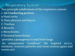
  4. THORACIC CAGE Conical in shape Has 2 apertures (openings): 1. Superior (thoracic outlet): narrow, open, continuous with neck 2. Inferior: wide, closed by diaphragm Formed of: 1. Sternum & costal cartilages: anteriorly 2. Twelve pairs of ribs: laterally 3. Twelve thoracic vertebrae: posteriorly

  5. ARTICULATIONS Costovertebral Sternocostal Costochondral

  6. ARTICULATIONS Costovertebral Costochondral Sternocostal

  7. RESPIRATORY MOVEMENTS A- MOVEMENTS OF DIAPHRAGM Inspiration Contraction (descent) of diaphragm Increase of vertical diameter of thoracic cavity Relaxation (ascent) of diaphragm) Expiration

  8. RESPIRATORY MOVEMENTS B- MOVEMENTS OF RIBS BUCKET HANDLE MOVEMENT Elevation of ribs PUMP HANDLE MOVEMENT Elevation of ribs Increase in lateral diameter of thoracic cavity Increase in antero-posterior diameter of thoracic cavity

  9. INSPIRATORY MUSCLES Diaphragm (most important muscle) Rib elevators: external intercostal muscles Accessory muscles (only during forced inspiration): 1. Muscles attaching cervical vertebrae to first & second rib: scalene muscles 2. Muscles attaching thoracic cage to upper limb: pectoralis major

  10. ORIGIN OF DIAPHRAGM 1) Costal: lower 6 costal cartilages 3) Sternal: xiphoid process of sternum 2) Vertebral: upper 3 lumbar vertebrae (right & left crus + arcuate ligaments) Medial arcuate ligament Lateral arcuate ligament Lateral arcuate ligament Medial arcuate ligament Lateral arcuate ligament Median arcuate ligament B. Posterior view Posterior view

  11. INSERTION OF DIAPHRAGM (CENTRAL TENDON)

  12. DIAPHRAGM A musculotendinous partition between thoracic & abdominal cavity Convex toward thoracic & concave toward abdominal cavity Attached to: sternum, costal cartilages,12th rib & lumbar vertebrae Fibers converge to join the central tendon Nerve supply: phrenic nerve (C3,4,5), penetrates diaphragm & innervates it from abdominal surface Action: contraction (descent) of diaphragm increase vertical diameter of thoracic cavity (essential for normal breathing)

  13. EXTERNAL INTERCOSTAL Attachments: from lower border of rib above to upper border of rib below Direction of fibers: downward & medially Nerve supply: intercostal nerves Action: rib elevators (inspiratory)

  14. SCALENE MUSCLES 5- Scalenus anterior 6. Scalenus medius 7. Scalenus posterior Origin: cervical vertebrae Insertion: 1st & 2nd ribs Action: elevates 1st & 2nd ribs (inspiratory) Cervical vertebrae 1st rib 2nd rib

  15. PECTORALIS MAJOR Action: increases antero- posterior diameter of thoracic cavity, when arm is fixed (inspiratory) Origin: sternum + costal cartilages Insertion: humerus

  16. EXPIRATORY MUSCLES Act only during forced expiration Rib depressors: 1. Internal intercostal 2. Innermost intercostal 3. Subcostals 4. Transversus thoracis Anterior abdominal wall muscles: 1. External oblique 2. Internal oblique 3. Transversus abdominis 4. Rectus abdominis

  17. RIB DEPRESSORS: REST OF INTERCOSTAL MUSCLES 3. Subcostal 4. Transversus thoracis Nerve supply: intercostal nerves (ventral rami of T1-T11) 1. Internal intercostal 2. Innermost intercostal Direction: upward & medially

  18. ANTERIOR ABDOMINAL WALL Internal oblique (middle layer) Direction: upward & medially External oblique (outer layer) Direction: downward & medially Linea alba

  19. ANTERIOR ABDOMINAL WALL Transversus abdominis (inner layer) Direction: transverse Rectus abdominis Direction: vertical Rectus abdominis Transversus abdominis

  20. Anterior abdominal wall Is formed of 3 layers of muscles of fibers running in different directions (to increase strength of anterior abdominal wall) The 3 muscles form a sheath in which a fourth muscles lies (rectus abdominis) Muscles are attached to: sternum, costal cartilages and ribs + hip bones The aponeurosis of the 3 muscles on both sides fuse in the midline to form linea alba Action (during forced expiration): Compression of abdominal viscera to help in ascent of diaphragm (during forced expiration) Nerve supply: lower intercostal nerves (T7 T11), subcostal nerve (T12) and first lumbar nerve.

  21. SUMMARY OF RESPIRATORY MOVEMENTS Inspiration Expiration Quiet Expiration (passive) 1. Elastic recoil of lung 2. Relaxation of diaphragm & external intercostal Quiet Inspiration (active) Contraction (Descent) Elevation of ribs of diaphragm (external intercostal) Contraction of anterior Depression of ribs abdominal wall muscles (rest of intercostal muscles) Compression of abdominal viscera Forced Expiration (active): Increase in vertical diameter - anteroposterior diameter - lateral diameter Increase in: Forced Inspiration (active) Accessory muscles of inspiration: 1. Pectoralis major 2. Scalene muscles Ascent of diaphragm

  22. QUESTIONS Are the following muscles have a respiratory role? If yes, what is it? 1. Levatores costarum. 2. Serratus posterior superior. 3. Serratus posterior inferior. 4. Pectoralis minor. 5. Serratus anterior. 6. Latissimus dorsi. 7. Quadratus lumborum. Why diaphragm is supplied by cervical nerves? Why right crus of diaphragm is larger than left crus?

  23. THANK YOU THANK YOU

More Related Content

giItT1WQy@!-/#giItT1WQy@!-/#giItT1WQy@!-/#giItT1WQy@!-/#giItT1WQy@!-/#giItT1WQy@!-/#