Bacterial Culturing Methods & Staining Techniques

Culturing methods
&
Bacterial staining
L
A
B
.
5
A
.
L
.
N
O
O
R
A
M
E
E
R
P
u
r
p
o
s
e
o
f
c
u
l
t
u
r
i
n
g
i
s
:
1- Isolation of microorganisms from a sample.
2- For counting the microorganism in the sample.
3- Obtain pure cultures.
4- To test for antibiotic sensitivity.
C
u
l
t
u
r
e
m
e
t
h
o
d
s
i
n
c
l
u
d
e
:
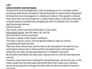
1- Streaking plates: Used for the isolation of bacteria in pure culture
from clinical specimens (to obtain pure colonies), include:
a- ABCD method: (by using loop).
b- Continuous streaking (by using loop).
2- Streaking of the slant: (by using loop).
3- Stab culture (by using needle).
4- Lawn culture: (by using swab).
5- Pour plate method.
6- Spread plate method (glass spreading rod)
B
a
c
t
e
r
i
a
l
s
t
a
i
n
i
n
g
The bacteria are stained for the following reasons:
1- To study their shapes
2- To differentiate the species of bacteria by using differential stain
3- To study the internal components of the bacterial cell
B
a
s
e
d
o
n
t
h
e
f
u
n
c
t
i
o
n
o
f
s
t
a
i
n
i
s
d
i
v
i
d
e
d
i
n
t
o
:
1- Simple stain: using of a single dye to staining the bacteria as
methylene blue, safranin, and crystal violet can be used to determine
cell shape, size, and arrangement.
2- Differential stain: using of more than one dye used to
differentiate between different bacteria
3- Selective stain: using of more than one dye to determine the
special structure such as spore, capsule, flagella, cell wall & nucleic
acid staining.
D
i
f
f
e
r
e
n
t
i
a
l
s
t
a
i
n
1- Gram staining: used to differentiate bacteria into 2 large groups
Gram positive which are blue- purple in color and Gram negative
bacteria are pink- red in color
- Include stains:
1- Primary stain (crystal violet) .
2- A mordant: (iodine solution).
3- A decolorizing agents: ( alcohol, acetone).
4- Counter stain: (safranin)
2- Acid- fast stain (Ziehl- Neelson stain): used to determine acid fast
organisms that causes tuberculosis (Mycobacterium tuberculosis)
which appear as pink bacilli - Include stains:
1- Primary stain: (carbol fuchsin).
2- A decolorizing agents: ( acid alcohol).
3- Counter stain: (methylene blue)
S
e
l
e
c
t
i
v
e
s
t
a
i
n
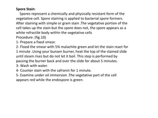
1- Endospore stain:
used to distinguish between the vegetative cells
and the endospores. A primary stain (malachite green) is used to
stain the endospores while the safranin used as counterstain, the
vegetative cells will appear pink and the spores will appear green. Ex:
Bacillus & Clostridium.
2- Capsule staining:
used to observe bacterial capsule of Klebsiella
pneumonia or any bacteria have a capsule by using primary stain
(crystal violet for 2 minutes), the encapsulsated cells will have a halo
appearance under the microscop
3- Flagella stain: flagella are fine, threadlike organelles and
usually invisible under light microscope to observe it must
be bind with chemical substances such as tannic acid &
potassium alum. to increasing the thickness of flagella and
then staining with basic fuchsin or silver nitrate .
Learn about culturing methods for isolating microorganisms and staining techniques to study bacteria's shapes, species, and internal components. Explore differential, selective, and simple staining methods like Gram staining and Acid-fast stain.

Download Presentation

Please find below an Image/Link to download the presentation.
The content on the website is provided AS IS for your information and personal use only. It may not be sold, licensed, or shared on other websites without obtaining consent from the author.If you encounter any issues during the download, it is possible that the publisher has removed the file from their server.
You are allowed to download the files provided on this website for personal or commercial use, subject to the condition that they are used lawfully. All files are the property of their respective owners.
The content on the website is provided AS IS for your information and personal use only. It may not be sold, licensed, or shared on other websites without obtaining consent from the author.
E N D
Presentation Transcript
Culturing methods & Bacterial staining LAB. 5 A.L. NOOR AMEER
Purpose of culturing is: 1- Isolation of microorganisms from a sample. 2- For counting the microorganism in the sample. 3- Obtain pure cultures. 4- To test for antibiotic sensitivity.
Culture methods include: 1- Streaking plates: Used for the isolation of bacteria in pure culture from clinical specimens (to obtain pure colonies), include: a- ABCD method: (by using loop). b- Continuous streaking (by using loop).
2- Streaking of the slant: (by using loop). 3- Stab culture (by using needle). 4- Lawn culture: (by using swab). 5- Pour plate method. 6- Spread plate method (glass spreading rod)
Bacterial staining The bacteria are stained for the following reasons: 1- To study their shapes 2- To differentiate the species of bacteria by using differential stain 3- To study the internal components of the bacterial cell
Based on the function of stain is divided into: 1- Simple stain: using of a single dye to staining the bacteria as methylene blue, safranin, and crystal violet can be used to determine cell shape, size, and arrangement. 2- Differential stain: using of more than one dye used to differentiate between different bacteria 3- Selective stain: using of more than one dye to determine the special structure such as spore, capsule, flagella, cell wall & nucleic acid staining.
Differential stain 1- Gram staining: used to differentiate bacteria into 2 large groups Gram positive which are blue- purple in color and Gram negative bacteria are pink- red in color - Include stains: 1- Primary stain (crystal violet) . 2- A mordant: (iodine solution). 3- A decolorizing agents: ( alcohol, acetone). 4- Counter stain: (safranin)
2- Acid- fast stain (Ziehl- Neelson stain): used to determine acid fast organisms that causes tuberculosis (Mycobacterium tuberculosis) which appear as pink bacilli - Include stains: 1- Primary stain: (carbol fuchsin). 2- A decolorizing agents: ( acid alcohol). 3- Counter stain: (methylene blue)
Selective stain 1- Endospore stain: used to distinguish between the vegetative cells and the endospores. A primary stain (malachite green) is used to stain the endospores while the safranin used as counterstain, the vegetative cells will appear pink and the spores will appear green. Ex: Bacillus & Clostridium. 2- Capsule staining: used to observe bacterial capsule of Klebsiella pneumonia or any bacteria have a capsule by using primary stain (crystal violet for 2 minutes), the encapsulsated cells will have a halo appearance under the microscop
3- Flagella stain: flagella are fine, threadlike organelles and usually invisible under light microscope to observe it must be bind with chemical substances such as tannic acid & potassium alum. to increasing the thickness of flagella and then staining with basic fuchsin or silver nitrate .


.")


: used to")