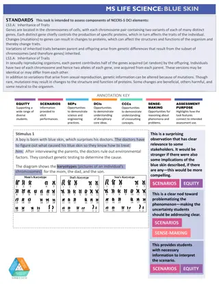
Chromosomal Karyotypes: An Overview

 undefined
Chromosomal Karyotypes
Dawn Adams
Cytogenetics
CDC’s 2003 Science Ambassador Program
Chromosomal Karyotypes


Overview
I. Chromosomes
A. Definition
B. Structure
C. Identification
II. Karyotypes
A. Definition
B. Methods
C. Staining
D. Importance
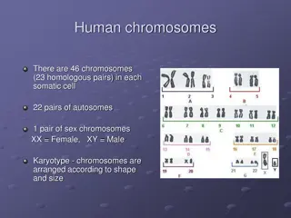
Chromosomes
Definition
Genetic structures of cells containing
DNA
Identification
Each chromosome has a characteristic
length and banding pattern
The breakdown of a Chromosome
Each autosome is numbered from 1-22,
sex chromosomes either X or Y
p arm
(short arm)
q arm
(long arm)
Centromere
Chromosome Labeling
Example - 1q2.4
The first chromosome, long arm, second
region of the chromosome, the fourth band of
that sub-region
Chromosome is
identified with
a number
ranging 1-22,
or X and Y
Each arm
divided into
sub-regions
and identified
by a number
Each sub-
region divided
into bands
identified with
a number
A Karyotype
Definition
A photographic
arrangement of
a complete set
of chromosomes
of a cell or
organism
Obtaining a Sample
Fetal samples for karyotypes are commonly
obtained in two ways
1.
Amniocentesis – sample taken from the
fluid of the amniotic sac
2.
Chorionic Villus Sampling – sample
taken from the fetal tissue that forms
part of the placenta
Obtaining a Karyotype
Chromosomes are stained for easy
visualization
Light microscope used to view chromosomes
in metaphase of mitosis
Chromosomes arranged into homologous
pairs based on size and banding patterns
Staining
Banding patterns can be visually identified on
chromosomes after staining.
Traditional Types
G-Banding – Giemsa stain
Q-Banding – Fluorescent stain
R-Banding – Reverse Giemsa stain
New Type
Fluorescence In Situ Hybridization techniques
Importance of Karyotypes
Karyotypes show the chromosomal
makeup of an individual. Knowing the
number of chromosomes is essential for
identifying chromosomal variations that
cause genetic disorders.
References
1.
Fairbanks, D. J., Anderson, W. R. Genetics: The Continuity
of Life.
Pacific Grove, CA: Brooks/Cole Publishing
Company; 1999.
2.
NIH. Amniocentesis [online]. 2004. [cited 2004 Feb 6].
Available from URL:
http://www.nlm.nih.gov/medlineplus/ency/article/00392
1.htm
.
3.
NIH. Chorionic villus sampling [online]. 2004. [cited 2004
Feb 6]. Available from URL:
http://www.nlm.nih.gov/
medlineplus/ency/article/003406.htm
.
4.
Campbell, N. A. Biology. 3
rd
ed. Redwood City, CA: The
Benjamin/Cummings Publishing Company, Inc.; 1993.
References (continued)
5.
On-line medical dictionary. G-banding: Banding pattern.
1997. [cited 2004 Feb 6]. Available from URL:
http://cancerweb.ncl.ac.uk/cgi-bin/omd?G-banding
.
6.
On-line medical dictionary. Q-banding. 2000. [cited 2004
Feb 6]. Available from URL:
http://cancerweb.ncl.ac.uk/cgi-bin/omd?query=q-
banding
.
7.
On-line medical dictionary. R-banding stain. 2000. [cited
2004 Feb 6]. Available from URL:
http://cancerweb.ncl.ac.uk/cgi-bin/omd?R-
banding+stain
.
8.
National Human Genome Research Institute, Fluorescence
In Situ Hybridization (FISH). 2004 [cited 2004 Feb 6].
Available from URL:
http://www.genome.gov/10000206
.
In this presentation, we will be looking into the field of cytogenetics, the study of genetics dealing with chromosomes and their genetic implications. In particular, we will be exploring karyotypes. Karyotypes are commonly used to investigate chromosomal number and banding, and are particularly helpful in looking for chromosomal abnormalities. (1)
Explore the world of chromosomal karyotypes with this detailed guide covering definitions, structures, identification methods, staining techniques, and the importance of karyotyping in genetic analysis. Learn about chromosome labeling, obtaining samples for karyotyping, and the process of arranging and visualizing chromosomes to create karyotypes. Discover the significance of banding patterns in identifying chromosomes and the various staining methods used in cytogenetics.

Download Presentation

Please find below an Image/Link to download the presentation.
The content on the website is provided AS IS for your information and personal use only. It may not be sold, licensed, or shared on other websites without obtaining consent from the author.If you encounter any issues during the download, it is possible that the publisher has removed the file from their server.
You are allowed to download the files provided on this website for personal or commercial use, subject to the condition that they are used lawfully. All files are the property of their respective owners.
The content on the website is provided AS IS for your information and personal use only. It may not be sold, licensed, or shared on other websites without obtaining consent from the author.
E N D
Presentation Transcript
Chromosomal Karyotypes Dawn Adams Cytogenetics CDC s 2003 Science Ambassador Program
Overview I. Chromosomes A. Definition B. Structure C. Identification II. Karyotypes A. Definition B. Methods C. Staining D. Importance
Chromosomes Definition Genetic structures of cells containing DNA Identification Each chromosome has a characteristic length and banding pattern
The breakdown of a Chromosome Each autosome is numbered from 1-22, sex chromosomes either X or Y p arm (short arm) q arm (long arm) Centromere
Chromosome Labeling Each arm divided into sub-regions and identified by a number Each sub- region divided into bands identified with a number Chromosome is identified with a number ranging 1-22, or X and Y Example - 1q2.4 The first chromosome, long arm, second region of the chromosome, the fourth band of that sub-region
A Karyotype Definition A photographic arrangement of a complete set of chromosomes of a cell or organism 1 2 3 4 5 6 7 8 9 10 11 12 13 14 15 16 17 18 19 20 21 22 X Y
Obtaining a Sample Fetal samples for karyotypes are commonly obtained in two ways 1.Amniocentesis sample taken from the fluid of the amniotic sac 2.Chorionic Villus Sampling sample taken from the fetal tissue that forms part of the placenta
Obtaining a Karyotype Chromosomes are stained for easy visualization Light microscope used to view chromosomes in metaphase of mitosis Chromosomes arranged into homologous pairs based on size and banding patterns
Staining Banding patterns can be visually identified on chromosomes after staining. Traditional Types G-Banding Giemsa stain Q-Banding Fluorescent stain R-Banding Reverse Giemsa stain New Type Fluorescence In Situ Hybridization techniques
Importance of Karyotypes Karyotypes show the chromosomal makeup of an individual. Knowing the number of chromosomes is essential for identifying chromosomal variations that cause genetic disorders.
References 1. Fairbanks, D. J., Anderson, W. R. Genetics: The Continuity of Life. Pacific Grove, CA: Brooks/Cole Publishing Company; 1999. 2. NIH. Amniocentesis [online]. 2004. [cited 2004 Feb 6]. Available from URL: http://www.nlm.nih.gov/medlineplus/ency/article/00392 1.htm. 3. NIH. Chorionic villus sampling [online]. 2004. [cited 2004 Feb 6]. Available from URL: http://www.nlm.nih.gov/ medlineplus/ency/article/003406.htm. 4. Campbell, N. A. Biology. 3rd ed. Redwood City, CA: The Benjamin/Cummings Publishing Company, Inc.; 1993.
References (continued) 5. On-line medical dictionary. G-banding: Banding pattern. 1997. [cited 2004 Feb 6]. Available from URL: http://cancerweb.ncl.ac.uk/cgi-bin/omd?G-banding. 6. On-line medical dictionary. Q-banding. 2000. [cited 2004 Feb 6]. Available from URL: http://cancerweb.ncl.ac.uk/cgi-bin/omd?query=q- banding. 7. On-line medical dictionary. R-banding stain. 2000. [cited 2004 Feb 6]. Available from URL: http://cancerweb.ncl.ac.uk/cgi-bin/omd?R- banding+stain. 8. National Human Genome Research Institute, Fluorescence In Situ Hybridization (FISH). 2004 [cited 2004 Feb 6]. Available from URL: http://www.genome.gov/10000206.


")