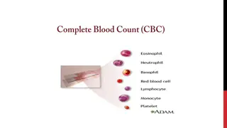
Planning Your Flow Cytometry Experiment: Building a Staining Panel
For successful flow cytometry experiments, it is crucial to plan your staining panel carefully by selecting appropriate markers and antibodies. Determine the goal of your experiment, research historical data for similar experiments, and choose markers specific to your cell type. Utilize resources for panel design, confirm your panel design with core personnel, adjust antibody concentrations through titration, and always include a viability dye. Understand laser and filter options based on instrument specifications. Make sure to fix samples when using BDLSR Fortessa for up to 16-color analysis.

Download Presentation

Please find below an Image/Link to download the presentation.
The content on the website is provided AS IS for your information and personal use only. It may not be sold, licensed, or shared on other websites without obtaining consent from the author.If you encounter any issues during the download, it is possible that the publisher has removed the file from their server.
You are allowed to download the files provided on this website for personal or commercial use, subject to the condition that they are used lawfully. All files are the property of their respective owners.
The content on the website is provided AS IS for your information and personal use only. It may not be sold, licensed, or shared on other websites without obtaining consent from the author.
E N D
Presentation Transcript
Flow Cytometry: Planning your experiment
Determine the goal of your experiment Search for papers historical info detailing a similar experiment and panel set-up to help plan your experiment Identify what markers/antibodies will answer your hypothesis. Pick markers that will express on your cell type and answer your hypothesis Make sure the marker is specific for your species of interest, as some markers are not interchangeable between mouse and human cells If data is available, know the expected expression of your markers of interest. Not having an idea of the expected expression of the targets of the antibodies adds a level of difficulty to your analysis.
Build a staining panel Useful panel builders can be found here for BD instruments (Fortessa, Aria, Celesta) https://www.bdbiosciences.com/en-us/resources/bd-spectrum-viewer Many of the companies (Biolegend, Tonbo, etc.) that sell Abs for use in flow cytometry have useful resource pages for panel design and other flow cytometry tools https://www.biolegend.com/en-us/web-tools-tab Double check the laser and filter options based on the charts and information for each instrument as follows. Email core personnel proposed panel to confirm it is a good experimental design. Research suggested antibody concentrations (Recommendations often found on vendor TDS) or best practice is to titrate antibodies prior to use Always include a viability dye! This is recommended as it gives more accurate and clean results. Many reviewers ask for this. Some are specific for non-fixed cells: DAPI, 7-AAD, etc. Others can be used on cells that are fixed: Zombie Dyes (Biolegend), live dead fixable dyes (ThermoFisher).
BDLSR Fortessa Excitation Laser Detection Filter Example 488 nm (blue) 695/40 (675-715 nm) PERCP/5.5, 7AAD, PERCP/EF710 515/20 (505-525 nm) AF488, GFP, FITC 561 nm (green) 780/60 (750-810 nm) PE-CY7 710/50 (685-735 nm) PE-CY5 670/30 (655-685 nm) PE-CY5.5 610/20 (600-630 nm) PE-TXRED, PI, MCHERRY, CF594 582/15 (574-590 nm) PE 633 nm (red) 780/60 (750-810 nm) APC-Cy7, APC-EF780 720/40 (700-740 nm) AF700 660/20 (650-670 nm) APC, AF647, EF660 407 nm (violet) 780/60 (750-810 nm) BV785, QD800 710/50 (685-735 nm) BV711 660/20 (650-670 nm) BV650, QD655 610/20 (600-630 nm) BV605, QD605 525/50 (500-550 nm) BV510, AMCYAN 450/50 (425-475 nm) DAPI, BV421, EF450, PACBLUE
BDLSRFortessa The Fortessa is a 16 color, 3 laser traditional flow cytometer. The samples are run by the core personnel and FCS files are created to be analyzed in FlowJo or similar software by the researcher. The Fortessa is commonly used for cell cycle, apoptosis, and cell surface and intracellular staining of up to 16 colors. All samples should be fixed to run on the Fortessa unless your assay prohibits (i.e. apoptosis). This allows for timing flexibility in running samples and eliminates biosafety concerns. A fixation protocol can be found here:
BD FACS Aria III Excitation Laser 488 nm (blue) Detection Filter 695/40 (675-715 nm) Example PERCP/5.5, 7AAD, EPRCP- EF710 AF488, GFP, FITC 515/20 (505 525 nm) 561 nm (green) 780/60 (750- 810 nm) PE-CY7 710/50 (685-735 nm) PE-CY5 670/30 (655-685 nm) PE-CY5.5 610/20 (600-630 nm) PE-TXRED, PI, MCHERRY, CF594 PE 582/15 (574-590 nm) 633 nm (red) 780/60 (750-810 nm) APC-Cy7, APC-EF780 720/40 (700-740 nm) AF700 660/20 (650-670 nm) APC, AF647, EF660 407 nm (violet) 710/50 (685-735 nm) BV711 660/20 (650-670 nm) BV650, QD655 610/20 (600-630 nm) BV605, QD605 525/50 (500-550 nm) BV510, AMCYAN 450/50 (425-475 nm) DAPI, BV421, EF450, PACBLUE
BD FACS Aria III The BD FACS Aria III is a sterile cell sorter. It can analyze up to 15 colors and sort up to 4 populations at one time. Samples are run by core personnel. The cell sorter is housed in a Biosafety hood allowing for the sorting of BSL-2 samples, including patient samples. No BSL-3 samples can be sorted. Samples do not need to be fixed to run on this instrument. However, source of the samples should be disclosed to the flow personnel.
BD Celesta Excitation Laser 488 nm (Blue) Detection Filter 780/60 (750-810 nm) Example PE-CY7 695/40 (675-715 nm) PERCP, PERCP5.5, 7AAD, PERCP-EF710 575/25 (562-588 nm) PE 530/30 (545-515 nm) FITC, GFP, AF488 633 nm (red) 780/60 (750-810 nm) APC-Cy7, APC-EF780 720/40 (700-740 nm) AF700 660/20 (650-670 nm) APC, AF647, EF660 407 nm (violet) 780/60 (750-810 nm) BV785 660/20 (650-670 nm) BV650 610/20 (600-630 nm) BV605 525/50 (500-550 nm) BV510, AMCYAN 450/50 (425-475 nm) DAPI, BV421, EF450, PACBLUE
BD Celesta The BD Celesta is a 12 color traditional flow cytometer. Samples are run by the researcher. Prior training and authorization is required to operate this instrument. This instrument is ideal for use when your experiment requires after hours analysis. FCS files are created that are then analyzed on FlowJo or similar software by the researcher.
Preparing Cells All cell types require different digestion, harvesting, and isolation techniques. It is recommended you find a protocol specific for your cell type that gives you a single cell suspension with optimal cell number recovery. Lyse out red blood cells! This step is very important. RBCs will overwhelm your samples and make finding cells of interest very difficult. COUNT YOUR CELLS! This step is very important. After cell collection and prior to staining, count your cells. This will help you determine antibody concentrations, as well as determine if your cell harvest was successful and you have enough cells to stain. Unless you are identifying very rare cell populations, 1-5 million cells is a sufficient number needed for staining. Make sure you have a single cell suspension. If you see clumps in your sample, these cannot be run on the cytometer. They can damage the instrument and cause inaccurate results. All samples should be filtered through a 40 m cell strainer topped 5 ml FACs tube before bringing the samples to the flow core.
Staining Protocol All cell types require different variations to the staining protocol, so it s best to find protocols specific to your assay/cell type. There are many protocols available on-line and on antibody manufacturer websites. It is highly recommended that you test your protocols on a small sample size prior to staining a large number of cells/samples on valuable cells. Keep it simple to start. Do not use your two month highly expensive or valuable experimental samples for your first flow run. A generic staining protocol can be found on my website here: https://medicine.uams.edu/mbim/research-cores/flow-cytometry-core- facility/sample-submission/sample-preparation/
Experimental Quality Controls Quality control samples are needed with each experiment. Best practice is to prepare these with each run. A multi-color experiment cannot be run without proper controls. For best practice, the controls needed for each experiment are as follows: Negative/unstained cells Single stain controls for each color FMOs (fluorescence minus one) for each color. If you have an 8 color panel you should have 1 unstained control, 8 individual color controls, and 8 FMO controls. If only staining/sorting for a single color, then just a negative unstained control is required If you are using the same panel for multiple days of an experiment then settings can be re-used but additional compensation may be needed within FlowJo These controls are needed to set voltages and compensate the instrument, while the FMOs are used to set the positive and negative gates for each color (fluorophore).
Experimental Quality Controls The single stain/compensation controls are used to set up the instrument and samples cannot be run without them. Example: A panel stained with FITC=CD3, PE=CD4, APC=B220, and EF780=viability dye Tube 1. Cells only Tube 2. Cells stained with FITC CD3 only Tube 3. Cells stained with PE CD4 only Tube4. Cells stained with APC B220 only Tube 5. Cells stained with ef780 viability only
Experimental Quality Controls FMO controls are used for analysis purposes. When drawing gates to determine which proportion of the cell population expresses a given marker, these controls are used to help set the positive and negative gates. While some populations have clear pos/neg expression, low expressing markers or markers that just cause a slight shift are easier to confidently gate on with FMO controls FMO example: Using the same panel listed on previous slide FMO controls would be: Tube 6: FITC FMO- Cells with all stains except FITC Tube 7: PE FMO- Cells with all stains except PE Tube 8: APC FMO- Cells with all stains except APC Tube 9: ef780 viability FMO- Cells with all stains except ef780
Experimental Quality Controls Some cells types are prohibitive in the number of cells available, making it difficult to generate control samples. In these instances other options are available. For example, many researchers will use splenocytes or other abundant tissue types to harvest cells to stain for controls. Beads can also be used. Make sure that the sample/cells chosen for the single stain controls will express your marker. There has to be a positive population for the marker to accurately compensate. If you are unsure that the marker you are using is expressed by the cells in your assay, then chose another marker that is expressed to make this control. For example: Many researchers use CD4 for all the single stain controls to ensure good expression. The marker can differ from the one in your panel as long as it s the same color and expressed by cells in your sample.
Booking an appointment Now that you have your panel designed, staining protocol, and tube template made, you re ready to book an appointment. Log into iLab: https://uams.corefacilities.org/ Reserve time on the instrument that meets your needs. Make sure you have received an appointment approval email before you begin your experiment. Most importantly, make sure to reserve time on the appropriate instrument before starting your experiment, especially if you have a time sensitive experiment. For analysis on Fortessa, if you want to collect all cells in a sample tube with a 200 l volume, estimate 2 minutes per sample plus 10-15 minutes of set up time. If it s a quick check with only needing 10,000 cells recorded, then 30 sec/sample is a good estimate. For sorting, we can sort through about 10 million cells in 20-30 min.
LSRFortessa Samples The recommended sample concentration is 10 million cells/ml and adjust according to your cell count with a minimum final volume of 200 ul. If cells are over diluted, it causes unnecessary long run times Filter cells prior to bringing to the core Include a form with samples indicating all necessary info: Lab name, your name, contact info, staining panel including markers & colors used, cell type, etc. This must be submitted with samples, or it can be emailed to core personnel ahead of time. Make sure tubes are numbered or labeled legibly to prevent mishandling of tubes out of order. Samples can be dropped off in core fridge or in afterhours fridge prior to appointment time. Data will be exported as FCS files and uploaded to a server for the user to download and analyze with FlowJo or similar software A FlowJo dongle is available for check out use from the core
FACSAria sorting samples Recommended sample concentration is 10 million cells/ml and adjust according to your cells count with a minimum final volume of 200 ul. For final volume, cells need to be diluted in sorting buffer. Recipe can be found here: If cells are over diluted, it causes unnecessarily long sort times and too concentrated samples causes poor sorting efficiency. You can bring extra sorting buffer with you so that cells can be diluted, if necessary, by core personnel. Filter cells through blue cap filter tubes (BDFalccon #352235) prior to bringing to the core for sorting. If any clumps are visible, then cells will not be sorted Bring collection tubes you want to sort live cells into. We can sort into 96 well plates, 5 ml FACS tubes, 15 ml conical tubes and 1.5 ml microcentrifuge tubes. If sorting more than 2 populations at once, 15 ml tubes cannot be used. Prepare collection tubes with 0.5-1 ml of media or solution of your choice in the collection tubes to preserve viability. It is recommended for low cell number to also coat the insides of the collection tubes to prevent cells sticking to sides and drying out. That protocol can be found here: After sorting, it is recommended the cells rest on ice for 30-60 minutes before moving to next step are the sorting procedure puts the cells under strain and stress.
Analyzing Data FCS data files are uploaded to the Flow cytometry server Data can be analyzed using FlowJo or a similar Flow Analysis software The Core has a FlowJo dongle available for check- out for free of charge It is preferred that the user and not the core staff analyze the data.