Gel Electrophoresis DNA Extension Activity in Modern Biology Lab
This lab activity uses agarose gel electrophoresis to determine the length of an unknown DNA fragment by comparing it to known DNA fragment lengths. The process involves gel preparation, loading samples, electrophoresis, staining, de-staining, and measurement of DNA bands. The activity was produced by Purdue University's Science Express Program.
Download Presentation

Please find below an Image/Link to download the presentation.
The content on the website is provided AS IS for your information and personal use only. It may not be sold, licensed, or shared on other websites without obtaining consent from the author.If you encounter any issues during the download, it is possible that the publisher has removed the file from their server.
You are allowed to download the files provided on this website for personal or commercial use, subject to the condition that they are used lawfully. All files are the property of their respective owners.
The content on the website is provided AS IS for your information and personal use only. It may not be sold, licensed, or shared on other websites without obtaining consent from the author.
E N D
Presentation Transcript
Science Express Science Express Gel Electrophoresis DNA Extension Gel Electrophoresis DNA Extension
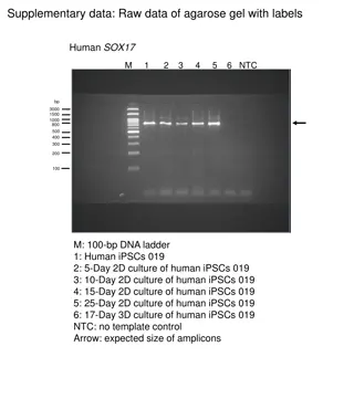
This gel electrophoresis DNA extension activity assumes the How Gel Electrophoresis Works and Gel Electrophoresis Dyes Lab videos have been watched. They can be found here: How Gel Electrophoresis Works : https://www.youtube.com/watch?v=mN5IvS96wNk Gel Electrophoresis Dyes Lab : https://www.youtube.com/watch?v=9OEOHQDiYuw&feature=youtu.be In this lab the Modern Biology Exp 301 The Length of DNA Molecules experiment was used. Since this was lab used real DNA, the agarose gel was stained with Carolina Blu stain to view the DNA bands. There are other fluorescing stains that show the DNA bands much better, but they are either cost-prohibitive are not legal to use in Indiana high schools. This laboratory investigation uses a sample of known DNA fragment lengths to determine the length of an unknown DNA fragment length.
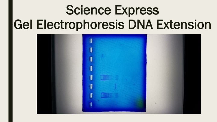
First, a 1.2% agarose gel is made. Then the gel is loaded as shown: Lane 1: Dyes Lane 2: Known DNA fragments Lane 3: Unkown DNA fragment Lane 4: Known DNA fragments The gel was then run at 170 volts for one hour. The gel was considered complete when the some of the dyes loaded in to lane 1 started to run off of the gel. The gel was then removed from the electrophoresis chamber and placed into gel staining container and covered with Carolina Blu stain and allowed to sit for 4 hours. After 4 hours the stain was removed and the gel was de-stained with a de-stain solution for 20 minutes. Then, the gel was ready for viewing on a light box, as shown below.
This angle shows the DNA bands better. This gel could have been run longer to get more separation, but the loading dyes included in the kit where starting run off of the gel, so it was removed from the chamber. Unknown DNA fragment Unknown DNA fragment Known DNA fragment lengths Known DNA fragment lengths
The DNA bands were then measured. The measurements were as follow: Known DNA bands: 1) 7mm, 2) 14mm, 3) 16mm, 4) 18mm, 5) 21mm, 6) 22mm Known DNA fragment band: 1) 20mm
The known DNA fragment lengths are as follows: 1 - 23.1kbp 2 9.4kbp 3 6.7kbp 4 4.4kbp 5 2.3kbp 6 2.0kbp
This activity was produced by: This activity was produced by: The Purdue University The Purdue University Science Express Program Science Express Program
