mmWave SAR
The world of millimeter wave SAR, including its operating frequencies, system design, and applications such as personal security, ground imaging, automotive imaging, and 3D imaging.
6 views • 15 slides
Evolution of Digital Radiography in Medical Imaging
Explore the evolution of digital radiography in medical imaging, from the invention of CMOS in 1963 to the development of Gadolinium-based flat-panel detectors in 1997. Learn about the advantages and challenges of digital imaging technology and its impact on the field of radiography.
0 views • 102 slides
Automated CT Perfusion Imaging in Acute Ischemic Stroke: Overview
This presentation delves into the significance of automated CT perfusion imaging in diagnosing and treating acute ischemic stroke. It covers essential information such as the burden of stroke in Ontario, the critical aspect of time in stroke treatment, hyperacute stroke treatment goals, and the impo
4 views • 41 slides
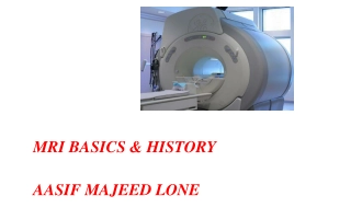
MRI BASICS & HISTORY AASIF MAJEED LONE
Magnetic Resonance Imaging (MRI) has revolutionized medical imaging, offering detailed views of the body without harmful radiation. This article delves into the historical development of MRI, highlighting key milestones such as the discoveries of the Rotating Magnetic Field by Nikola Tesla and the w
9 views • 49 slides
Chest Imaging Interpretation for Tuberculosis Screening
Chest imaging plays a crucial role in screening for pulmonary tuberculosis (TB). This activity, led by Dana Kissner from Wayne State University Detroit Tuberculosis Clinic, focuses on interpreting chest imaging results to aid in the diagnosis and management of TB cases. The content includes informat
3 views • 22 slides
Premier Radiology: Quality Imaging Provider in Middle Tennessee
Premier Radiology, operating in Middle Tennessee under various names, offers a range of imaging services including X-Ray, CT, Mammography, and Ultrasound. The company does not plan to add MRI services to its Lebanon location but will acquire an existing unit. Detailed projections, utilization data,
0 views • 24 slides
Understanding Diffusion Weighted Imaging (DWI) in Neuroimaging Research
This content delves into the significance of Diffusion Weighted Imaging (DWI) in studying the motion of water molecules in brain tissue. It explains how water diffusion varies in different brain matter types and explores techniques like Diffusion Tensor Imaging (DTI). The impact factors on diffusion
4 views • 25 slides
Role and Challenges of PET Imaging in Proton Therapy
PET imaging plays a crucial role in proton therapy by aiding in range verification and monitoring treatment delivery. Proton therapy offers precise tumor targeting but is sensitive to uncertainties, making accurate imaging essential. Positron emitters generated during therapy can be detected using P
1 views • 17 slides
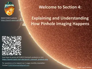
Understanding Pinhole Imaging in NASA's PUNCH Mission
Delve into the world of pinhole imaging with NASA's PUNCH mission. Explore how small, lens-less holes create images of the Sun and discover the fascinating process behind pinhole projection. Follow along a playful learning adventure with Bhanu guiding you through sections explaining pinhole imaging
1 views • 53 slides
Basic Principles of MRI Imaging
MRI, or Magnetic Resonance Imaging, is a high-tech diagnostic imaging tool that uses magnetic fields, specific radio frequencies, and computer systems to produce cross-sectional images of the body. The components of an MRI system include the main magnet, gradient coils, radiofrequency coils, and the
2 views • 49 slides
Radiographic Imaging Methods of the Respiratory System
Radiographic imaging plays a crucial role in the evaluation and diagnosis of thoraco-mediastino-pleuro-pulmonary conditions. Techniques like radioscopy, digital radiography, computer tomography, magnetic resonance imaging, conventional pulmonary angiography, and hybrid imaging methods offer detailed
10 views • 21 slides
Radiological Investigation of Hepatobiliary System and Imaging Modalities Overview
Exploring the hepatobiliary system through radiology, this lecture covers the anatomy, modalities like X-ray, Ultrasound, CT scan, MRI, and nuclear scan used for imaging, their advantages, disadvantages, and indications. It delves into the significance of liver, gallbladder, and biliary duct imaging
2 views • 37 slides
SALIVARY GLAND IMAGING
Salivary gland imaging plays a crucial role in diagnosing and monitoring diseases of the major salivary glands, including the parotid, submandibular, and sublingual glands. Different imaging techniques such as computed tomography, MRI, and ultrasound are used to visualize these glands and aid in dif
1 views • 12 slides
Diagnostic Reference Levels in Medical Imaging and Radiation Protection
The concept of Diagnostic Reference Levels (DRLs) is crucial in evaluating the amount of ionizing radiation used in medical imaging procedures. DRLs help determine if radiation levels are appropriate and need optimization. Authorized bodies establish numerical DRL values as advisory guidelines. Loca
1 views • 26 slides
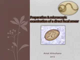
Direct Fecal Smear Preparation and Microscopic Examination Procedure
Direct fecal smears are useful for quick screenings to detect intestinal parasites. This article outlines the preparation and microscopic examination procedure for direct fecal smears. It covers advantages, disadvantages, fecal collection methods, and steps for preparing and examining fecal smears u
0 views • 16 slides
Australian Imaging Biomarkers and Lifestyle Study: Progress and Focus
The Australian Imaging Biomarkers and Lifestyle Study of Ageing (AIBL) commenced in late 2006, with a cohort size and follow-up progress detailed. The study includes assessments and biomarker imaging, such as MRI, amyloid PET, and tau PET scans, with ongoing review cycles and data additions. Current
0 views • 12 slides
Understanding Histological Techniques for Tissue Preparation
Histological technique involves preparing tissue for microscopic examination, aiming to preserve the microscopic anatomy and structure of tissues. The process includes fixation, dehydration, cleaning, embedding, cutting, and staining. Key steps like fixation using fixative materials help prevent aut
0 views • 18 slides
Understanding Paramoecium Caudatum: A Microscopic Organism with Unique Characteristics
Paramoecium caudatum is a typical ciliate protozoan characterized by the presence of cilia as a locomotor organelle, nuclear dimorphism, and a unique form of sexual reproduction called conjugation. Commonly found in freshwater habitats, this microscopic organism has a distinct structure with a pelli
3 views • 10 slides
Enhancing Stroke Treatment Response Through Advanced Imaging Management Center Strategy
This information details the strategy proposed by the Imaging Management Center to improve stroke treatment response. It emphasizes the importance of timely uploading of brain imaging data within specified timeframes using the AMBRA platform. The process involves modalities like NCCT, CTA, MRI, and
0 views • 6 slides
Understanding Ionizing Radiation in Medical Imaging
Radiation in medical imaging plays a critical role in diagnosis and treatment but also poses risks to patients and healthcare workers. Ionizing radiation, used in various imaging modalities, can cause harm to healthy cells if not managed properly. This article discusses the implications of radiation
0 views • 54 slides
Fungal Microbiology Laboratory Procedures
This content provides detailed guidelines on fungal culture purification, subculturing process, preparation of microscopic slides for fungal micromorphology assessment, and isolation of human samples for diagnosing fungal infections. The procedures include steps for identifying characteristics of in
1 views • 9 slides
Sparse Millimeter-Wave Imaging Using Compressed Sensing and Point Spread Function Calibration
A novel indoor millimeter-wave imaging system based on sparsity estimated compressed sensing and calibrated point spread function is introduced. The system utilizes a unique calibration procedure to process the point spread function acquired from measuring a suspended point scatterer. By estimating
2 views • 26 slides
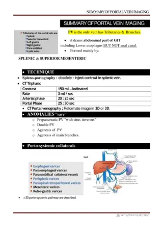
Portal Vein Imaging Techniques and Anomalies Overview
Portal vein imaging is crucial for evaluating conditions affecting the abdominal part of the gastrointestinal tract. Techniques such as spleno-portography and CT triphasic contrast imaging are used to visualize the portal vein and diagnose anomalies like portal-systemic collaterals and porto-systemi
4 views • 7 slides
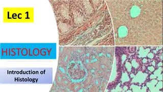
Introduction to Histology and Microscopy in Anatomy
Histology is the study of tissue structure and function in animals and plants, focusing on the microscopic structure of cells, tissues, and organs. It helps in understanding the relationship between structure and function, intertwining disciplines like cell biology, pathology, physiology, and bioche
0 views • 5 slides
Introduction to Radiology: Imaging Modalities and Techniques
Radiology is a medical specialty that utilizes various imaging modalities such as X-Ray, MRI, CT, and Ultrasound to diagnose and treat patients. This field involves the supervision, performance, and interpretation of imaging studies, with findings reported to referring physicians. Radiology also inv
0 views • 10 slides
Advanced Microscopy Techniques in EUV Lithography: SHARP Overview
SHARP utilizes Fresnel zone plate lenses to achieve diffraction-limited quality in EUV lithography, offering a range of NA values and image magnifications. The system allows emulation of mask-side imaging conditions with hundreds of lenses available. Coherence control and engineering are provided th
0 views • 18 slides
Insights into Multi-View Imaging System Optimization
Delve into the simulation and calibration of a multi-view imaging system using differentiable ray tracing and gradient-based optimization. Explore the challenges of ambiguity in results and the impact of angular offset on imaging accuracy. Discover how the system handles errors and maintains precise
0 views • 6 slides
Understanding PET-CT Imaging in Adrenal Tumors at Tehran University
Tehran University of Medical Sciences' Research Center for Nuclear Medicine specializes in PET-CT imaging for adrenal tumors. Dr. Arman Hassanzadeh-Rad, MD, explains the principles of PET imaging, hybrid imaging combining PET and CT, F-18 FDG use, SUV measurement, and more. PET-CT allows for both at
0 views • 21 slides
Exploring Microscopic Life Through a Microscope
Journey into the microscopic world by examining Elodea leaves and Paramecium under a microscope. Discover the intricate details of these tiny organisms, observe their movement, and learn about their structures. Engage in hands-on activities to explore the fascinating realm of microscopic life.
0 views • 16 slides
Microscopic Structure of Blood Vessels and Their Layers
The lecture covers the microscopic structure of the wall of blood vessels including large veins, elastic arteries, muscular arteries, and blood capillaries. The three concentric layers of blood vessel walls are discussed, which include the tunica intima, tunica media, and tunica adventitia. Detailed
0 views • 11 slides
Simulation Study of Photoacoustic Imaging for Bragg Peak Reconstruction
This study explores the use of simulation in Photoacoustic Imaging for reconstructing the Bragg peak in proton therapy. It discusses the concept of Photoacoustic Imaging, the generation of acoustic waves from pulsed electromagnetic radiation, and the experimental setups for characterizing proton dos
0 views • 13 slides
Overview of Radiography and Ultrasound Imaging Techniques
This content provides a comprehensive overview of conventional radiography, digital radiography, and ultrasound imaging techniques. It covers equipment details, safety issues, advantages vs. limitations, as well as the interpretation, magnification, and integration capabilities of these imaging moda
0 views • 28 slides
Understanding Magnetic Resonance Imaging (MRI)
Magnetic Resonance Imaging (MRI) is an imaging technique based on nuclear magnetic resonance principles. It was first developed in the 1970s by Paul Lauterbur and Peter Mansfield. MRI uses the interaction between protons in the body and magnetic fields to create detailed images. This technology has
0 views • 77 slides
Fascinating Microscopic World: Leech Feeding and Grasshopper Exoskeleton
Explore the intricate details of a leech feeding on beef kidney and a Grasshopper's cleaned-out exoskeleton by The E. obscura in these captivating microscopic images.
0 views • 7 slides
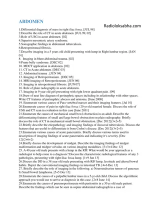
Comprehensive Radiological Imaging Review in Abdominal and Adrenal Pathologies
This extensive radiological imaging review covers a wide range of topics in abdominal and adrenal pathologies, discussing differential diagnoses, imaging modalities such as CT and USG, specific syndromes like prune belly syndrome, and conditions like retroperitoneal fibrosis. It delves into various
0 views • 39 slides
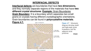
Understanding Interfacial Defects and Microscopy in Materials Science
Interfacial defects are boundaries separating regions with different crystal structures, like grain boundaries in polycrystalline materials. Microscopic examination distinguishes between macroscopic and microscopic dimensions, analyzed through optical and electron microscopy methods. Electron micros
0 views • 9 slides
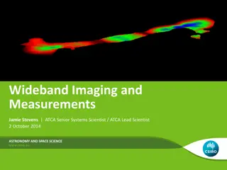
Wideband Imaging and Measurements in Astronomy
Wideband imaging and measurements play a crucial role in astronomy and space science. This involves utilizing wide bandwidths to make accurate measurements and create images for continuum mapping and spectral-line observations. Increasing bandwidth enables lower thermal noise levels, quicker identif
0 views • 32 slides
Overview of DICOM WG21 Multi-Energy Imaging Supplement
The DICOM WG21 Multi-Energy Imaging Supplement aims to address the challenges and opportunities in multi-energy imaging technologies, providing a comprehensive overview of imaging techniques, use cases, objectives, and potential clinical applications. The supplement discusses the definition of multi
0 views • 33 slides
Understanding Correlation of Normal Clinical and Microscopic Features in Gingival Health
The correlation of normal clinical and microscopic features in gingival health involves factors such as color, size, contour, shape, consistency, surface texture, stippling, position, and continuous tooth eruption. These elements depend on various aspects like vascular supply, thickness of epitheliu
0 views • 13 slides
Understanding Initial Asymptotic Acoustic RTM Imaging Results in Salt Model
Acquire insights into the initial asymptotic acoustic RTM imaging results for a salt model in Xinglu Lin, San Antonio. This study delves into the concept of Reverse Time Migration (RTM), showcasing the methodology, workflow, and imaging conditions involved in this innovative seismic imaging techniqu
0 views • 22 slides


