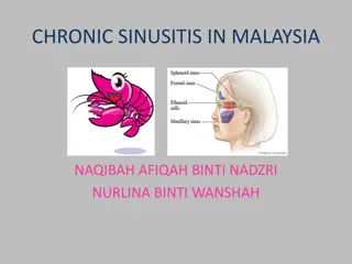
Understanding Cryptococcosis: Causes, Symptoms, and Diagnosis
Cryptococcosis is a fungal infection caused by Cryptococcus neoformans, affecting immunocompromised individuals. The disease presents with various clinical features, including pulmonary and extrapulmonary manifestations. Laboratory diagnosis involves examining specimens such as serum and CSF for the presence of budding yeast cells. Understanding the pathogenesis, virulence factors, and clinical types of cryptococcosis is crucial for effective management and treatment.

Download Presentation

Please find below an Image/Link to download the presentation.
The content on the website is provided AS IS for your information and personal use only. It may not be sold, licensed, or shared on other websites without obtaining consent from the author. Download presentation by click this link. If you encounter any issues during the download, it is possible that the publisher has removed the file from their server.
E N D
Presentation Transcript
Opportunistic Mycoses & Miscellaneous Mycoses MBI 531
Opportunistic Systemic Mycoses Produced by relatively non-pathogenic or contaminant fungi in a host whose immunological defense mechanisms are weakenedby Endogenous causes - cancer, leukemia Exogenous causes - immuno suppressive therapy, AIDS. Important cause of morbidity & mortality in hospitalized patients.
OpportunisticMycoses Includes -Candidiasis - Cryptococcosis - Aspergillosis - Zygomycosis /Mucormycosis - Pneumocystosis - Penicilliosis - Miscellaneous opp. mycoses.
CRYPTOCOCCOSIS Caused by an encapsulated yeast Cryptococcus neoformans, pathogenic to man &animals. 2nd candidiasis in HIV - infectedindividuals. most common fungal infection after Isolated from pigeon nests, droppings, old buildings & nitrogenous soil - Creatine favour the growth.
Virulence factors Capsule Inhibitsphagocytosis. Melanin production by the enzyme phenol oxidase. L- DOPA Melanin Melanin -Antioxidant - protects the organism from intracellular killing by phagocytes
Pathogenesis Infection occurs by inhalation, but sometimes through skin ormucosa. Weakening of immune system leads to reactivation & dissemination to CNS and other sites. Visceral forms simulate tuberculosis & cancer clinically. Cutaneous form varies form small ulcers to granulomas.
ClinicalFeatures Seen in HIV +ve patients when CD4+ count falls below 200 cells / mm3 Extra pulmonary cryptococcosis is one of the AIDS defining disease. Clinical types : 1. Pulmonary 2. Extrapulmonary CNS, viscera, bones & skin Cryptococcal meningitis is the most serious type of infection, resembles TB and is often seen in AIDS patients.
Laboratory Diagnosis Specimen Serum, CSF, body fluids Direct examination Wet mount: India ink - budding yeast cells 5-20 , with adistinct halo Gram s stain - Gram +ve budding yeastcells
Fungal Culture & Serology SDA - highly mucoid, cream to buff colored colonies. Birdseed (Niger seed) agar selective media - Brown colored colonies due to melanin production. Crypto LA test Ag detection in Serum, CSF &Urine - Titer > 1 8 is significant.
ASPERGILLOSIS Systemic infection in immunocompromised as well as immunocompetent individuals Infection occurs by - Inhalation of conidia - Direct entry through wounds or during surgery. Imp. species A. niger, A. fumigatus, A. flavus
Clinicaltypes PulmonaryAspergillosis 3 categories depending upon whether the host is atopic or immunocompromised. Allergicaspergillosis 1. Aspergillus asthma - Atopic individuals - Inhalation of spores A.
2. Allergic bronchopulmonary aspergillosis(ABPA) - Repeated & heavy exposure tospores - Breathlessness, fever &malaise 3. Obstructiveaspergillosis -Plugs of entangled mycelia & mucus block segments of lung tissue & even entirelobe. - Productive cough: contains aspergillushyphae.
B. Aspergilloma Noninvasive Colonization in a pre- existing cavity (tubercular) Compact mass of fungal mycelia surrounded by dense fibrous walls : FUNGUS BALL Usually solitary 8-10 cm C. InvasiveAspergillosis Important cause of morbidity & mortality May disseminate to kidneys & brain.
Extrapulmonary Aspergillosis CNS, Paranasal sinus, skin, endocardium (in prior cardiacsurgery) Miscellaneousforms - Eye(oculomycosis) - Ear(otomycosis) - Nails (onychomycosis)
Laboratory Diagnosis Common lab contaminant hence repeated isolation from specimen is mandatory. Specimen sputum, BAL, tracheobronchial biopsy. Direct Examination Wet mount 10% KOH, CFW - hyaline, septate hypha, 3-6 with acute angled (45 ) branching.
Fungal Culture SDA - Grows easily & quickly on routinemedia. Identification of species by inductionof sporulation on special media: - Czapek Dox agar - 2% malt extractagar Colony characteristics & the morphology help in identifying thespecies.
Aspergillus Culturalcharacteristics A.niger A.fumigatus A.flavus
MUCORMYCOSIS Invasive disease caused by lower Zygomycetes Found in food items, soil &air. Common labcontaminants fungi Reproduction Asexual Sporangiospores within sporangium. Sexual single, dark thick walled spores called Zygospores. 1. 2.
Introduction Includes following genera - Mucor - Rhizopus - Absidia - Rhizomucor Usually occurs in diabetic patients with ketoacidosis. * high glucose & acidotic condition favours their growth. distinguished on the basis of morphology.
Clinicaltypes 1 infection occurs in URT or nose by inhalation Rhinocerebral commonest, fulminant type Nasalmucosa Turbinatebones - Paranasal sinus - Orbit, Palate & Brain Pulmonary Cutaneous Disseminated lungs, kidney, brain, heart & GIT.
Laboratory Diagnosis Specimen nasal discharge, sputum, biopsy DirectExamination KOH irregular broad, aseptate ribbon like hyphae with wide angle (90 ) branching at irregular intervals. Special stains must - HE, GMS
Fungal Culture Cottony, dense & floccose colony. LPCB mount for microscopic details & species identification.
MUCOR ABSIDIA RHIZOPUS
Treatment & Prophylaxis A life threatening condition 4 concomitant approaches : Rapid correction of underlying predisposing conditions like diabetic ketoacidosis. Surgical debridement of necrotizing tissue. Antifungals (not azoles) Adjunctive therapy with hyperbaric oxygen. 1. 2. 3. 4. Ideal treatment = surgical debridement + I.V. AMB
PENICILLIOSIS Caused by Penicillium marneffei, a dimorphic facultative intracellularfungi. Only species of genus Penicillium which causes infection. 3rdmost common AIDS - related opportunistic infection after TB & Cryptococcosis.
Epidemiology Route of transmission - Inhalation of conidia - Ingestion (eating rats, China) - Direct inoculation of skin. Bamboo rat harbors P. marneffei in their internal organs. Isolated from - feces - soil samples from their burrows.
Pathogenesis & Pathology RES is the 10 site ofinfection. 3 histologic patterns ofdisease: Granulomatous granulomas Suppurative multiple abscesses - lungs, skin & subcutaneous tissue of immunocompetent individuals. Necrotizing - immunocompromised pt. 1. 2. 3.
Laboratory diagnosis Clinical diagnosis difficult as symptoms are very similar to other fungal pathogens like H.capsulatum Presumptive diagnosis - yeast forms with cross- walls in biopsy. - CD4+count < 50 cells /mm3 Definitive diagnosis : direct demonstration & isolation of organisms in culture
Direct Examination Wrights s, Giemsa stain of skin, biopsies septate yeast like cells. Tissues PAS, GMS Peripheral blood smears AIDS patient.
Fungal Culture Isolated from blood, skin, BM, sputum, LNs, pleural fluid, urine & BAL SDA & BA at 250C : woolly pigmented colonies, reverse is bright rose color. Microscopy:- short, septate hyphae with branching. - Brush like conidiophores bearing conidia.
Culture At 370 C - SDA, BHIA, 5% sheep BA & Pine s medium Colony white chalk like LPCB pleomorphic yeast cells with transversesepta. Treatment - Amphotericin B & Itraconozole
PNEUMOCYSTOSIS Infection by inhalation of droplet nuclei leads to interstitial cell pneumonia, primarily involves alveoli. Caused by Pneumocystis carinii, a unicellular eukaryote. Initially it was considered as a protozoa but now considered as a fungi - shares biological features of both groups fungi & protozoa.
Features of Pneumocystis carinii Stained with the fungal stains like GMS. Does not grow on fungal culture media but requires tissue culture or cell lines. Susceptible to anti-protozoan agents like Pentamidine & TMP-SMZ. Insensitive to antifungals because of lack of ergosterols. Produceschitin. Cyst wall ultrastructure similar to Ascomycetes. Hence classified as Atypical fungus.
Life Cycle Divided into 3 stages : - Trophozoite (asexual): fill the alveoli - Cyst(sexual) - Sporozoite (Intracystic body) The transition phase between trophic & cystic stage is called Precyst (SPOROCYST)
Clinicalfeatures Incubation period : 4 to 8 weeks. Pulmonary manifestations non productive cough, dyspnoea, fever, 20 infections, cyanosis (late sign) Extrapulmonary manifestations in advance HIV infection - LNs, BM, spleen, liver, stomach, small intestine, pancreas, thyroid & eyes. - CNS (late complication ofAIDS)
Radiodiagnosis Ground glass OR Honey comb like appearance classical finding in PCP. Laboratorydiagnosis CD4+ count < 200 cells / mm3 Clinical specimen sputum, BAL or lung biopsy.
Direct Examination Selective stains GMS, Toluidine blue. Lung biopsy - frothy edema fluid inalveoli. Immunofluorescence
Culture Cannot be cultured on fungal media. Requires tissue culture : A- 549, a continuous cell line derived from human lung adenocarcinoma cells. 1. Human embryonic lung fibroblasts (WI-38) 2.
Treatment & Prophylaxis Combination of TMP & SMZ I.V. drug of choice Pentamidine Isothionate I.V. who cannot tolerate TMP-SMZ. Dapsone
MiscellaneousMycoses Oculomycoses Fungal disease of eye. Can be : Mycotic keratitis follows corneal trauma, overuse of corticosteroids. Endophthalmitis Infections of ocular adnexa 1. 2. 3. Causative agents Aspergillus, Fusarium, C. albicans Treatment : Local AMB, Nystatin
A picture of a Gram stain of scrapings from a corneal ulcer. This was from a farmer who had a piece of vegetable matter embedded in his cornea. The culture grew a pure culture of Aspergillus fumigatus.
Otomycoses Externalear. Usually caused by Aspergillus species. Secondary to bacterial infection, injury or excessive accumulation of cerumen (wax).
MycoticPoisoning 2 types : Mycetism fungus which is eaten, itself causes toxic effects. - GI disease, dermatitis or death. e.g. Psilocybe species 1. Mycotoxicosis food contaminated by fungal toxins. - Aflatoxin produced by A.flavus. 2.
ANTIFUNGAL AGENTS Ergosterol present in the cytoplasmic membrane of only fungi. -most important site of action of most antifungals. Classification - Antifungal antibiotics - Syntheticantifungals - Miscellaneous antifungals


")