Understanding Articulations: Types and Functions
Articulations, also known as joints, play a crucial role in allowing movement and providing strength in the human body. They can be classified structurally into fibrous, cartilaginous, and synovial joints, and functionally into synarthroses, amphiarthroses, and diarthroses. Each type serves specific purposes and varies in flexibility and strength. Understanding the different types of articulations is essential for comprehending how our skeletal system functions effectively.

Download Presentation

Please find below an Image/Link to download the presentation.
The content on the website is provided AS IS for your information and personal use only. It may not be sold, licensed, or shared on other websites without obtaining consent from the author. Download presentation by click this link. If you encounter any issues during the download, it is possible that the publisher has removed the file from their server.
E N D
Presentation Transcript
Articulations A. Joints (articulations) wherever two bones meet B. Functions dependent on need for strength and mobility 1. Joints in skull are very strong & immovable 2. Joints in appendages are more flexible but not very strong (surrounding muscles provide the strength)
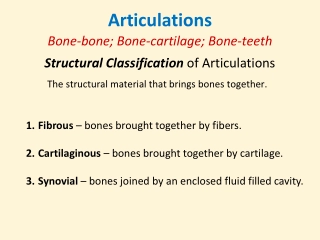
Articulations C. There are 3 structural classifications: 1. Fibrous joints composed of fibrous tissue with no cavity 2. Cartilaginous joints articulating bones are united by cartilage and no cavity present 3. Synovial joints articular bones are separated by a fluid-filled joint cavity
Articulations D. There are 3 functional classifications: 1. Synarthroses immovable joints (sternocostal, tibiofibular, sutures) 2. Amphiarthroses slightly movable joints (intervertebral joint and pubic symphysis) 3. Diarthroses freely movable joints (most appendicular joints)
Articulations E. Fibrous Joints 1. Characteristics: A) Bones are joined by fibrous tissue B) No joint cavity is present C) Most are immovable (synarthrotic) but some are slightly moveable (amphiarthrotic) 2. Three types of fibrous joints
Articulations A) Sutures contain dense fibrous connective tissue until adulthood when they ossify 1) Examples are the skull sutures B) Syndesmoses bones are connected by a filamentous sheet or cord (ligament or interosseous membrane) 1) Movement can range from slight to considerable 2) Examples include the tibiofibular & radioulnar (between the shafts)
Articulations C) Gomphoses articulation of tooth with alveolar margin 1) Possesses a fibrous connection called the periodontal ligament F. Cartilaginous joints 1. Characteristics: A) Articulating surfaces are united by cartilage B) No joint cavity
Articulations 2. Two main types of cartilaginous joints: A) Synchondroses hyaline cartilage unites bones; usually temporary joints (sites of bone growth) 1) Cartilage is replaced by bone and the joints become synarthrotic 2) Examples are the epiphyseal plate and the first rib and the manubrium
Articulations B) Symphyses articular surface of bone covered by hyaline cartilage fused to an intervening pad or plate of fibrocartilage 1) It is compressible, resilient and functionally amphiarthrotic 2) Examples are the pubic symphysis and the intervertebral joints G. Synovial Joints 1. Articular bones are separated by a fluid-filled joint cavity
Articulations 2. Most common joint in the body 3. Diarthrotic 4. Five distinct features of synovial joints A) Articular cartilage hyaline cartilage forms a glassy smooth surface over the opposing ends of bones B) Synovial (joint) cavity small space between the bones C) Synovial fluid largely derived from blood; has a viscous, egg-white consistency
Articulations D) Articular capsule 2 parts 1) Fibrous capsule (external) 2) Synovial membrane (internal) E) Reinforcing ligaments 1) Intrinsic (capsular) parallel bundles of fibers within the fibrous capsule 2) Extracapsular extend bone to bone 3) Intracapsular located inside the cavity
Articulations 5. Movement of synovial joints: A) Axis of motion 1) Non-axial motion slipping movements (intercarpal & intertarsal) 2) Uniaxial motion movement in one plane (interphalangeal, ulna/humerus)
Articulations 3) Biaxial motion movement in two planes (occipital bone/atlas) 4) Multiaxial motion movement in more than two planes (scapula/humerus and coxal bone/femur)
Articulations B) Types of motion 1) Gliding bones displaced in relation to one another (intercarpal, intertarsal, and intervertebral joints) 2) Angular changing the angle between two bones a) Flexion bending or decreasing the joint angle
Articulations b) Extension stretching or increasing the joint angle c) Abduction moving away from the midline d) Adduction moving towards the midline e) Circumduction a limb creates a conical (cone) shape in space f) Rotation turning movement of a bone around its own axis
Articulations 6. Special movements A) Supination of the hand turning the palm forward or facing up B) Pronation of the hand turning the palm backward or facing down C) Inversion (supination) of the foot movement of the sole inward D) Eversion (pronation) of the foot movement of the sole outward
Articulations E) Dorsiflexion upward movement of foot/toes F) Plantar flexion downward movement of foot/toes G) Protraction movement of the mandible forward H) Retraction movement of the protracted part back to its start position
Articulations I) Elevation lifting a body part superiorly J) Depression moving the elevated part inferiorly K) Opposition touching your thumb to the tips of other fingers 7. Types of synovial joints A) Plane joints articular surface is flat and only allows for short gliding movements (intercarpal and intertarsal)
Articulations B) Hinge joints cylindrical projection of one bone fits into a trough-shaped surface on another bone (elbow & knee) C) Pivot joints rounded end of one bone protrudes into a sleeve or ring composed of bone or ligament (radius to ulna and axis to atlas) D) Condyloid joints oval articular surface of one bone fits into a complementary depression in another (metacarpophalanges knuckles)
Articulations E) Saddle joints each articular surface has a concave and convex area (carpometacarpal joint of the thumb) F) Ball and socket joints the spherical end of one bone articulates with a cuplike socket of another bone (shoulder or hip joints)
Articulations H. Common Joint Injuries 1. Sprain stretching or tearing of a ligament 2. Luxation bones are forced out of their normal position (a.k.a. dislocation) A) Subluxation partial dislocation 3. Bursitis inflammation of the bursa A) Bursa flattened sacs that contain a thin film of synovial fluid; located where ligaments, muscles, and tendons rub against bone
Articulations 4. Tendonitis inflammation of the tendon 5. Arthritis inflammatory or degenerative disease of the joint where synovial membranes thicken and fluid production decreases resulting in friction and pain A) Osteoarthritis degenerative B) Rheumatoid arthritis autoimmune disease C) Gouty arthritis uric acid accumulation 6. Synovitis inflammation of synovial membranes