Neuroanatomy Atlas Figures - Haines & Blumenfeld
Explore a collection of detailed neuroanatomy images from the Haines Atlas and Blumenfeld, showcasing structures and areas of the brain associated with language processing, sensory functions, and more. Learn about Broca and Wernicke aphasia, cerebral hemispheres, major cisterns, and midline structures through these informative visuals.

Download Presentation

Please find below an Image/Link to download the presentation.
The content on the website is provided AS IS for your information and personal use only. It may not be sold, licensed, or shared on other websites without obtaining consent from the author. Download presentation by click this link. If you encounter any issues during the download, it is possible that the publisher has removed the file from their server.
E N D
Presentation Transcript
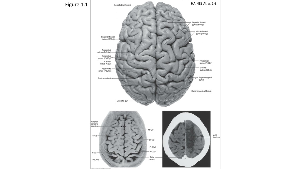
HAINES Atlas 2-8 Figure 1.1 HAINES 2-8
Figure 1.2 HAINES 2-11 HAINES Atlas 2-11
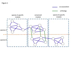
Figure 1.3 HAINES Atlas 2-6 Lateral (A) view of the cerebral hemisphere showing the more commonly described Brodmann areas . A lesion that is located primarily in areas 44 and 45 (shaded) of the inferior frontal gyrus will give rise to what is called a Broca aphasia, also called motor, expressive, or nonfluent aphasia . Lesions in the shaded area of the inferior parietal lobule (Brodmann s areas 40 and 39 sometimes extending into area 22), will give rise to what is known as Wernicke aphasia, also sometimes called sensory, receptive, or fluent aphasia. HAINES 2-8
Figure 1.4 HAINES Atlas 2-14 HAINES 2-8
Figure 1.5 HAINES Atlas 2-26
Figure 1.6 HAINES 2-39 HAINES 2-39
Figure 1.7 Blumenfeld Figures 18.2a and 18.2b
Figure 1.8 HAINES Atlas 2-29
Figure 1.9 HAINES Atlas 2-30
Figure 1.10 HAINES 2-36 HAINES Atlas 2-36
Figure 1.11 Figure 1.12 HAINES 2-17 HAINES Atlas 2-17 HAINES Atlas 2-20
Figure 2.1 Figure 2.2 HAINES 4-10 HAINES 4-10 HAINES 4-6 (7th ed) HAINES Atlas 2-30 HAINES Atlas 4-10
Figure 2.3 Figure 2.4 A median sagittal MRI (T2 weighted) of the brain showing positions of major cisterns associated with midline structures. HAINES Atlas 4-6 A Human Anatomy Carpenter and Sutin 8th edition
Figure 2.6 Figure 2.5 HAINES Atlas 4-1
Figure 2.7 Figure 2.8