Understanding Embolism and Venous Thrombosis in Mechanism of Disease
Embolism, defined as a detached intravascular mass carried through the blood, poses serious risks like pulmonary embolism. Venous thrombosis, particularly deep vein thrombosis (DVT), can lead to embolization, emphasizing the importance of understanding these mechanisms in medical practice.

Download Presentation

Please find below an Image/Link to download the presentation.
The content on the website is provided AS IS for your information and personal use only. It may not be sold, licensed, or shared on other websites without obtaining consent from the author. Download presentation by click this link. If you encounter any issues during the download, it is possible that the publisher has removed the file from their server.
E N D
Presentation Transcript
Ministry of higher Education and Scientific Researches UNIVERSITY OF BASRAH AL-ZAHRAA MEDICAL COLLEGE The module: Mechanism of Disease Haemostasis and Thrombosis II Module staff: Session 6 Dr. Ihsan Mardan Dr. Sadiq K. Ali Dr. Ghada Lateef Dr. Wasan Mansur Robbin s Basic pathology 9th edition Hoffbrands Essential Haematology 7thedition
Learning objectives: 3. Embolism; Definition Thromboembolism particularly DVT and pulmonary embolism Other types of embolism 4. Anti-coagulant therapy; Heparin Warfarin Prophylaxis in general 5. Other disorders of coagulation; Haemophilia Disseminated intravascular coagulation Thrombocytopenia Thrombophilia
3- EMBOLISM An embolus is a detached intravascular solid, liquid, or gaseous mass that is carried by the blood to a site distant from its point of origin. Virtually 99% of all emboli represent some part of a dislodged thrombus, hence the term thromboembolism. Less common types of emboli include fat droplets, bubbles of air or nitrogen, atherosclerotic debris (cholesterol emboli), tumor fragments, bits of bone marrow, and amniotic fluid.
Clinical Correlations: Venous versus Arterial Thrombosis Thrombi are significant because they cause obstruction of arteries and veins and are potential sources of emboli. Venous thrombi can cause congestion and oedema in vascular beds distal to an obstruction, but they are most worrisome for their capacity to embolize to the lungs and cause death. Conversely, while arterial thrombi can embolize and even cause downstream tissue infarction, their role in vascular obstruction at critical sites (e.g., coronary and cerebral vessels) is much more significant clinically.
Venous Thrombosis (Phlebothrombosis) Most venous thrombi occur in either the superficial or the deep veins of the leg.
Venous Thrombosis (Phlebothrombosis) Superficial venous thrombi usually arise in the saphenous system, particularly in the setting of varicosities; these rarely embolize but can be painful and can cause local congestion and swelling from impaired venous outflow, predisposing the overlying skin to development of infections and varicose ulcers. Deep venous thromboses (DVTs) in the larger leg veins at or above the knee joint (e.g., popliteal, femoral, and iliac veins) are more serious because they are prone to embolize.
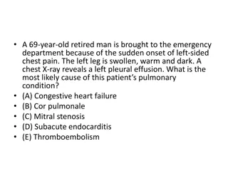
Venous Thrombosis (Phlebothrombosis) Consequently, DVTs are entirely asymptomatic in approximately 50% of patients and are recognized only after they have embolized. Venous thromboembolism (VTE), which comprises deep vein thrombosis (DVT) and pulmonary embolism (PE), with two-thirds presenting as DVT and one-third as PE.
Deep veins thrombosis Predisposing factors: 1. Immobility and bed rest. 2. Pregnancy and post-partum state. 3. Post-operative. 4. Severe burns. 5. Heart failure. 6. Disseminated cancer.
Pulmonary Thromboembolism Pulmonary embolism has an incidence of 20 to 25 per 100,000 hospitalized patients. Although the rate of fatal pulmonary emboli (as assessed at autopsy) has declined from 6% to 2% over the last quarter century, pulmonary embolism still causes about 200,000 deaths per year in the United States.
In greater than 95% of PE cases, venous emboli originate from thrombi within deep leg veins proximal to the popliteal fossa; embolization from lower leg thrombi is uncommon. The patient who has had one pulmonary embolus is at high risk of having more. Most pulmonary emboli (60% to 80%) are clinically silent because they are small.
Clinical presentation of PE depends on the size, location and number of emboli, and the patient s underlying cardiorespiratory reserve. The classic triad of chest pain, haemoptysis and dyspnoea is present in less than 20% of patients, but nearly all will have at least one of pleuritic chest pain, dyspnoea or tachypnoea. A large embolus that blocks a major pulmonary artery can cause sudden death. Sudden death, right ventricular failure (cor-pulmonale), or cardiovascular collapse occurs when 60% or more of the pulmonary circulation is obstructed with emboli.
Systemic Thromboembolism Systemic thromboembolism refers to emboli in the arterial circulation. Most (80%) arise from intracardiac mural thrombi, two-thirds of which are associated with left ventricular wall infarcts and another third with dilated left atria (e.g., secondary to mitral valve disease). By contrast with venous emboli, which lodge primarily in the lung, arterial emboli can travel virtually anywhere.
Systemic Thromboembolism Common arteriolar embolization sites include the lower extremities (75%) and central nervous system (10%); intestines, kidneys, and spleen are less common targets. The consequences of embolization depend on the caliber of the occluded vessel, the collateral supply, and the affected tissue s vulnerability to anoxia. Arterial emboli often lodge in end arteries and cause infarction.
Fat Embolism Microscopic fat globules can be found in the circulation after fractures of long bones (which contain fatty marrow) or after soft-tissue trauma. Fat enters the circulation by rupture of the marrow vascular sinusoids or rupture of venules in injured tissues. fat embolism syndrome characterized by pulmonary insufficiency, neurologic thrombocytopenia, and a diffuse petechial rash, which is fatal in 10% of cases. symptoms, anaemia,
Amniotic Fluid Embolism Amniotic fluid embolism is an uncommon, grave complication of labor and the immediate postpartum period (1 in 40,000 deliveries). The mortality rate approaches 80%, making it the most common cause of maternal death in the developed world. The underlying cause is entry of amniotic fluid (and its contents) into the maternal circulation via tears in the placental membranes and/or uterine vein rupture.
Air Embolism Gas bubbles within the circulation can obstruct vascular flow (and cause distal ischemic injury) almost as readily as thrombotic masses can. Air may enter the circulation during obstetric procedures or as a consequence of chest wall injury. Generally, more than 100 mL of air are required to produce a clinical effect; bubbles can coalesce to form frothy masses sufficiently large to occlude major vessels. Decompression sickness is caused by sudden changes in atmospheric pressure. caisson disease in which recurrent or persistent gas emboli in the bones lead to multifocal ischemic necrosis.
Embolism (summary) An embolus is a solid, liquid, or gaseous mass carried by the blood to a site distant from its origin; most are dislodged thrombi. Pulmonary emboli derive primarily from lower-extremity deep vein thrombi; their effects depend mainly on the size of the embolus and the location in which it lodges. Consequences may include right-sided heart failure, pulmonary haemorrhage, pulmonary infarction, or sudden death. Systemic emboli derive primarily from cardiac mural or valvular thrombi, aortic aneurysms, or atherosclerotic plaques; whether an embolus causes tissue infarction depends on the site of embolization and the presence or absence of collateral circulation.
4- ANTI-COAGULANT THERAPY Heparin: Unfractionated heparin (UFH) is a naturally occurring glycosaminoglycan produced by mast cells. For most clinical indications there has been a move from UFH to low-molecular-weight heparin (LMWH). Heparins are only active when administered parenterally. The APTT is used for routine monitoring of therapeutic doses of UFH Falls in platelet count (>50% compared with baseline) after 5 days of treatment should raise the possibility of heparin-induced thrombocytopenia (HIT)
4- ANTI-COAGULANT THERAPY Heparin: The anticoagulant action of heparin is primarily a result of its ability to bind to antithrombin (AT), thereby accelerating and enhancing the latter s rate of inhibition of the major coagulation enzymes (i.e., factors IIa and Xa and to lesser extents IXa, XIa and XIIa). The two main effects of heparin, the antithrombin and the anti-Xa effects, are differentially dependent on the size of the heparin molecule. Heparin also produces some anticoagulant effect by promoting the release of tissue factor pathway inhibitor (TFPI) from the endothelium.
4- ANTI-COAGULANT THERAPY Warfarin: Warfarin is vitamin K antagonist, that inhibit synthesis of normal FII, FVII, FIX and FX and so result in anticoagulation. Vitamin K is needed for the post-translational modification of the vitamin-K-dependant coagulation factors (II, VII, IX, X, protein C and protein S). The anticoagulant effect varies between individuals and over time and needs to be monitored by the International Normalized Ratio (INR).
4- ANTI-COAGULANT THERAPY Warfarin: After warfarin is given, factor VII levels fall considerably within 24 hours but prothrombin has a longer plasma half life and only falls to 50% of normal at 3 days; so if immediate anticoagulation is needed, heparin is given initially.
4- ANTI-COAGULANT THERAPY Prophylaxis in general: The decision as to whether to continue anticoagulant treatment indefinitely after a first proximal DVT or PE is dependent on the risk of recurrence without continued treatment and the perceived risk of bleeding on anticoagulation. Long-term anticoagulation is usually given after recurrent unprovoked VTE. For patients who receive long-term anticoagulation the risk benefit ratio of continued treatment should be reassessed at regular intervals.
4- ANTI-COAGULANT THERAPY Prophylaxis in general: Following an episode of venous thrombosis, patients are 40 times more likely to suffer an event in the future than patients who have not previously suffered from venous thrombosis. Patients with VTE provoked by surgery have an annualized recurrence rate of <1%. Patients with VTE provoked by a non-surgical factor have an annualized risk of 4% and patients with unprovoked VTE have an annualized event rate of 7%.
5- Other disorders of coagulation Haemophilia: Haemophilia A (factor VIII deficiency): It is the most common of the hereditary clotting factor deficiencies. Haemophilia B (Christmas disease, factor IX deficiency): The incidence is one fifth that of haemophilia A. The inheritance is sex linked but up to one third of patients have no family history and these cases result from recent mutation.
5- Other disorders of coagulation Haemophilia: The characteristic bleeding pattern of haemophilia, which is both delayed after trauma and much prolonged. Replacement by intravenous infusion of the deficient factor can normalize the haemostatic mechanism. Recurrent haematomas dominate the clinical course of severely affected patients and, if inadequately treated, lead to progressive joint deformity and disability painful haemarthroses and muscle
5- Other disorders of coagulation Disseminated intravascular coagulation: Widespread inappropriate intravascular deposition of fibrin with consumption of coagulation factors and platelets occurs as a consequence of many disorders that release procoagulant material into the circulation or cause widespread endothelial damage or platelet aggregation. It may be associated with a fulminant haemorrhagic or thrombotic syndrome with organ dysfunction or run a less severe and more chronic course. The main clinical presentation is with bleeding but 5 10% of patients manifest thrombotic lesions.
Disseminated intravascular coagulation: The key event underlying DIC is increased activity of thrombin in the circulation that overwhelms its normal rate of removal by natural anticoagulants. The pathogenesis of disseminated intravascular coagulation and the changes in clotting factors, platelets and fibrin degradation products (FDPs) that occur in this syndrome
5- Other disorders of coagulation Thrombocytopenia: Thrombocytopenia defines a subnormal number of platelets in the circulating blood, usually below 100 109/L. Thrombocytopenia, if severe, also causes skin and mucous membrane bleeding. It has a wide range of causes including: (i) failure of platelet production from a congenital cause, drugs or viral infection or a general bone marrow failure; (ii) increased consumption of platelets. This may be acute or chronic autoimmune, drug induced, caused by disseminated intravascular coagulation or thrombotic thrombocytopenic purpura.
5- Other disorders of coagulation Thrombophilia: There is no internationally accepted definition of thrombophilia. The term is sometimes used to describe disorders of haemostasis detected in the laboratory that appear to predispose to venous thrombosis. A clinical definition of heritable thrombophilia describes an inherited tendency for venous thrombosis, i.e. deep vein thrombosis (DVT) with or without associated pulmonary embolus (PE).
5- Other disorders of coagulation Thrombophilia: Prevalence of heritable thrombophilias shown to be associated with at least a two fold increased risk of venous thrombosis in patients and controls


")
")
")
")


")