Understanding Hemodynamics Pathology: Causes and Consequences of Various Embolisms
Hemodynamics pathology lecture covers fat embolism, air embolism, amniotic fluid embolism, ischemia, and infarction. Each type of embolism has distinct causes and clinical features, leading to serious consequences. Understanding these conditions is crucial for proper management and prevention in medical practice.

Download Presentation

Please find below an Image/Link to download the presentation.
The content on the website is provided AS IS for your information and personal use only. It may not be sold, licensed, or shared on other websites without obtaining consent from the author. Download presentation by click this link. If you encounter any issues during the download, it is possible that the publisher has removed the file from their server.
E N D
Presentation Transcript
Hemodynamics pathology Lecture 3 MAYS IBRAHIM, ARAB BOARD OF PATHOLOGY, CABP UNIVERSITY OF AL-MUSTANSIRIYAH, COLLEGE OF MEDICINE
Fat embolism Causes 1. Fracture of long bones in which there is an injury to the fatty marrow lead to small hemorrhages in the brain. 2. Burn 3. Acute pancreatitis. Clinical features: Symptoms start within 1 to 3days after injury, the embolism syndrome characterized by pulmonary insufficiency, nurological symptoms, anemia, & thrombocytopenia Small bleeding in the neck , conjuctiva, armpit. fatal in about 10% of cases.
Air Embolism Is the presence of air bubbles in the circulation. Causes: neck wounds cardiothoracic injury venous or arterial catheterization Decompression sickness 1. 2. 3. 4. Caisson disease where air bubble in the bone lead to necrosis 5. Outcome (results): Small volume of air is harmless More than 100 ml will cause symptoms and signs like acute dyspnea, tachypnea, headach seizure and sometimes syncope.
Amniotic Fluid Embolism It is a rare obstetrical complication with a mortality rate of 80% , it is unpredictable, unpreventable event. The patient (the mother) present with dyspnea , cyanosis , shock , convulsions , coma. Pathogenesis: Fetal epithelial cells and fat migrate to maternal pulmonary circulation and cause mechanical obstruction and vasoconstriction of the pulmonary vessels and intravascular coagulation DIC.
Ischemia Defined as deficient blood supply to the tissue which is either: 1. Complete deficiency: caused by obstruction by a thrombus or an embolus which cause infarction. 2. Partial deficiency: caused by an atheroma or spasm.
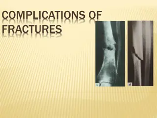
Infarction Area of ischemic necrosis caused by occlusion of either arterial supply or venous drainage in particular tissue. Pathogenesis: 1. 90 % results from thrombotic or embolic events & almost all result from arterial occlusion. 2. Local vasospasm 3. Extrinsic compression of vessels e.g. by tumor. 4. Torsion of arteries and veins (e.g. volvulus, ovarian torsion)
Types of infarction: 1. Red (hemorrhagic) 2. White (anemic)
Infarct white infarc in a kidney White Infarct occur with: Arterial occlusion. Solid organs (heart, spleen, kidneys) where solidity of tissues limits the amount of hemorrhage into ischemic necrosis. solid organs with end-arterial circulations (i.e., few collaterals). 1. 2. 3. The few of extravasated RBCs lysed & hemoglobin released which remain in form of hemosidrin, while in spongy organs, the hemorrhage is too extensive.
Infarct Red (haemorrhagic) infarcts occur due to venous occlusion or embolism in organs with a dual blood supply Red Infarct occur with: 1. Venous occlusion like ovarian torsion. 2. Loose tissues e.g. lung that allow blood to collect in infarcted zone. 3. Tissues with dual circulation e.g. lung & small intestines. 4. Tissues that previously congested because of sluggish venous outflow. 5. When flow re-established to a site of previous arterial occlusion & necrosis (fragmentation of occlusive embolus).
Histologically: The infracted area shows ischemic coagulative necrosis (most organs). Liquifactive necrosis in brain If infarction occur minutes or hours before death, no demonstrable histological changes. Inflammatory response begin within few hours along margin & becomes well defined in 1-2 days caused by necrotic tissues then gradual degradation of dead tissues with phagocytosis by inflammatory cells. Reparative response begin in margin & most infarction replaced by scar tissues.
Factors Influence Development of an Infarct (1) Nature of Vascular Supply: Organs with dual blood supply( lung, liver)are less susceptible than those with end arterial circulation(spleen and kidney) (2)Rate of Development of Occlusion: Slowly developing occlusion less likely to cause infarction because it provides time for alternative pathway to develop like anastomosing vessels in coronary arteries in case of atherosclerosis (3) Vulnerability to hypoxia: Neurons undergo irreversible damage when deprived of their blood supply 3-4 minutes, myocardial cells 20-30 mins (4) Oxygen content of blood: Partial obstruction in anemic patient may lead to infarction, while no effect in normal conditions.
Shock widespread hypoperfusion of cells and tissue due to reduced blood volume, cardiac output or vascular tone. It is a state in which the supply of blood to the tissues is inadequate to meet the metabolic demands (either real loss or relative decrease in blood volume).
Types of shock 1. Cardiogenic shock 2. Hypovolemic shock 3. Septic shock 4. Neurogenic shock
1- Hypovolemic shock In which there is a real decrease in blood volume. Causes: Hemorrhage more than 1 liter of blood or 20% of blood Fluid loss as in severe vomiting, diarrhea & burns. 1. 2. Mechanism of development: is inadequate blood or plasma volume with low cardiac output.
Cardiogenic shock There is relative decrease in blood volume (pooling of blood). Causes: Myocardial infarction. Rupture of the heart. Arrhythmias. Cardiac temponade. 1. 2. 3. 4. Mechanism of development: It is failure of myocardial pump due to intrinsic myocardial damage or extrinsic pressure or obstruction to outflow.
3-Septic shock tissue hypoperfusion and multi organ dysfunction despite initially preserved or even increased cardiac output Causes: Overwhelming bacterial infection (gram negative septicemia or endotoxic shock) or gram positive septicemia. Mechanism of development: Release of endotoxins (gram ve) result in production of TNF, IL-1, IL-6. The mortality rate Peripheral vasodilatation & pooling of blood with hypotension. Cell membrane injury by direct microbial injury. Endothelial cell injury with DIC (disseminated intravascular coagulopathy) Insulin resistance and adrenal insufficiency 1. 2. 3. 4. 5.
4- Neurogenic shock Causes: Anesthesia & spinal cord injury . Mechanism: peripheral vasodilatation.
Stages of shock 1- Non progressive phase: which is the compensatory phase .In this stage a compensatory mechanisms operate to maintain cardiac output &blood pressure near normal levels .The compensatory mechanisms include: a- Arteriolar constriction leading to increase blood pressure. b- Increase heart rate &cardiac output. c- Retention of fluid through increase secretion of ADH & activation of rennin angiotensin aldosteron axis to retain fluid. 2- Progressive phase :When an additional factor is added like extensive burn complicated by bacterial infection .In this stage ,despite the compensatory mechanisms ,there is progressive decline in blood pressure &cardiac output .Clinically observed increase in respiratory rate &decrease in urine output reflecting pulmonary &renal hypoperfusion . 3. Irreversible Phase: result from irreversible injury to the cell membrane as manifested by paralysis of sodium-potassium pump & defect in cell membrane so cell contents go to outside .The reduction in blood flow to the vital organs such as brain, heart, kidney lead to ischemic cell death in these organs .
Pathological Changes: Brain: Ischemic encephalopathy. Heart: coagulation necrosis. Kidneys: Extensive tubular ischemic injury (acute tubular necrosis) which lead to oliguria or anuria & electrolytes disturbances. Lungs: diffuse alveolar damage. Adrenals: cortical cell lipid depletion &use stored lipids for synthesis of steroid GIT: patchy mucosal hemorrhage &necrosis. Liver: fatty change.
Clinical course: In hypovolemic &cardiogenic shock: patient present with hypotension, weak rapid pulse, tachypnea, cool skin In septic shock: the skin may initially be warm &flushed because of peripheral vasodilatation . Prognosis: In hypovolemic shock: 80-90 of young patients survive. Cardiogenic shock associated with extensive myocardial infarction and gram-negative shock, 75% died .