Distal Femur Fracture Diagnosis and Management Overview
Maryam Haider, a 45-year-old male, presented with a severe distal femur fracture following a traumatic incident. This case includes initial history and physical examination findings, differential diagnosis considerations, relevant imaging studies depicting the extent of the injuries, and key findings from the X-rays of the femur and tibia/fibula. The patient was admitted for further management by orthopedic trauma specialists and pain management.

Download Presentation

Please find below an Image/Link to download the presentation.
The content on the website is provided AS IS for your information and personal use only. It may not be sold, licensed, or shared on other websites without obtaining consent from the author. Download presentation by click this link. If you encounter any issues during the download, it is possible that the publisher has removed the file from their server.
E N D
Presentation Transcript
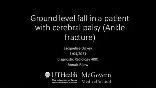
Distal Femur Fracture Due to Trauma Maryam Haider Date: 1/5/2021 RAD 4013- ER Radiology Dr. Ron Bilow
Initial H&P- 12/28/20 45yo M with no significant PMH/PSH who presented as a Level 1 trauma to MHH ED s/p auto ped accident. Presented with bilateral LE deformities due to a forklift running over his legs multiple times. On arrival, pt was HDS with a GCS of 15. Physical exam: VS: WNL Pertinent findings: 2+ radial pulses bilat, DP palpable b/l, deformity of RLE and L thigh, doppler signal b/l, ABIs 0.9 b/l Pertinent lab findings: elevated wbc 16 Pertinent imaging: XRays: L femur series + R tibia/fibula series Pt was admitted to the trauma floor w/plan to consult ORS trauma and with MMPC for pain management.
Differential Diagnosis Fracture 2/2 trauma Compartment syndrome Soft tissue injury Hematoma Hip dislocation Muscle contusion
XR L distal femur- AP view 12/28/20 Femur Distal comminuted and displaced L femur fx Femoral condyles Patella Fibula Intercondylar eminence Tibia Not for diagnostic use. Link: https://theradiologictechnologist.com/normal-knee-xray-knee-joint- anatomy-knee-replacement-surgery/
XR L distal femur- lateral view Femur Medial femoral condyle Lateral femoral condyle Not for diagnostic use. Link:https://theradiologictechnologist.com/normal-knee-xray- knee-joint-anatomy-knee-replacement-surgery/
XR L knee- lateral view Oblique fx in distal fragment of femur possibly extending into knee joint
XR- R tib fib AP view tibia Fibula Displaced transverse fx of R tibia Minimally displaced fx of distal fibula Lateral malleolus Medial malleolus
Key imaging findings XR Femur series: Distal metadiaphyseal comminuted and displaced fracture of the L femur. Proximal component overrides the distal fragment by approx. 7 cm and the proximal part is displaced anteriorly and medially relative to the distal fragment. Oblique oriented fx extending longitudinally through the distal fragment of the femur- likely extends into the knee joint. XR Tibia fibula series: Displaced transverse fx of the mid diaphyseal R tibia. Second oblique minimally displaced fx of the distal metadiaphyseal fibula w/o involvement of the ankle joint.
Distal Femoral Fractures Typically involve the femoral condyles and the metaphyseal region and are often the result of high energy trauma like MVC or a fall from height. Elderly- may occur because of a domestic accident Young pts, typically males are more affected and in the elderly- more women Presents with pain on weight bearing, swelling and bruising, deformity, and in the context of polytrauma. Some complications include dislocations, meniscal or vascular injuries Dx is typically with plain radiographs, however CT is helpful as most of these fx s are intra-articular. MRI can be useful if there is concern for a ligamentous or meniscal injury
Distal Femoral Fx Cont. Radiological description includes: Fracture lines and plane location of fracture in relation to the joint Extra-articular, partial, or completely articular Involvement of the condylar weight-bearing surfaces or the notch Simple, fragmentary, or multi-fragmentary Common example is Hoffa fracture- distal condylar femoral fx that is intra-articular and in the coronal plane Tx typically includes surgery depending on the severity of the fx- options include simple screw fixation, intramedullary nailing, or ORIF
Examples 1 https://www.researchgate.net/figu re/Radiograph-showing-coronal- plane-Hoffa-fracture-of-the- medial-femoral- condyle_fig2_282896930 Hoffa fracture
Examples 2 https://ota.org/for-patients/find-info-body-part/3716
Final Diagnosis + Treatment Pt s injuries included L distal femur fx that is comminuted and both anteriorly and medially displaced Second fx extending through the distal femur fragment and is possibly intra- articular Transverse R tibia fx Second oblique fx of the distal fibula Pt s treatment course included initial LLE traction splinting + plaster splinting of R tib-fib 12/30- OR: underwent IMN of R tibia and L femur Monitored on the trauma floor; PT/OT consulted
Total Cost of MHH Imaging (Inpatient) CXR 1V: $274.68 Ankle XR 2V: $410.33 Ankle XR 3V: $261 L hip XR 2V: $265.32 L femur XR 2V: $237.31 L knee XR 3V: $237.31 L tibia-fibula XR 2V: $237.31 L foot XR 3V: $239.11 XR Abdomen/pelvis 1V: $206.50 CTA LE with or w/o contrast: $2008.44 MRI Pelvis w/o contrast: $2114.71 Total: $6492.02 https://www.memorialhermann.org/patients-caregivers/pricing-estimates-and-information
Take Home Points Pt presents with blunt force trauma- include trauma radiography series of the injured area May need multiple imaging modalities to confirm different types of injuries Distal femoral fx s are quite common in the setting of high intensity trauma
References https://radiopaedia.org/articles/hoffa-fracture?lang=us https://radiopaedia.org/articles/distal-femoral-fracture?lang=us https://www.orthobullets.com/trauma/1041/distal-femur-fractures https://ota.org/for-patients/find-info-body-part/3716


")