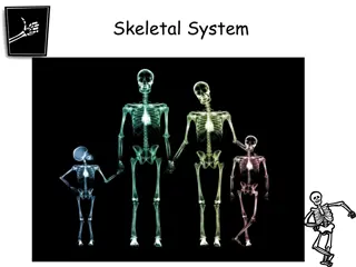
Sex Determination Through Bone Analysis in Forensic Anthropology


P
r
o
j
e
c
t
1
.
2
.
3
:
S
t
u
d
e
n
t
D
a
t
a
S
h
e
e
t
S
E
X
D
E
T
E
R
M
I
N
A
T
I
O
N
The most valuable bones in sex
determination are the pelvis and the skull,
although the femur, tibia and the humerus
provide unique measurements that often
assist in determining identity. Once you
have determined your result for each trait,
circle or highlight male or female on the
data sheet.
P
e
l
v
i
s
(
s
e
x
)
P
e
l
v
i
s
(
s
e
x
)
P
e
l
v
i
s
(
s
e
x
)
P
e
l
v
i
s
(
s
e
x
)
Male Female
Coccyx
Sacrum
S
k
u
l
l
(
s
e
x
)
Square
Round
S
k
u
l
l
(
s
e
x
)
External
auditory
meatus
S
k
u
l
l
(
s
e
x
)
Crest
May need to compare to other
S
k
u
l
l
(
s
e
x
)
Frontal
S
k
u
l
l
(
s
e
x
)
Mandible
Ramus
F
e
m
u
r
(
s
e
x
)
(
I
f
a
v
a
i
l
a
b
l
e
)
Femur Head
Bicondylar
T
i
b
i
a
(
s
e
x
)
(
I
f
a
v
a
i
l
a
b
l
e
)
Proximal
Distal
H
u
m
e
r
u
s
(
s
e
x
)
Vertical
Transverse
Epicondylar
F
I
N
A
L
S
E
X
D
E
T
E
R
M
I
N
A
T
I
O
N
________________________________________________
R
A
C
E
D
E
T
E
R
M
I
N
A
T
I
O
N
Forensic anthropologists generally use a
three-race model to characterize skeletal
remains: White (European), Asian and
Black (African). The skull is often the most
valuable bone in tracing origins, and
various measurements around the face
reveal information about ancestry and
heritage.
S
k
u
l
l
(
r
a
c
e
)
Nasal width:
_______ mm
Nasal height:
_______ mm
Height
Width
Use caliper to measure
S
k
u
l
l
(
r
a
c
e
)
Nasal Index= width mm / height mm
Nasal spine= hold pencil at base of nose and see how smoothly you can raise it up
Nasal silling= Feel base on nasal cavity to see if it is sharp, rounded, or no ridge
S
k
u
l
l
(
r
a
c
e
)
Prognathism= jaw thrust out. To test, hold pencil vertically at the nasal spine
and point down to chin, does it go straight down , at a slight angle, or does it
angel too far out.
See previous results of orbital opening.
S
k
u
l
l
(
r
a
c
e
)
Use the three skull photographs found at station #5 to calculate the nasal
index for each racial group. Compare these values to those listed above and
if needed, describe what could account for any inconsistencies.
W
h
i
t
e
s
k
u
l
l
:
Nasal width ______ mm ÷ Nasal height ______ mm = Nasal index
_______
A
s
i
a
n
s
k
u
l
l
:
Nasal width ______ mm ÷ Nasal height ______ mm = Nasal index
_______
B
l
a
c
k
s
k
u
l
l
:
Nasal width ______ mm ÷ Nasal height ______ mm = Nasal index
_______
F
e
m
u
r
(
r
a
c
e
)
White- fingers can fit under the curvature of the
femur
Black- fingers cannot fit under the curvature of the
femur
Lay femur on table so that the lesser trochanter forms an arch.
Now lay palm of hand flat on table and see if your finger can slide under arch.
F
I
N
A
L
R
A
C
E
D
E
T
E
R
M
I
N
A
T
I
O
N
_
_
_
_
_
_
_
_
_
_
_
_
_
_
_
_
_
_
_
_
_
_
_
_
_
_
_
_
H
E
I
G
H
T
D
E
T
E
R
M
I
N
A
T
I
O
N
The height of the individual is most often determined by
examining the long bones, such as the femur, tibia, or humerus.
The accuracy of these calculations is improved if two or more
bones are used. Forensic anthropologists have compared bone
length to height and have calculated formulas that describe this
relationship (broken down by racial group and by gender). Using
information you have gathered about the gender and ethnicity of
the skeleton, choose the appropriate equations and calculate a
possible height range. As there is no listed formula for using the
maximum length of the femur to estimate height in an Asian
female, use only the equation listed for the other available bone.
F
e
m
u
r
(
h
e
i
g
h
t
)
(
I
f
a
v
a
i
l
a
b
l
e
)
Maximum Length of the Femur (MLF) _______ mm = _______ cm
T
i
b
i
a
(
h
e
i
g
h
t
)
(
I
f
a
v
a
i
l
a
b
l
e
)
Maximum Length of Tibia (MLT) _______ mm = _________ cm
H
u
m
e
r
u
s
(
h
e
i
g
h
t
)
Maximum Length of the Humerus (MLH) _______ mm = _______ cm
F
I
N
A
L
H
E
I
G
H
T
D
E
T
E
R
M
I
N
A
T
I
O
N
To determine the probable height range of the individual, refer to the
height tables you filled in above and record the minimum and maximum
value of the calculated height ranges in the space below. Convert each
value to feet and inches and show the final height range.
Minimum value = __________ cm ÷ 2.54 = _______ inches = ____ feet _____ inches
Maximum value = __________ cm ÷ 2.54 = _______ inches = ____ feet _____ inches
A
G
E
D
E
T
E
R
M
I
N
A
T
I
O
N
Make sure to refer to the photograph of the
pelvic bones while completing your
analysis. Remember, you are looking for
landmarks that occur as a human ages. At
birth, humans have about 405 bones.
These bones will eventually fuse and form
the 206 bones we see in an adult skeleton
P
e
l
v
i
s
(
a
g
e
)
F
e
m
u
r
(
a
g
e
)
(
I
f
a
v
a
i
l
a
b
l
e
)
T
i
b
i
a
(
a
g
e
)
(
I
f
a
v
a
i
l
a
b
l
e
)
H
u
m
e
r
u
s
(
a
g
e
)
Final Ending=
Find your missing person
F
I
N
A
L
M
I
N
I
M
U
M
A
G
E
D
E
T
E
R
M
I
N
A
T
I
O
N
(
r
a
n
g
e
)
_
_
_
_
_
_
_
_
_
_
_
_
_
_
y
e
a
r
s
In forensic anthropology, the pelvis and skull are crucial in sex determination, with additional assistance from femur, tibia, and humerus measurements. This data sheet guides on circling male or female based on traits like sub-pubic angle, pelvic cavity shape, eye orbit sharpness, zygomatic process, nuchal crest, and mandible shape. Images provide visual aid for understanding each trait's result for sex determination.

Download Presentation

Please find below an Image/Link to download the presentation.
The content on the website is provided AS IS for your information and personal use only. It may not be sold, licensed, or shared on other websites without obtaining consent from the author. Download presentation by click this link. If you encounter any issues during the download, it is possible that the publisher has removed the file from their server.
E N D
Presentation Transcript
Project 1.2.3: Student Data Sheet
SEX DETERMINATION The most valuable bones in sex determination are the pelvis and the skull, although the femur, tibia and the humerus provide unique measurements that often assist in determining identity. Once you have determined your result for each trait, circle or highlight male or female on the data sheet.
Pelvis (sex) Trait Result Female > 90 Male 90 Sub-Pubic Angle
Pelvis (sex) Trait Result Female Male Pubis Body Width ~ 40 mm 25-30 mm
Pelvis (sex) Trait Result Female > 68 Male < 68 Greater Sciatic Notch
Pelvis (sex) Trait Result Female Male Pelvic Cavity Shape Circular and wide, showing mainly coccyx Heart-shaped, showing sacrum and coccyx Sacrum Coccyx Male Female
Skull (sex) Trait Trait Result Result Female Female Male Male Sharp Upper Edge of Eye Orbit Shape of Eye Orbit Blunt Round Square Square Round
Skull (sex) Trait Result Female Male Zygomatic Process Not expressed beyond external auditory meatus Expressed beyond external auditory meatus External auditory meatus
Skull (sex) Trait Result Female Male Smooth Nuchal Crest (Occipital Bone) External Occipital Protuberance Rough and Bumpy Generally absent Generally Present Crest May need to compare to other
Skull (sex) Trait Result Female Male Frontal Bone Round, globular Low, slanting Frontal
Skull (sex) Trait Result Female Male Rounded, V-shaped Mandible Shape Square, U-shaped Ramus of Mandible Slanting Straight Ramus Mandible
Femur (sex) (If available) Trait Result Female < 43.5 Indeterminate Sex 43.5- 44.5 Male > 44.5 Vertical (maximum) Diameter of Femoral Head (mm) Bicondylar Width (mm) <74 74- 76 >76 Maximum Length (mm) <405 405-430 >430 Femur Head Bicondylar
Tibia (sex) (If available) Measurement Maximum Epiphyseal Breadth of Proximal Tibia (mm) Maximum Epiphyseal Breadth of Distal Tibia (cm) Result Average Female 70.26 Average Male 79.40 46.31 52.48 Proximal Distal
Humerus (sex) Trait Result Average Female 37.0- 39.0 Average Male 42.7- 44.7 Transverse Diameter of Humeral Head (mm) Vertical Diameter of Humeral Head (mm) Maximum Length (mm) 42.7 48.8 305.9 339.0 Epicondylar Width (mm) 56.8 63.9 Vertical Transverse Epicondylar
FINAL SEX DETERMINATION ________________________________________________
RACE DETERMINATION Forensic anthropologists generally use a three-race model to characterize skeletal remains: White (European), Asian and Black (African). The skull is often the most valuable bone in tracing origins, and various measurements around the face reveal information about ancestry and heritage.
Skull (race) Nasal width: Nasal height: _______ mm _______ mm Height Width Use caliper to measure
Skull(race) Trait Result White < .48 Asian .48- .53 Somewhat prominent spine Rounded ridge Black > .53 Nasal Index Nasal Spine Prominent spine Very small spine Nasal Sharp ridge (silling) No ridge (guttering) silling/Guttering Nasal Index= width mm / height mm Nasal spine= hold pencil at base of nose and see how smoothly you can raise it up Nasal silling= Feel base on nasal cavity to see if it is sharp, rounded, or no ridge
Skull (race) Trait Result White Straight Asian Variable Black Prognathic Prognathism Shape of the Orbital Openings Rounded, somewhat square Rounded, somewhat circular Rectangular Prognathism= jaw thrust out. To test, hold pencil vertically at the nasal spine and point down to chin, does it go straight down , at a slight angle, or does it angel too far out. See previous results of orbital opening.
Skull (race) Use the three skull photographs found at station #5 to calculate the nasal index for each racial group. Compare these values to those listed above and if needed, describe what could account for any inconsistencies. White skull: Nasal width ______ mm Nasal height ______ mm = Nasal index _______ Asian skull: Nasal width ______ mm Nasal height ______ mm = Nasal index _______ Black skull: Nasal width ______ mm Nasal height ______ mm = Nasal index _______
Femur (race) White- fingers can fit under the curvature of the femur Black- fingers cannot fit under the curvature of the femur Lay femur on table so that the lesser trochanter forms an arch. Now lay palm of hand flat on table and see if your finger can slide under arch.
FINAL RACE DETERMINATION ____________________________
HEIGHT DETERMINATION The height of the individual is most often determined by examining the long bones, such as the femur, tibia, or humerus. The accuracy of these calculations is improved if two or more bones are used. Forensic anthropologists have compared bone length to height and have calculated formulas that describe this relationship (broken down by racial group and by gender). Using information you have gathered about the gender and ethnicity of the skeleton, choose the appropriate equations and calculate a possible height range. As there is no listed formula for using the maximum length of the femur to estimate height in an Asian female, use only the equation listed for the other available bone.
Femur (height) (If available) Maximum Length of the Femur (MLF) _______ mm = _______ cm Male Female Regression Formula 2.32 (MLF) + 65.53 3.94 2.15 (MLF) + 72.57 3.80 2.10 (MLF) + 72.22 3.91 Height Range (cm) Regression Formula 2.47 (MLF) + 54.10 3.72 Formula not available 2.28 (MLF) + 59.76 3.41 Height Range (cm) Caucasoid (White) Mongoloid (Asian) Negroid (Black)
Tibia (height) (If available) Maximum Length of Tibia (MLT) _______ mm = _________ cm Male Female Regression Formula 2.42 (MLT) + 81.93 4.00 2.39 (MLT) + 81.45 3.27 2.19 (MLT) + 85.36 3.91 Height Range (cm) Regression Formula 2.90 (MLT) + 61.53 3.66 2.68 (MLT) + 67.05 3.68 2.45 (MLT) + 72.56 3.70 Height Range (cm) Caucasoid (White) Mongoloid (Asian) Negroid (Black)
Humerus (height) Maximum Length of the Humerus (MLH) _______ mm = _______ cm Male Female Regression Formula 2.89 (MLH) + 78.10 4.57 2.68 (MLH) + 83.19 4.16 2.88 (MLH) + 75.48 4.23 Height Range (cm) Regression Formula 3.36 (MLH) + 57.97 4.45 3.22 (MLH) + 51.32 4.35 3.08 (MLH) + 64.67 4.25 Height Range (cm) Caucasoid (White) Mongoloid (Asian) Negroid (Black)
FINAL HEIGHT DETERMINATION To determine the probable height range of the individual, refer to the height tables you filled in above and record the minimum and maximum value of the calculated height ranges in the space below. Convert each value to feet and inches and show the final height range. Minimum value = __________ cm 2.54 = _______ inches = ____ feet _____ inches Maximum value = __________ cm 2.54 = _______ inches = ____ feet _____ inches
AGE DETERMINATION Make sure to refer to the photograph of the pelvic bones while completing your analysis. Remember, you are looking for landmarks that occur as a human ages. At birth, humans have about 405 bones. These bones will eventually fuse and form the 206 bones we see in an adult skeleton
Pelvis (age) Developmental Occurrence Approximate Age 7 - 8 The pubis bone and the ischium are almost completely united by bone. (Figure 6) The illium, ishium, and pubis bones are joined together. (Figure 6) 13 -14 The two lowest segments of the sacral vertebrate become joined together. (Figure 8) The illium, ischium, and pubis bones become fully ossified with no evidence of epiphyseal unions (indicated by cartilaginous lines). All segments of the sacrum are united with no evidence of epiphyseal unions. 18 20-25 25-30
Femur (age) (If available) Developmental Occurrence Approximate Age 4 The greater trochanter first appears. The lesser trochanter first appears. 13 -14 The head, greater trochanter, and lesser trochanter first join the shaft. The condyles first join the shaft. 18 20
Tibia (age) (If available) Developmental Occurrence Approximate Age 18 The lower epiphysis joins the shaft The upper epiphysis joins the shaft 20
Humerus (age) Developmental Occurrence Approximate Age 6 The head and the tuberosities join to become a single large epiphysis. The radial head, trochlea, and external condyle blend and unite with the shaft. The internal condyle unites with the shaft. The upper epiphysis unites with the shaft. 16-17 18 20
Final Ending= Find your missing person FINAL MINIMUM AGE DETERMINATION (range) ______________ years


")
")
")
")
")
")
")
")
")
 (If available)")
 (If available)")
")


")
")
")
")
")


 (If available)")
")
")


")
")
")
")