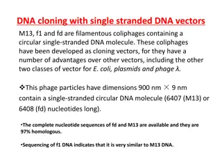
Cloning Other Genes and Recombinant DNA Technology
The recombinant vector with a kanamycin resistance gene can be used to clone other genes by inserting human DNA fragments and selecting transformed E. coli cells. This technique has enabled the production of various human proteins for therapeutic purposes, such as insulin, growth hormones, and clot-dissolving agents. Additionally, the Polymerase Chain Reaction (PCR) allows for the rapid cloning of DNA in vitro, offering a method to amplify specific DNA sequences for research and medical applications.

Download Presentation

Please find below an Image/Link to download the presentation.
The content on the website is provided AS IS for your information and personal use only. It may not be sold, licensed, or shared on other websites without obtaining consent from the author. Download presentation by click this link. If you encounter any issues during the download, it is possible that the publisher has removed the file from their server.
E N D
Presentation Transcript
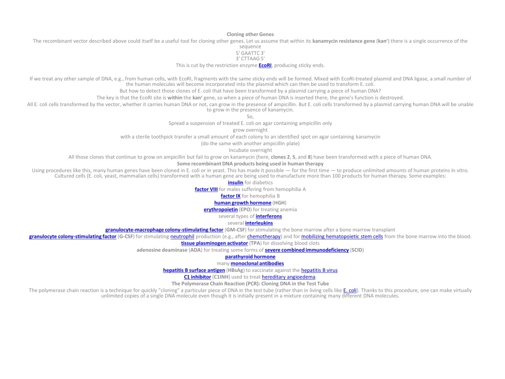
Cloning other Genes The recombinant vector described above could itself be a useful tool for cloning other genes. Let us assume that within its kanamycinresistance gene(kanr) there is a single occurrence of the sequence 5' GAATTC 3' 3' CTTAAG 5' This is cut by the restriction enzyme EcoRI, producing sticky ends. If we treat any other sample of DNA, e.g., from human cells, with EcoRI, fragments with the same sticky ends will be formed. Mixed with EcoRI-treated plasmid and DNA ligase, a small number of the human molecules will become incorporated into the plasmid which can then be used to transform E. coli. But how to detect those clones of E. coli that have been transformed by a plasmid carrying a piece of human DNA? The key is that the EcoRI site is within the kanrgene, so when a piece of human DNA is inserted there, the gene's function is destroyed. All E. coli cells transformed by the vector, whether it carries human DNA or not, can grow in the presence of ampicillin. But E. coli cells transformed by a plasmid carrying human DNA will be unable to grow in the presence of kanamycin. So, Spread a suspension of treated E. coli on agar containing ampicillin only grow overnight with a sterile toothpick transfer a small amount of each colony to an identified spot on agar containing kanamycin (do the same with another ampicillin plate) Incubate overnight All those clones that continue to grow on ampicillin but fail to grow on kanamycin (here, clones 2, 5, and 8) have been transformed with a piece of human DNA. Some recombinant DNA products being used in human therapy Using procedures like this, many human genes have been cloned in E. coli or in yeast. This has made it possible for the first time to produce unlimited amounts of human proteins in vitro. Cultured cells (E. coli, yeast, mammalian cells) transformed with a human gene are being used to manufacture more than 100 products for human therapy. Some examples: insulin for diabetics factor VIIIfor males suffering from hemophilia A factor IX for hemophilia B human growth hormone(HGH) erythropoietin(EPO) for treating anemia several types of interferons several interleukins granulocyte-macrophage colony-stimulating factor (GM-CSF) for stimulating the bone marrow after a bone marrow transplant granulocyte colony-stimulating factor (G-CSF) for stimulating neutrophil production (e.g., after chemotherapy) and for mobilizing hematopoietic stem cells from the bone marrow into the blood. tissue plasminogenactivator (TPA) for dissolving blood clots adenosine deaminase (ADA) for treating some forms of severe combined immunodeficiency(SCID) parathyroid hormone many monoclonal antibodies hepatitis B surface antigen (HBsAg) to vaccinate against the hepatitis B virus C1 inhibitor (C1INH) used to treat hereditary angioedema. The Polymerase Chain Reaction (PCR): Cloning DNA in the Test Tube The polymerase chain reaction is a technique for quickly "cloning" a particular piece of DNA in the test tube (rather than in living cells like E. coli). Thanks to this procedure, one can make virtually unlimited copies of a single DNA molecule even though it is initially present in a mixture containing many different DNA molecules.
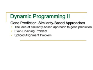
The procedure In order to perform PCR, you must know at least a portion of the sequence of the DNA molecule that you wish to replicate. You must then synthesize primers: short oligonucleotides (containing about two dozen nucleotides) that are precisely complementary to the sequence at the 3' end of each strand of the DNA you wish to amplify. The DNA sample is heated to separate its strands and mixed with the primers. If the primers find their complementary sequences in the DNA, they bind to them. Synthesis begins (as always 5' -> 3') using the original strand as the template. The reaction mixture must contain all four deoxynucleotide triphosphates (dATP, dCTP, dGTP, dTTP) a DNA polymerase. It helps to use a DNA polymerase that is not denatured by the high temperature needed to separate the DNA strands. Polymerization continues until each newly-synthesized strand has proceeded far enough to contain the site recognized by the other primer. Now you have two DNA molecules identical to the original molecule. You take these two molecules, heat them to separate their strands, and repeat the process. Each cycle doubles the number of DNA molecules. Using automated equipment, each cycle of replication can be completed in less than 5 minutes. After 30 cycles, what began as a single molecule of DNA has been amplified into more than a billion copies (230= 1.02 x 10⁹). With PCR, it is routinely possible to amplify enough DNA from a single hair follicle for DNA typing. Some workers have successfully amplified DNA from a single sperm cell. The PCR technique has even made it possible to analyze DNA from microscope slides of tissue preserved years before. However, the great sensitivity of PCR makes contamination by extraneous DNA a constant problem. Genetic Recombination in Bacteria Bacteria have no sexual reproduction in the sense that eukaryotes do. The have no alternation of diploid and haploid generations no gametes no meiosis But the essence of sex is genetic recombination, and bacteria do have three mechanisms to accomplish that: transformation conjugation transduction Transformation Many bacteria can acquire new genes by taking up DNA molecules (e.g., a plasmid) from their surroundings. The ability to deliberately transform the bacterium E. coli has made possible the cloning of many genes including human genes and the development of the biotechnology industry. The first demonstration of bacterial transformation was done with Streptococcus pneumoniae and led to the discovery that DNA is the substance of the genes. The path leading to this epoch-making discovery began in 1928 with the work of an English bacteriologist, Fred Griffith. The cells of S. pneumoniae (also known as the pneumococcus) are usually surrounded by a gummy capsule made of a polysaccharide. When grown on the surface of a solid culture medium, the capsule causes the colonies to have a glistening, smooth appearance. These cells are called "S" cells. Streptococcus pneumoniae (pneumococci) growing as colonies on the surface of a culture medium. Left: The presence of a capsule around the bacterial cells gives the colonies a glistening, smooth (S) appearance. Right: Pneumococci lacking capsules have produced these rough (R) colonies. (Courtesy of Robert Austrian, J. Exp. Med. 98:21, 1953.) However, after prolonged cultivation on artificial medium, some cells lose the ability to form the capsule, and the surface of their colonies is wrinkled and rough ("R"). With the loss of their capsule, the bacteria also lose their virulence. Injection of a single S pneumococcus into a mouse will kill the mouse in 24 hours or so. But an injection of over 100 million (100 x 106) R cells is entirely harmless. Encapsulated (left) and nonencapsulated (right) pneumococci. The encapsulated forms produce smooth colonies (above). (Courtesy of Robert Austrian, J. Exp. Med. 98:21, 1953.) The reason? The capsule prevents the pneumococci from being engulfed and destroyed by scavenging cells neutrophils and macrophages in the body. The R forms are completely at the mercy of phagocytes. Pneumococci also occur in over 90 different types: I, II, III and so on. The types differ in the chemistry of their polysaccharide capsule. Unlike the occasional shift of S -> R, the type of the organism is constant. Mice injected with a few S cells of, say, Type II
pneumococci, will soon have their bodies teeming with descendant cells of the same type. However, Griffith found that when living R cells (which should have been harmless) and deadS cells (which also should have been harmless) were injected together, the mouse became ill and living S cells could be recovered from its body. Furthermore, the typeof the cells recovered from the mouse's body was determined by the type of the dead S cells. In the experiment shown, injection of living R-I cells and dead S-II cells produced a dying mouse with its body filled with living S-II pneumococci. The S-II cells remained true to their new type. Something in the dead S-II cells had made a permanent change in the phenotype of the R-I cells. The process was named transformation. Oswald Averyand his colleagues at The Rockefeller Institute in New York City eventually showed that the "something" was DNA. In pursuing Griffith's discovery, they found that they could bring about the same kind of transformation in vitro using an extractof the bacterial cells. Treating this extract with enzymes to destroy all polysaccharides (including the polysaccharide of the capsule) a lipase to destroy any lipids proteases to destroy all proteins RNase to destroy RNA did notdestroy the ability of their extracts to transform the bacteria. But treating the extracts with DNase to destroy the DNA in them did abolish their transforming activity. So DNA was the only material in the dead cells capable of transforming cells from one type to another. DNA was the substance of genes. View an electron micrograph showing DNA entering a pneumococcus. Although the chemical composition of the capsule is determined by genes, the relationship is indirect. DNA is transcribed into RNA and RNA is translated into proteins. The phenotype of the pneumococci the chemical composition of the polysaccharide capsule is determined by the particular enzymes (proteins) used in polysaccharide synthesis. Conjugation Some bacteria, E. coli is an example, can transfer a portion of their chromosome to a recipient with which they are in direct contact. As the donor replicates its chromosome, the copy is injected into the recipient. At any time that the donor and recipient become separated, the transfer of genes stops. Those genes that successfully made the trip replace their equivalents in the recipient's chromosome. Features: Can only occur between cells of opposite mating types. The donor (or "male") carries a fertility factor (F+). The recipient ("female") does not (F ). F is a set of genes originally acquired from a plasmid and now integrated into the bacterial chromosome; establishes the origin of replication for the chromosome. A portion of F is the "locomotive" that pulls the chromosome into the recipient cell. The rest of it is the "caboose". In E. coli, about one gene gets across each second that the cells remain together. (So, it takes about 100 min for the entire genome (4377 genes) to make it. But, the process is easily interrupted so it is more likely that host genes close behind the leading F genes ("locomotive") will make it than those farther back The "caboose" seldom makes it so failing to receive a complete F factor, the recipient cell continues to be "female" The DNA that makes it across finds the homologous region on the female chromosome and replaces it (by a double crossover). By deliberately separating the cells (in a kitchen blender) at different times, the order and relative spacing of the genes can be determined. In this way, a genetic map equivalent to the genetic maps of eukaryotes can be made. But here the map intervals are seconds, not centimorgans (cM). Demonstration The figure shows the mechanism of conjugation in E. coli cells where The "male" lacks functional genes needed to synthesize the vitamin biotin and the amino acid methionine (Bio , Met ) so these must be added to its culture medium. The "female" has those genes (Bio+, Met+) but has nonfunctional (mutant) genes that prevent it from being able to synthesize the amino acids threonine and leucine (Thr , Leu ) so these must be added to its culture medium). When cultured together, some female cells receive the functional Thr and Leugenes from the male donor. A double crossover enables them to replace the nonfunctional alleles. Now the cells now can grow on a "minimal" medium containing
only glucose and salts. Transduction Bacteriophagesare viruses that infect bacteria. In the process of assembling new virus particles, some host DNA may be incorporated in them. The virion head can hold only so much DNA so these viruses while still able to infect new host cells may be unable to lyze them. Instead the hitchhiker bacterial gene (or genes) may be inserted into the DNA of the new host, replacing those already there and giving the host an altered phenotype. This phenomenon is called transduction. Significance of genetic recombination in bacteria. Transformation, conjugation, and transduction were discovered in the laboratory. How important are these mechanisms of genetic recombination in nature? We don't really know, but Some thoughts: The completion of the sequence of the entire genome of a variety of different bacteria (and archaea) suggest that genes have in the past moved from one species to another. This phenomenon is called lateral gene transfer (LGT). The remarkable spread of resistance to multiple antibiotics may have been aided by the transfer of resistance genes within populations and even between species. Many bacteria have enzymes that enable them to destroy foreign DNA that gets into their cells. It seem unlikely that these would be needed if that did not occur in nature. In any case, these restriction enzymeshave provided the tools upon which the advances of molecular biology and the biotechnology industry depend. Reductionism The understanding of complex systems almost always has to await unraveling the details of some simpler system. You may feel that trying to find out how one type of pneumococcus could be converted into another was an exceedingly specialized and esoteric pursuit. But Avery and his coworkers realized the broader significance of what they were observing and, in due course, the rest of the scientific world did as well. By electing to work with a well-defined system: the conversion of R forms of one type into S forms of a different type, these researchers made a discovery that has revolutionized biology and medicine. Attempting to understand the workings of complex systems by first understanding the workings of their parts is called reductionism. Some scientists (and many nonscientists) question the value of reductionism. They favor a holistic approach emphasizing the workings of the complete system. But the record speaks for itself. From skyscrapers to moon walks, to computer chips to the advances of modern medicine, progress comes from first understanding the properties of the parts that make up the whole. The late George Wald, who won the 1967 Nobel Prize in Physiology or Medicine for his discoveries of the molecular basis of detecting light [Link], once worried that his work was overly specialized studying not vision, not the eye, not the whole retina, not even their rods and cones, but just the chemical reactions of their rhodopsins. But he came to realize "it is as though this were a very narrow window through which at a distance one can see only a crack of light. As one comes closer, the view grows wider and wider, until finally through this same window one is looking at the universe. I think this is the way it always goes in science, because science is all one. It hardly matters where one enters, provided one can come closer...." Gene Therapy I Many human diseases are caused by defective genes. A few common examples: Disease Genetic defect hemophilia A absence of clotting factor VIII cystic fibrosis defective chloride channel protein muscular dystrophy defective muscle protein (dystrophin) sickle-cell disease defective beta globin hemophilia B absence of clotting factor IX severe combined immunodeficiency (SCID) any one of several genes fail to make a protein essential for T and B cell function All of these diseases are caused by a defect at a single gene locus. (The inheritance is recessive so both the maternal and paternal copies of the gene must be defective.) Is there any hope of introducing functioning genes into these patients to correct their disorder? Probably. Other diseases also have a genetic basis, but it appears that several genes must act in concert to produce the disease phenotype. The prospects of gene therapy in these cases seems far more remote. Case study: severe combined immunodeficiency (SCID) SCID is a disease in which the patient has neither
cell-mediated immune responses nor is able to make antibodies. It is a disease of young children because, until recently, the absence of an immune system left them prey to infections that ultimately killed them. About 25% of the cases of SCID are the result of the child being homozygous for a defective gene encoding the enzyme adenosine deaminase (ADA). The normal catabolism of purines is deficient, and this is particularly toxic for T cells and B cells. Treatment Options: Raise the child in a strictly germfree environment: all food, water, and air to be sterilized. David, the "bubble boy" from Houston, survived this way until he was 12 years old. Give the child a transplant of bone marrowfrom a normal, histocompatible, donor. Ideally, this would give the child a continuous source of ADA+T and B cells. However, even though the child cannot reject the transplant (the child has no immune system), T cells in the transplant (unless the donor was an identical twin) can attack the cells of the child producing graft- versus-host disease. the donor cells may be infected with a virus which could overwhelm the recipient before his or her immune system was restored. (David received a bone marrow transplant from his sister, but she, like many people, had been infected earlier with the Epstein-Barr virus (the cause of "mono")). The virus was still present in the cells she donated, and killed her brother. Give injections of ADA (the enzyme is currently extracted from cows). When conjugated with polyethylene glycol (PEG) to delay its breakdown in the blood, ADA-PEG injections have kept SCID patients reasonably healthy. But just like the insulin injections of a diabetic, they must be repeated at frequent intervals. So, what about giving the patient functioning ADA genes; that is, gene therapy? Gene Therapy: requirements The gene must be identified and cloned.This has been done for the ADA gene. It must be inserted in cells that can take up long-term residence in the patient. So far, this means removing the patient's own cells, treating them in tissue culture, and then returning them to the patient. It must be inserted in the DNA so that it will be expressed adequately; that is, transcribed and translated with sufficient efficiency that worthwhile amounts of the enzyme are produced. All these requirements seem to have been met for SCID therapy using a retrovirusas the gene vector. Retroviruses have several advantages for introducing genes into human cells. Their envelope protein enables the virus to infect human cells. RNA copies of the human ADA gene can be incorporated into the retroviral genome using a packaging cell. Packaging cells are treated so they express: an RNA copy of the human ADA gene along with a packaging signal (P) needed for the assembly of fresh virus particles inverted repeats ("R") at each end; to aid insertion of the DNA copies into the DNA of the target cell. an RNA copy of the retroviral gag, pol, and envgenes but with no packaging signal (so these genes cannot be incorporated in fresh viral particles). Treated with these two genomes, the packaging cell produces a crop of retroviruseswith: the envelope protein needed to infect the human target cells an RNA copy of the human ADA gene, complete with R sequences at each end reverse transcriptase, needed to make a DNA copy of the ADA gene that can be inserted into the DNA of the target cell none of the genes (gag, pol, env) that would enable the virus to replicate in its new host. Once the virus has infected the target cells, this RNA is reverse transcribed into DNA and inserted into the chromosomal DNA of the host. What to use for target cells? T cells The first attempts at gene therapy for SCID children (in 1990), used their own T cells (produced following ADA-PEG therapy) as the target cells. The T cells were: placed in tissue culture stimulated to proliferate (by treating them with the lymphokine, Interleukin 2 (IL-2) infected with the retroviral vector returned, in a series of treatments, to the child The children developed improved immune function but: the injections had to be repeated because T cells live for only 6 12 months in the blood the children also continued to receive ADA-PEG so the actual benefit of the gene therapy was unclear Stem cells
Blood ("hematopoietic") stem cells: produce (by mitosis) all the types of blood cells, including T and B lymphocytes produce (by mitosis) more stem cells, thus ensuring an inexhaustible supply In June of 2002, a team of Italian and Israeli doctors reported on two young SCID patients that were treated with their own blood stem cells that had been transformed in vitro with a retroviral vector carrying the ADA gene. After a year, both children had fully-functioning immune systems (T, B, and NK cells) and were able to live normal lives without any need for treatment with ADA-PEG or immune globulin (IG). The doctors attribute their success to first destroying some of the bone marrow cells of their patients to "make room" for the transformed cells. Nine years later (August 2011) these two patients are still thriving and have been joined by 28 other successfully-treated children most of whom no longer need to take ADA-PEG. Gene Therapy for X-linked SCID Gene therapy has also succeeded for 20 baby boys who suffered from another form of severe combined immunodeficiencycalled X-linked SCID because it is caused by a mutated X- linked gene encoding a subunit called c (gamma-c) of the receptorfor several interleukins, including interleukin-7(IL-7). IL-7 is essential for converting blood stem cells into the progenitors of T cells. [View]. Boys with X-linked SCID can make normal B cells, but because B cells need T-helper cells to function, these boys could make neither cell-mediated nor antibody-mediated immune responses and had to live in a sterile bubble before their treatment. Their doctors isolated blood stem cells from the bone marrow of each infant; treated the cells with a retroviral vector containing the normal gene for the c interleukin receptor subunit; returned the treated cells to each donor. The results: Now after as long as 11 years, 19 of these boys are able to live normal lives at home instead of inside a sterile "bubble"; have normal (with some exceptions*) numbers of T cells of both the CD4 and CD8 subsets; have responded to several childhood immunizations, including diphtheria, tetanusand polio by producing both T cells and antibodies specific for these agents. Antibody production is sufficiently good that most of the boys have no need for periodic infusions of immune globulin (IG). * Five of the little boys developed leukemia (one has died): in one case caused by a proliferating clone of T cells in which the vector has inserted itself in a gene (on chromosome 11) implicated in some cases of acute lymphoblastic leukemia (ALL). in a second case, the leukemia was of T cells. Gene Therapy for -thalassemia -thalassemia is an inherited disease. The most severe cases result from mutations in both copies of the gene encoding the beta chain of hemoglobin. Many causative mutations have been identified, and most lead to a failure to make any beta chains. The resulting hemoglobin functions poorly and the victim requires frequent blood transfusions. In the 16 September 2010 issue of Nature, Cavazzana-Calvo (and many colleagues) report a single case of successful gene therapy for this disorder. Their patient was an 18-year old male. Their procedure: Harvest blood stem cells from the patient. Expose them to a retroviral vector that contained a human gene for beta-hemoglobin complete with its promoter, enhancer, and other control elements; alterations to the vector to make it safe. After sufficient chemotherapy to "make room" for them, the patient was injected with these cells. The result: Almost three years later, the patient is well and no longer requires periodic blood transfusions. One-third of his hemoglobin is now manufactured by the red-cell precursors descended from the gene-altered stem cells. Adenovirus Vectors Adenoviruses are human pathogens responsible for some cases of the human "cold". Modified versions of two strains are currently being used as vectors in gene therapy trials. Advantages: Unlike retroviral vectors, they do not integrate into the host genome and thus should not be able to disrupt host genes (It was such disruption that caused some X-linked SCID patients treated with a retroviral vector to develop leukemia). They can infect nondividing cells with high efficiency. Disadvantages: They elicit a powerful immune response, both by T cells and by B cells (antibodies) so repeated doses soon lose their effectiveness. Many people already have antibodies against the virus from earlier "colds", and these can inactivate the vector at the outset. A recent trial of an HIV vaccine using an adenovirus as the vector was halted when it was found not only not to be effective but, in people with preexisting high levels of anti-adenovirus antibodies, may have even increased their susceptibility to HIV.