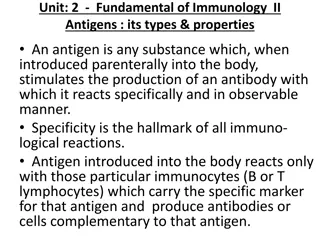
Understanding Antigen-Antibody Precipitation Reaction in Microbiology

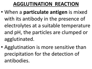
Antigen-antibody precipitation reaction involves the formation of insoluble products when a soluble bivalent antibody interacts with a soluble antigen. This reaction leads to the formation of a visible precipitate known as a lattice. The mechanism of precipitation, including the prozone phenomenon, zone of equivalence, and postzone, is explained through the lattice hypothesis. By understanding the molecular interactions between antigens and antibodies, scientists can perform quantitative precipitation reactions to study immune responses.

Download Presentation

Please find below an Image/Link to download the presentation.
The content on the website is provided AS IS for your information and personal use only. It may not be sold, licensed, or shared on other websites without obtaining consent from the author. Download presentation by click this link. If you encounter any issues during the download, it is possible that the publisher has removed the file from their server.
E N D
Presentation Transcript
B.Sc. III: Semester B.Sc. III: Semester- -VI: Microbiology Microbiology- -P P- -I I VI: UNIT- III ANTIGEN-ANTIBODY REACTION I PRECIPITATION REACTION Prof. D.A.Chouhan M.Sc., NET JRF (CSIR), NET JRF ( UGC), GATE, DMLT
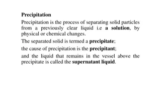
PRECIPITATION PRECIPITATION Reaction Precipitation reaction is the reaction, in which a soluble bivalent antibody reacts with a soluble antigen to give an insoluble product or the precipitate. Soluble antibodies that aggregate soluble antigens are called precipitins. Soluble antigen that induces the formation of a specific precipitin is called a precipitinogen. The antigens, which have two or more epitopes per molecule are cross-linked by the bivalent antibodies, a lattice formation occurs which ultimately develops into a visible precipitate. For the lattice to be formed, the bivalent antibody will bind to epitopes on two different antigens so that the complex is formed. The complex continues to grow and when it is sufficiently large, it becomes insoluble and can be visible as a precipitate. Reaction : :
Mechanism Mechanism of of Precipitation Marrack in 1934 proposed the lattice hypothesis to explain the prozone phenomenon. Marrack s hypothesis is based on the assumptions that each antibody molecule must have at least two binding sites, and antigen must be multivalent. In the zone of equivalence where optimum precipitation occurs, the number of multivalent sites of antigen and antibody are approximately equal. In this zone, precipitation occurs as a result of random, reversible reactions whereby each anti-body binds to more than one antigen and vice versa, forming a stable network or lattice. As they combine, it results in a multimolecular lattice that increases in size until it precipitates out of solution. A quantitative precipitation reaction can be performed by placing a constant amount of antibody in a series of tubes and adding increasing amounts of antigens to the tubes. Plotting the amount of precipitate against increasing antigen concentrations yields a precipitation curve. Precipitation reaction reaction ( ( Lattice Lattice hypothesis) hypothesis)
Mechanism Mechanism of of Precipitation Prozone Prozone (zone of antibody excess) in which the antigen concentration is very low and that of the antibody is relatively high, as a result of which precipitation is inhibited, formation of small complexes occur and residual antibodies will remain in the supernatant. The The second second zone zone is is the the equivalence equivalence zone precipitation in which antigen and antibody form large insoluble complexes and there is neither antigen nor antibody present in the supernatant. The third zone is the zone of antigen excess or postzone antigen concentration is very high, and therefore with increasing the amounts of antigen, the lattice size becomes too small to precipitate as a result of which precipitation is inhibited and binding of antigen-antibody is absent in the supernatant. Precipitation reaction reaction ( ( Lattice Lattice hypothesis) hypothesis) zone, also the zone of maximal postzone, , in which the
Precipitation tests Precipitation tests
Precipitation tests Precipitation tests
Precipitation tests Precipitation tests
VDRL Slide Flocculation Test Result The precipitation Ring Test Slide Slide test Examples Examples: : test The VDRL (Venereal Disease Research Laboratory) test (Treponema pallidum). This test will detect antibodies against Treponema pallidum, and these appear in 4 to 6 weeks. VDRL is a nontreponemal test which detects reagin and antibodies that act against cardiolipin as an antigen. VDRL may show the negative result is late . for syphilis antibodies Ring Ring test Examples Examples: : Ascoli s thermo-precipitin test and the streptococci by the Lancefield technique. test Tube Tube test Examples Examples: : The kahn test for syphilis is an example of a tube flocculation test. test grouping of
a. a. SIMPLE SIMPLE IMMUNODIFFUSION IMMUNODIFFUSION: : Simple Immunodiffusion refers to the diffusion of antigens and antibodies without any external forces. This technique further divided into two types namely Single immunodiffusion and double immunodiffusion. Single Single immunodiffusion immunodiffusion: : In this type of immunodiffusion, only antigen or antibody diffuses in gel. Single Single diffusion diffusion in in one one dimension dimension ( (Oudin antibody is incorporated in agar gel in a test tube and the antigen solution is layered over it. The antigen diffuses downward through the agar gel, forming a line of precipitation that appears to move downwards. This is due to the precipitation formed at the advancing front of the antigen and is dissolved as the concentration of antigen at the site increases due to diffusion. The number of bands indicates the number of different antigens present. Oudin Procedure) Procedure): : The
Single Single immunodiffusion immunodiffusion) ): : diffusion diffusion in in two two dimensions dimensions (Radial (Radial Here the antiserum is incorporated in agar gel poured on a flat surface of slide or Petri dish. The antigen is added to the wells cut on the surface of the gel. The antigen diffuses radially from the well and forms ring shaped bands of precipitation (halos) concentrically around the well. The diameter of the halo gives an estimate of the concentration of the antigen. This method has been employed for the estimation of the immunoglobulin classes in sera. It has also been used for screening sera for antibodies to influenza viruses. This method is also otherwise known as Mancini method. The Mancini method is routinely used to quantitate serum levels of IgM, IgG and IgA by incorporating class-specific anti-isotype antibody into the agar. The technique is also applied to determine the concentrations of complement components in serum. The Mancini method cannot detect antigens present in concentrations below 5- 10ug/ml; this moderate sensitivity is the major limitation of the radial immunodiffusion method. This method is used to determine alpha fetoprotein occurring in certain liver tumor. Mancini procedure of single diffusion Mancini procedure of single diffusion in two direction in two direction ( Radial Immunodiffusion) ( Radial Immunodiffusion)
DOUBLE DOUBLE IMMUNODIFFUSION IMMUNODIFFUSION: : In double immunodiffusion, double refers to the diffusion of both antigens and antibodies. Under this heading, simple double immunodiffusion, Ouchterlony procedure and immunoelectrophoresis are studied. . Simple Simple double double immunodiffusion It is double immunodiffusion in one dimension. Here, the antibody is incorporated in gel, above which is placed a column of plain agar. The antigen is layered on top of this. The antigen and antibody move towards each other through the intervening column of plain agar and form a band of precipitate where they meet at optimum proportion. This technique usually used to identify the presence of either antigen or antibody immunodiffusion (Oakley (Oakley Fulthorpe Fulthorpe procedure) procedure): : Oakley Oakley Fulthorpe Fulthorpe procedure of double diffusion in one procedure of double diffusion in one dimension dimension
Ouchterlony Ouchterlony Procedure It is double diffusion in two dimensions. Agar gel is poured on a slide and wells are cut using a template. The antiserum is placed in the central well and different antigens in the surrounding well. If two adjacent antigens are identical, the lines of precipitate formed by them will fuse and prove V shaped curve. If they are unrelated, the lines will cross each other and provide X shaped curve. Cross reaction or partial identify is indicated by spur formation and provide Y shaped curve. This method was a routine technique for the diagnosis of smallpox. When extracts of smallpox lesions are tested against the antiserum, precipitation lines can be seen within 2-6 hours. A special variety of double diffusion in two dimensions is the Elk test for toxigenicity in diphtheria bacilli. When diphtheria bacilli are streaked at right angles to a filter paper strip carrying the antitoxin implanted on a plate of suitable medium, arrowhead shaped lines of precipitation appear on incubation, if the bacillus is toxigenic. Procedure: :
Immunoelectrophoresis Immunoelectrophoresis: : In Immunoelectrophoresis, , the antigen mixture is first electrophoresed to separate its components by charge. Troughs are then cut into the agar gel parallel to the direction of the electric field, and antiserum is added to the troughs. Antibody and antigen then diffuse toward each other and produce lines of precipitation where they meet in appropriate proportions. Immunoelectrophoresis is used in clinical laboratories to detect the presence or absence of proteins in the serum. A sample of serum is electrophoresed, and the individual serum components are identified with antisera specific for a given protein or immunoglobulin class. This technique is useful in determining whether a patient produces abnormally low amounts of one or more isotypes, characteristic of certain immunodeficiency diseases. It can also show whether a patient overproduces some serum protein, such as albumin, immunoglobulin, or transferrin. Because immunoelectrophoresis is a strictly qualitative technique that only detects relatively high antibody concentrations (greater than several hundred ug/ml).
ELECTROIMMUNODIFFUSION ELECTROIMMUNODIFFUSION: : The development of precipitin lines can be speeded up by electrically driving the antigen and antibody. Various methods have been described combining electrophoresis with diffusion. In this technique, charge of antibody and antigen plays vital role. Of these, A. One dimensional double electro-immunodiffusion (Counter current Immuno-electrophoresis) B. One dimensional single electro diffusion (rocket electrophoresis) C. Two dimensional immune-electrophoresis are used frequently in the clinical laboratory.
Countercurrent Countercurrent Immunoelectrophoresis Immunoelectrophoresis (CIE) This involves simultaneous electrophoresis of the antigen and antibody in gel in opposite directions resulting in precipitation at a point between them. This method produces visible precipitation lines within thirty minutes and is ten times more sensitive than the standard double diffusion techniques. The clinical applications are for detecting various antigens such as alpha fetoprotein in serum and specific antigens of cryptococccus and meningococcus in cerebrospinal fluid (CIE): :
Rocket Rocket electrophoresis electrophoresis: : The antiserum to the antigen to be quantified is incorporated in agarose and gelled on the glass slide. The antigen, in increasing concentrations, is placed in wells punched in the set gel. The antigen is then electrophoresed into the agarose. The pattern of immune-precipitation resembles a rocket and hence the name. The main application of this technique is for quantitative estimation of antigens. This method is useful determination of proteins transferrin, etc.; one electrophoresis is the need for the antigen to be negatively charged for electrophoretic movement within the agar matrix. antibody containing in the quantitative as albumin, of such limitation rocket
Two Two dimensional dimensional immune Several antigens in a complex mixture can be quantitated simultaneously with a modification of rocket electrophoresis called two-dimensional immune- electrophoresis. In this technique antigen is first separated into components by electrophoresis. The gel is then laid over another agar gel containing antiserum and electrophoresis is repeated at right angles to the first direction, forming precipitin peaks similar to those obtained with rocket electrophoresis. Measurement of the size of the peaks allows quantification of a number of proteins in a complex antigen mixture. immune- -electrophoresis electrophoresis: :