Understanding Immunoassay of Digoxin in Pharmaceutical Sciences
Immunoassay is an analytical method utilizing specific antibody-antigen reactions to determine reactant amounts. This method involves competitive binding between labeled and unlabeled analytes and specific antibodies. Antibodies, antigens, labels, and separation matrices play crucial roles in immunoassay development.

Download Presentation

Please find below an Image/Link to download the presentation.
The content on the website is provided AS IS for your information and personal use only. It may not be sold, licensed, or shared on other websites without obtaining consent from the author. Download presentation by click this link. If you encounter any issues during the download, it is possible that the publisher has removed the file from their server.
E N D
Presentation Transcript
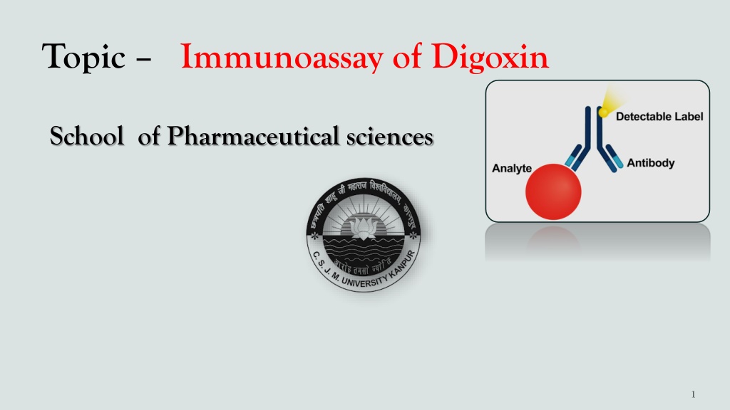
Topic Immunoassay of Digoxin School of Pharmaceutical sciences
Contents 1. 2. 3. 4. 5. 6.
Immunoassay Immunoassay is an analytical method based on the specific immuno- reaction between the antibody(Ab) and antigen(Ag) for the determination of the amount of either reactant in the solution. An antigen-antibody complex is known as an immuno-complex. An immune assay is a test that uses antibody and antigen complexes as a means of generating a measurable result.
Principle of Immunoassay Immuno assay methods are based on competitive binding between a fixed amount of labeled form of an analyte and an available amount of unlabeled sample analyte for a limited amount of binding sites on a highly specific anti-analyte antibody. Reagents required immunoassay development. Antibodies Antigen Signal-generating labels Separation matrices
Antibodies Antibodies are also known as Immunoglobulin, Y-shaped molecules of glycoproteins manufactured by the body that help fight against foreign substances called antigens. They have high specificity and affinity towards the antigen. There are two types of antibodies. Monoclonal and Polyclonal.
Antigen: Antigens are any substances that stimulate the immune to produce antibodies Antigens can be Bacteria, viruses, or fungi that cause infection or disease. The area of antigen where the antibody binds is called an epitope.
Labels All immunoassays require the use of labeled material in order to measure the amount of antigen or antibody present. The labeled component of immunoassay is sometimes called Tracer Examples of a label A radioactive compound, An enzyme that causes a change of color in a solution. or a substance that produces light.
Separation matrices They are used for the separation of the immune complexes that formed as a result of immuno analytical reactions including polyethylene, glycol, charcoal, a second antibody, microbeads, or the most useful 96- well microwell plate; each well of the plate serves as a separate reaction tube. One component of the reaction(antibody or analyte) is coated onto the surface of the bottom of the plate wells and the immune complex is formed on the surface of the wells.
Competitive immunoassay These are reagent (ag) excess immunoassay. In this assay a fixed amount of antibody and fixed amount of labeled antigen and an unlabeled antigen variable amount was taken. Here labeled and unlabeled antigens both compete with each other for binding to a fixed amount of antibody.
Non-competitive immunoassay These are antibody-excess immunoassays. In this, a fixed amount of antigen and a variable amount of unlabeled antibody and a fixed amount of labelled antibody were taken. The unknown analyte in the sample binds with labelled antibodies. The unbound, labelled antibodies are washed away and the bound labelled antibodies are measured. The intensity of the signal is directly proportional to the amount of unknown analyte. The amount of labelled antibody on the site is then measured. It will be directly proportional to the concentration of the analyte because the labelled antibody will not bind if the analyte is not present in the unknown sample.
. Homogeneous competitive immunoassay In this, the unlabeled analyte in A sample competes with the labelled analyte to bind an antibody. The amount of labelled unbound analyte was then measured. The amount of labelled unbound analyte is proportional to the amount of analyte in the sample. Heterogeneous competitive immunoassay In this assay, an unlabeled analyte in a sample competes with the labelled analyte to bind an antibody. The unbound analyte is separated or washed away, and the remaining labelled, bound analyte is measured.
Digoxin Digoxin is the primary cardiac glycoside extracted from the foxglove plant, Digitalis lanata. It is used in the: treatment of CHF because of its inotropic effects on the myocardium. treatment of atrial fibrillation because of its chronotropic effects. treatment of atrial flutter because of its positive inotropic effects. treatment paroxysmal atrial tachycardia because of its positive inotropic effects. Pharmacological Class :- Cardiac Glycoside Therapeutic Class :- Inotrope, antiarrhythmic.
Need of Digoxin immunoassay Digoxin has a narrow range of safe and effective concentrations in serum. Although the therapeutic and toxic concentrations overlap, measurement of digoxin levels helps to maintain effective concentrations and to diagnose and prevent overdosage. Monitoring serum digoxin concentrations, along with careful clinical assessment, is the most effective means of ensuring safe and effective therapy for several reasons: Studies have shown a relationship between serum digoxin concentrations and clinical signs of toxicity. Clinical manifestations of digoxin toxicity (cardiac disturbances, gastrointestinal problems, and central nervous system disorders) can mimic those of disease processes. Concomitant use of other drugs, particularly quinidine, can markedly alter serum digoxin concentrations
Analytical methods for immunoassay of Digoxin Radioimmunoassay (RIA) Enzyme Immunoassay ( EIA) Fluorescence Polarisation Immunoassay (FPIA) Enzyme Multiplied Immunoassay (EMIT)
Radioimmunoassay (RIA) RIA is an analytical technique used to determine the concentration of antigens present in the blood or serum with help of a radiolabeled tracer. Principle It is based upon the competitive binding between radiolabeled antigen and unlabeled antigen with selective antibody, then radioactivity is determined.
Radioimmunoassay (RIA) Process
Enzyme Immunoassay ( EIA) The general principle of this technique is based on the binding of conjugated enzyme molecules with specific antibodies to detect and quantify the presence of either antigens or antibodies in the test sample. In this method, the label on the tracer is an enzyme. The catalytic properties of enzymes allow the detection and quantitation of small quantities of the drug.
Fluorescence Polarization Immunoassay (FPIA) The principle of the assay is that a fluorescent dye (attached to an antigen) can be excited by plane-polarized light at the appropriate wavelength. The patient sample is incubated with a known quantity of the fluorescent-labeled drug and an antibody specific to the drug. The labeled and unlabeled drugs compete for the binding sites of the antibody. Polarized light is emitted at certain angles depending on whether the fluorescent-labeled drug is bound to antibodies or not. Since this is a competitive assay, the greater the amount of drug in the sample, the lower the amount of fluorescence. The intensity of polarized light is a measure of the concentration of the analyte.
Enzyme Multiplied Immunoassay (EMIT) Commonly used analytical method for immunoassay of digoxin. Methodology: Enzyme multiplied immunoassay is manufactured using recombinant DNA technology, used for the analysis of digoxin and its active metabolites in serum or plasma. The assay is based on competition between the drug in the sample and the drug labeled with recombinant glucose-6-phosphate dehydrogenase (rG6PDH) for antibody binding sites. Enzyme activity decreases upon binding to the antibody, so the drug concentration in the sample can be measured in terms of enzyme activity.
The general procedure of Immunoassay of Digoxin Specimen Collection and Preparation : Each assay requires serum or plasma. Sample volume is instrument-dependent. Use fresh samples. If samples are to be tested within 8 hours of collection, they may be stored at room temperature (20 25 C). For transporting, maintain the sample temperature at 2 8 C. Samples can be stored refrigerated at 2 8 C for up to 7 days or stored frozen (- 20 C) for up to 6 months. Repeated freeze-thaw cycles should be avoided. If the samples contain particulate matter, or fibrous material then Vigorously mix the sample in a vortex for at least 30 seconds or Centrifuge the sample at 2000 rpm for 15 minutes.
Conti Reagents: Sample( serum containing digoxin) Digoxin tracer reagent( digoxin-HRP in a buffer dye) Rabbit serum antibody(digoxin-ab) Washing buffer Signal agent( luminol in buffer)
Procedure: 96-well microplate Add 0.025ml (25 l) of the appropriate serum specimen into the assigned well. Add 0.050 ml (50 l) of Digoxin Enzyme Reagent( HRP) to all the wells. Swirl the microplate gently for 20-30 seconds to mix. Add 0.050 ml (50 l) Digoxin antibody( rabbit serum) to all wells Swirl the microplate gently for 20-30 seconds to mix. Cover and incubate for 30 minutes at room temperature. Add 0.350ml (350 l) of washing buffer & wash for 3 times.
Cont Add 0.100 ml (100 l) of working signal reagent to all wells. DO NOT SHAKE THE PLATE AFTER THIS ADDITION. Incubate at room temperature for fifteen (15) minutes. Read the relative light unit with a chemiluminescence microplate reader. The results should be read within thirty (30) minutes of adding the signal reagent.
References 1. A.F.Davidson, H.L. Walton, D. J. Pinto, R.E.Immunoassay,New York ,1988, pp.60. 2. R.Keller, J.M.Mermet, Analytical Chemistry,1998, pp. 405-429 3. W.C.Smith, J.W. Lee,J.Pharma.Biomed.Anal.21 (2000), pp. 1249-1273 4. A.F.Davidson, H.L.Walton, D. J. Pinto, R.E.Immunoassay,NewYork,1988, pp.60.


")
")
")
")
")

")