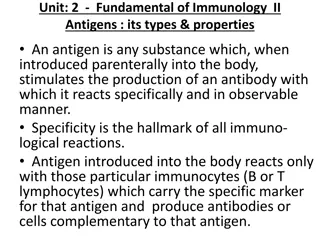
Understanding Antigen-Antibody Interactions and Diagnostic Tests

Antigen-antibody interactions are essential in immunology, determining disease presence, blood types, and more. Diagnostic tests such as agglutination and precipitation reactions play a crucial role. Specimen collection involves obtaining blood samples for analysis, while precipitation reactions form visible precipitates. Different types of precipitation tests are used for quantitative measurement.

Download Presentation

Please find below an Image/Link to download the presentation.
The content on the website is provided AS IS for your information and personal use only. It may not be sold, licensed, or shared on other websites without obtaining consent from the author. Download presentation by click this link. If you encounter any issues during the download, it is possible that the publisher has removed the file from their server.
E N D
Presentation Transcript
Antigen Antibody interaction Principles and applications
Antigen-Antibody Interactions: Principles and Applications Antigen-Antibody Interactions: Principles and Applications The Ag-Ab interaction is a biomolecular association similar to enzyme-substrate interaction but with few differences as: 1. It does not lead to irreversible chemical alteration in either the Ag or the Ab. 2. It is a noncovalent interaction which include hydrogen bond, ionic bond, hydrophobic bond and van der Waals interaction. Strong Ag-Ab interaction depend on a very close fit between the Ag & Ab.
Types of diagnostic tests Many types of diagnostic tests are performed in the immunology laboratory. Most of these tests can be designed to determine the presence of either the Ag or the Ab. Major uses of serologic (Ab based ) tests: 1. Diagnosis of infectious diseases: When the organism cannot be cultured as in syphilis, hepatitis A,B, and C. When the organism is too dangerous to culture e.g. rickettsial disease. When culture technique are not readily available, e.g. HIV. When the organism takes long time to grow, e.g. Mycoplasma. 2. Diagnosis of autoimmune disease, e.g. Ab against DNA in SLE. 3. Determination of blood type and HLA.
Antigen-Antibody Interaction Agglutination reaction Labeled Ags OR Abs Chromatography Immunoassay Precipitation reaction Direct : Indirect (passive) -Blood group -Bacterial aggl. PPT in Soluble PPT in gel Immunoelectrophoresis PPT in Agar with electric field Single Radial diffusion Counter immuno electroph. Double diffusion Rocket electrophoresis
Specimen collection and preparation Obtain a sample of venous blood from the patient and allow a clot to form. Centrifuge clotted blood sample and collect clear serum .
1. Precipitation reactions Antibody and soluble Ag interacting in aqueous solution form a lattice that eventually develops into a visible precipitate. The Ab that aggregate soluble Ag are called precipitins. PPT depend on: 1. Ab must be bivalent, i.e. PPT will not develop with monovalent Fab fragment. 2. Ag must be either bivalent or polyvalent.
Types of PPt test: 1. PPT in solution. This reaction can be made quantitative. 2. PPT in agar. Of many types, which are: *. Single diffusion (radial immunodiffusion, the Mancini method). In it Ab is incorporated into agar & Ag is measured into a well. As the Ag diffuses with time ,precipitation rings form depending on the Ag concentration. Such radial immunodiffusion is used to measure IgG, IgM, Complement component and other substance in the serum but not IgE because its concentration is to low.
* 2. Double diffusion(Ouchterlony method). In it the Ag & Ab are placed in different wells in agar and allowed to diffuse and form concentration gradiants, with line of precipitate form. This method indicate whether Ag are identical, related but not identical, or not related. So it is a qualitative way.
3. PPT in agar with electric field.Which are either - Immunoelectrophoresis, of which a serum is placed in a well in agar on a glass slide, then a current is passed through the agar, and a protein move in the electric field according to their charge and size. Then a trough is cut into the agar and filled by Ab. As they move toward each other, they form a series of arcs of ppt.used in diagnosis of human myeloma protein. - Counter-immunoelectrophoresis. It relies on movement of Ag toward the cathod and Ab toward the anod with meeting visibile in 30-60 min. used detection of bacterial and fungal Ag in CSF. - Rocket electrophoresis. It is a quantitative technique, of which , it permit measurement of Ag levels. In it the negatively charged Ag is electrophoresed in a gel containing Ab. The ppt form a shape of rocket, the height of which is proportional to the concentration of the Ag.
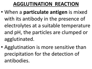
Agglutination Reactions It is an interaction between Ab & particulate Ag resulting in visible clumping called (agglutination). The Ab that produce such a reaction is called (agglutinin).This reaction need a polyvalent Ag. Just as an excess of Ab inhibit ppt reaction, it will also inhibit agglutination and this is called (Prozone effect). Causes of Prozone effect: 1.High Ab concentration. This will result in univalent binding of the Ab to Ag, so no cross-linking of one Ag to another. To overcome this you most dilute the serum . 2.Incomplete Ab (often of IgG). This Ab may occupy most of the Agic site, this block the access by IgM, which is a good agglutinin.
Assay procedure : 1- Qualitative method : 1- Allow kit and patient serums to come to room temperature . 2- Transfer one drop (50 l) of patient s serum on a test slide. 3- Shake the reagent then using the dropper and add one drop to the test slide and mix and gently rotate the mixture for 4 minutes.
2- Semi-quantitative method : 1- Using isotonic saline prepare serial dilution of the patient serum (1/2, 1/4, 1/8, 1/16, 1/32, 1/64, and so on ). 2- Transfer one drop (50 l) of each serum dilution to the test circle slide . 3- Shake the reagent, add one drop of suspension to the test slide then mix and gently rotate the test slide for 4 minutes .
- Results : A positive result is indicated by the obvious agglutination pattern of the suspension on the test slide. A negative result is indicated by no change in the suspension on the test slide . Agglutination of the Ag with undiluted sample indicates the presence of Ab at a concentration of greater than or equal to 25 IU/ml . A comparison between samples taken 10-14 days apart may be of value in acute illness .
Types of agglutination reactions Direct agglutination (Bacterial agglutination) [1] Rose-Bengal test: -Principle of the test: The test depends on the ability of the Ab in the patient s serum to agglutinate the stained bacterial Ag. When this occurs the aggregates become clearly visible to the naked eye . Ag : stained bacterial Ag (killed suspension of Brucella abortus ) Ab : Abs to Brucella in the patients serum .
2- Widal Test : - Principle of the test : The test depends on the ability of Ab in the patient s serum to agglutinate the stained bacterial Ags. When this occurs the aggregates become clearly visible to the necked eye. *Ags :Suspension of bacteria stained and killed. *Salmonella O-Group A(Paratyphi A-O). *Salmonella O-Group B(Paratyphi B-O). *Salmonella H-Group a(Paratyphi A-H). *Salmonella H-Group b(Paratyphi B-H). *Salmonella O-Group d(Typhi O). *Salmonella H-Group d(Typhi H). *Ab : Ab to bacterial Ags in patient s serum .
Indirect ( Passive ) agglutination : Is useful with soluble Ag. This test done with the use of Ag coated RBC, or the use of synthetic particales, such as latex beads. [1] Qualitative determination of anti-streptolysin O (ASO). -Clinical finding : Streptolysin O is toxic immunogenic exoenzyme produced by hemolytic streptococci of group A, C, and G. Measuring the ASO Abs are useful for the diagnostic of rheumatoid fever, acute glomerulonephritis and streptococcal infections. Rheumatic fever is an inflammatory disease affecting connective tissue from several parts of human body as skin, heart, joints, etc .and acute glomerulonephritis is a renal infection that affects mainly to renal glomerulus.
[2] Qualitative determination of Rheumatoid Factors (RF): -Clinical significance : Rheumatoid factors are a group of Abs directed to determinants in the FC portion of the immunoglobulin G molecule . Although rheumatoid factors are found in a number of rheumatoid disorders, such as, Systemic Lupus Erythematosus (SLE) ,and Sj gren s syndrome, as well as in nonrheumatoid arthritis (RA), utility as an aid in the diagnosis of rheumatoid arthritis (RA).
[3]A rapid latex slide test for the detection of CRP in serum : Tissue-damaging associated with inflammatory diseases, infection and neoplasms are associated with a major accute phase response of the C-reactive protein (CRP) and other acute phase reactants. The CRP response frequently precedes clinical symptoms, including fever, Measuring changes in the concentration of CRP provides useful diagnostic information about how acute and how serious a disease is. It also allows the assessment of complications during the disease and judgment about the disease genesis.
Chromatographic immunoassay : Pregnancy test strip (Urine/Serum): Human chorionic gonadotropin (hCG) is a glycoprotein hormone produced by the developing placenta shortly after fertilization. In normal pregnancy, hCG can be detected in both urine and serum as early as 7 to 10 days after conception. -Principle: The hCG one step pregnancy test strip (Urine/serum) is a rapid chromatographic immunoassay for the qualitative detection of hCG to aid in the early detection of pregnancy .
C C C C C T T T T T Positive Negative Invalid


. In it the Ag & Ab")


 agglutination :")