Understanding Peptic Ulcers: Causes, Prevalence, and Treatment

Peptic ulcers are circumscribed ulcerations in the gastrointestinal mucosa, commonly caused by Helicobacter pylori infection. They have a higher prevalence in developing countries and are associated with factors like socioeconomic status, poor hygiene, and genetic links. While peptic ulcers naturally tend to heal and recur, medical treatment focuses on reducing stomach acid secretion to promote healing. Gastric and duodenal ulcers differ in incidence rates, risk factors, and treatment approaches. Understanding these distinctions is crucial for effective management of peptic ulcers.

Download Presentation

Please find below an Image/Link to download the presentation.
The content on the website is provided AS IS for your information and personal use only. It may not be sold, licensed, or shared on other websites without obtaining consent from the author. Download presentation by click this link. If you encounter any issues during the download, it is possible that the publisher has removed the file from their server.
E N D
Presentation Transcript
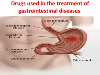
A circumscribed ulceration of the gastrointestinal mucosa 3mm or greater occurring in areas exposed to acid and pepsin most often caused by Helicobacter pylori infection.
Higher prevalence in developing countries H. Pylori is sometimes associated with socioeconomic status and poor hygiene In the US: Lifetime prevalence is ~10%. PUD affects ~4.5 million annually. Hospitalization rate is ~30 pts per 100,000 cases. Mortality rate has decreased dramatically in the past 20 years approximately 1 death per 100,000 cases
: 12 duodenal sites are 4x as common as gastric sites most common in middle age peak 30-50 years Male to female ratio 4:1 Genetic link: 3x more common in 1stdegree relatives more common in patients with blood group O associated with increased serum pepsinogen H. pylori infection common up to 95% smoking is twice as common
These have a natural tendency to heal and to recur- in a cyclical fashion Relapse after medical treatment with significant problems in relation to work or social life The feature common to all methods of treatment that prolong the healing phase is reduction to the amount of acid secreted by the stomach
: common in late middle age incidence increases with age Male to female ratio 2:1 More common in patients with blood group A Use of NSAIDs - associated with a three- to four-fold increase in risk of gastric ulcer Less related to H. pylori than duodenal ulcers about 80% 10 - 20% of patients with a gastric ulcer have a concomitant duodenal ulcer
Many gastric ulcers will heal. An unhealed ulcer has a 5-10 % chance of being malignant. Though there are a number of operations available, the most satisfactory treatment is a Billroth I partial gastrectomy. The most commonly performed operation, subtotal gastrectomy, does nothing more subtle than remove the ulcer and the ulcer-prone area of the stomach. There is no clear association ulceration and acid secretion, but it has been suggested by some that enhancing gastric drainage (by pyloroplasy) will promote healing. between gastric
50-60 12 30-50 4 4 : 1 0 H. Pylori (95%) Heal & Recur Reduction of the amount of acid secreted by the stomach ( : ) 2 : 1 NSAID H. Pylori (80%) Heal & Unheal Remove the ulcer Remove the ulcer- prone area Enhancing gastric drainage
The most important contributing factors are H pylori, NSAIDs, acid, and pepsin. Additional aggressive factors include smoking, ethanol, bile acids, aspirin, steroids, and stress. Important protective factors are mucus, bicarbonate, mucosal blood flow, prostaglandins, hydrophobic layer, and epithelial renewal. When an imbalance occurs, PUD might develop
, : 90% , 90% , 90% 1 . 10% . Johnson, : , , , - . , , . , ( 2-3cm ), . V, . . V, ( . NSAID).
Pain Duodenal ulcers: occurs 1-3 hours after a meal and may awaken patient from sleep. Pain is relieved by food, antacids, or vomiting. Gastric ulcers: food may exacerbate the pain while vomiting relieves it. Nausea, vomiting, belching, dyspepsia, bloating, chest discomfort, anorexia, hematemesis, &/or melena may also occur. nausea, vomiting, & weight loss more common with Gastric ulcers
Neoplasm of the stomach Pancreatitis Pancreatic cancer Diverticulitis Nonulcer dyspepsia (also called functional dyspepsia) Cholecystitis Gastritis GERD MI not to be missed if having chest pain
Stool for fecal occult blood Labs: CBC (R/O bleeding), liver function test, amylase, and lipase. H. Pylori can be diagnosed by urea breath test, blood test, stool antigen assays, & rapid urease test on a biopsy sample. Upper GI Endoscopy: Any pt >50 yo with new onset of symptoms or those with alarm markings including anemia, weight loss, or GI bleeding. Preferred diagnostic test b/c its highly sensitive for dx of ulcers and allows for biopsy to rule out malignancy and rapid urease tests for testing for H. Pylori.
Consider prophylactic therapy for the following patients: Pts with NSAID-induced ulcers who require daily NSAID therapy Pts older than 60 years Pts with a history of PUD or a complication such as GI bleeding Pts taking steroids or anticoagulants or patients with significant comorbid medical illnesses Prophylactic regimens that have been shown to dramatically reduce the risk of NSAID-induced gastric and duodenal ulcers include the use of a prostaglandin analogue or a proton pump inhibitor. Omeprazole 20-40 mg PO every day Lansoprazole 15-30 mg PO every day
: H. PYLORI (+) Medications: Triple therapy for 14 days is considered the treatment of choice. Proton Pump Inhibitor + clarithromycin and amoxicillin Omeprazole (Prilosec): 20 mg PO bid for 14 d or Lansoprazole (Prevacid): 30 mg PO bid for 14 d or Rabeprazole (Aciphex): 20 mg PO bid for 14 d or Esomeprazole (Nexium): 40 mg PO qd for 14 d plus Clarithromycin (Biaxin): 500 mg PO bid for 14 and Amoxicillin (Amoxil): 1 g PO bid for 14 d Can substitute Flagyl 500 mg PO bid for 14 d if allergic to PCN In the setting of an active ulcer, continue qd proton pump inhibitor therapy for additional 2 weeks. Goal: complete elimination of H. Pylori. Once achieved reinfection rates are low.
: H. PYLORI (-) Medications treat with Proton Pump Inhibitors or H2 receptor antagonists to assist ulcer healing H2: Tagament, Pepcid, Axid, or Zantac for up to 8 weeks PPI: Prilosec, Prevacid, Nexium, Protonix, or Aciphex for 4-8 weeks.
H pylori (+) NSAID (+): 7-14 ( s) H2 4 . H pylori (+) NSAID (-): . 95% . H pylori (-)NSAID (+): s NSAID, . H pylori (-) NSAID (-): . , PPIs . , , ( . , Crohn ).
Bleeding occurs in 25% to 33% of cases and accounts for 25% of ulcer deaths. Perforation & Penetration into pancreas, liver and retroperitoneal space Peritonitis Gastric outflow obstruction, & Pyloric stenosis Gastric CA
People who do not respond to medication People with positive for AdenoCa biopsy People who develop complications Vagotomy Antrectomy Pyloroplasty
DUMPING SYNDROME The stomach is a reservoir and the pylorus meters food rendered iso-osmotic with plasma into the small bowel for further digestion and absorption. Consequently, ablation of gastric areas plus, as is always the case, loss or bypass of the pylorus allows the entry of hyperosmolal, large volume loads into the jejunum. Two things follow: The bulk stimulates peristalsis and results in pain, rapid transit and thus occasionally diarrhea. The hyperosmolity draws fluid into the gut lumen which aggravates the bulk problem and may also reduce blood volume so creating vasomotor instability-the patient feels faint and tremulous after a meal. These features constitute the dumping syndrome which is aptlynamed because it does result from dumping a large volume of hypertonic liquid into the jejunum.
ANEMIA Partial gastrectomy interferes with duodenal absorption of iron and a macrocytic anemia may result More removed to cause failure of release of intrinsic factor and thus a macrocytic anemia rarely, sufficient stomach has been Malnutrition may contribute to both.
DIARRHEA Apart vagotomies except highly selective ones seem to cause diarrhea from the dumping syndrome, all Matters are made worse if cholecystectomy has been done or is subsequently done
: 25.000 & 16.000 / 2 3 13 : 65% 3- 4 80% (+) / 30% 2:1 ( 100.000 ) UK/ 52.6 6.3 16-20
2. EGC 1. + 2. + 3. EGC, 30mm, , 500 m (SM1) Oda , et al. Gastric Cancer 2006; 9: 262 270
H >90% 80% 100% ( ) 8% 1-7% 2-4% 3 99% 5 96%
3. 1 80% . 1 < 20% Suzuki H, et al. Gastric Cancer 2006;9:315-319 1a : 1 : 2% 15% Roukos DH. Cancer Treat Rev 2000;26:243 245 Mm & ITC 10% Morgagni P, et al. World J Surg 2003;27:558-561 1 : 2 : 50% Sasako M, et al. Br J Surg 1995; 82:346-351 Mm & ITC 52.6% Fukagaw , et al. Ann Surg Oncol. 2009;16:609-613 2 : 1 : : Skip metastases 2.8% Skip metastases 17.4% Li C, et al. J Gastrointest Surg 2008;12:550-554
In 1997, the AJCC/UICC guidelines were revised, requiring the pathological examination of at least 15 LN for accurate gastric cancer staging. Sobin LH, Wittekind Ch (eds). TNM classification of malignant tumors. 6th edition, New York, John Wiley & Sons, 2002. nly 29% of gastric cancer patients had more than 15 LN examined in the West. Coburn NG, et al. Cancer 2006;107:2143-2151. D2-gastrectomy clearly offers the mean number of required LN for pathological examination, even in cases of Western pathologists. Bunt AM, et al. J Clin Oncol 1996;14:2289-2294.
4. 1 1. 2. 3. 4. 5. - 6. -
2 7. . 8. . 9. 10. 11. . 12. 12
3 13. 14. (SMV) 15.
Bozzetti F. Principles of surgical radicality in the treatment of gastric cancer. Surg Oncol Clin N Am 2001; 10: 833-854 At least a 6-cm tumor-free (based on the frozen section result) proximal resection margin from the most proximal macroscopic border of the tumor should be achieved in all gastric resections.
Kasakura Y, Fujii M, Mochizuki F, et al.: Is there a benefit of pancreaticosplenectomy with gastrectomy for advanced gastric cancer? Am J Surg 2000;179:237 242. o significant difference in 5-year survival for stage II, III, or IV tumors was detected. Thus, splenectomy and distal pancreatectomy do not have an impact on survival and are associated with an increased incidence of complications.
Kodera Y, Sasako M, Yamamoto S, et al. Gastric Cancer Surgery Study Group of Japan Clinical Oncology Group Identification of risk factors for the development of complications following extended and superextended lymphadenectomies for gastric cancer. Br J Surg 2005; 92:1103-1109 Multivariate analysis Risk factors for postoperative complications: Pancreatectomy RR=5.62 Prolong operative time RR=2.65 BMI > 25 RR=2.18 Age > 65 years
esection of adjacent organs in conjunction with gastrectomy is still acceptable to achieve R0 resection in a highly selected patient population with histological confirmation of organ invasion but negative lymph nodes. Considering the high postoperative morbidity, the procedure of more than two organ resection with gastrectomy is not an optimal choice.
Cho BC, Jeung HC, Choi HJ, et al.: Prognostic impact of resection margin involvement after extended (D2/D3) gastrectomy for advanced gastric cancer: A 15-year experience at a single institute. J Surg Oncol 2007;95:461 468. Node (-), Margin (-) pts Median survival 147 months Node (-), Margin (+) pts Median survival 37 months Node ( ), Margin (-) pts Median survival 37 months Node ( ), Margin (+) pts Median survival 33 months More aggressive treatment, including reoperation, should be considered only for node-negative patients with positive microscopic margins Nodal status may be a more important determinant of survival than R0 resection


")
")
 NSAID (+): Η θεραπεία")