Understanding Leishmaniasis: Types, Symptoms, and Treatment Options
Leishmaniasis is a parasitic disease caused by Leishmania parasites transmitted through infected sand flies. It comes in three forms - cutaneous, mucocutaneous, and visceral - each with distinct characteristics and treatment approaches. Cutaneous leishmaniasis causes skin ulcers, mucocutaneous affects mucous membranes, and visceral damages internal organs and is fatal if left untreated.

Download Presentation

Please find below an Image/Link to download the presentation.
The content on the website is provided AS IS for your information and personal use only. It may not be sold, licensed, or shared on other websites without obtaining consent from the author. Download presentation by click this link. If you encounter any issues during the download, it is possible that the publisher has removed the file from their server.
E N D
Presentation Transcript
Haemoflagellates Leishmania Dr. Ibrahim Alkhalife
Leishmaniasis a parasitic disease caused by theLeishmaniaparasite. This parasite typically lives in infected sand flies. You can contract leishmaniasis from a bite of an infected sand fly. The sand flies that carry the parasite typically reside in tropical and subtropical environments. have occurred in areas of Asia, East Africa, and South America.
Promastigotes of Leishmania Amastigote of Leishmania
Leishmania Parasites and Diseases Disease SPECIES Leishmania tropica* Leishmania major* Leishmania aethiopica Leishmania mexicana Cutaneous leishmaniasis Mucocutaneous leishmaniasis Leishmania braziliensis Leishmania donovani* Leishmania infantum* Leishmania chagasi Visceral leishmaniasis * Endemic in Saudi Arabia
What are the types of leishmaniasis? Leishmaniasis comes in three forms: cutaneous, visceral, and mucocutaneous. Different species of the Leishmania parasite are associated with each form. Experts believe that there are about 20 Leishmania species that can transmit the disease to humans. 1. Cutaneous leishmaniasis Cutaneous leishmaniasis causes ulcers on your skin. It s the most common form of leishmaniasis. Treatment may not always be necessary depending on the person, but it can speed healing and prevent complications. 2. Mucocutaneous leishmaniasis A rare form of the disease, mucocutaneous leishmaniasis is caused by the cutaneous form of the parasite and can occur several months after skin ulcers heal.
With this type of leishmaniasis, the parasites spread to your nose, throat, and mouth. This can lead to partial or complete destruction of the mucous membranes in those areas. Although mucocutaneous leishmaniasis is usually considered a subset of cutaneous leishmaniasis, it s more serious. It doesn t heal on its own and always requires treatment. 3. Visceral leishmaniasis Visceral leishmaniasis is sometimes known as systemic leishmaniasis or kala azar. It usually occurs two to eight months after being bitten by a sand fly. It damages internal organs, such as your spleen and liver. It also affects your bone marrow, as well as your immune system through damage to these organs. The condition is almost always fatal if it s not treated.
World distribution of Visceral Leishmaniasis
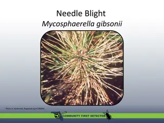
Clinical types of cutaneous leishmaniasis Leishmania major: Zoonotic cutaneous leishmaniasis, wet lesions with severe reaction Leishmania tropica: Anthroponotic cutaneous leishmaniasis, dry lesions with minimal ulceration Oriental sore (most common) classical self-limited ulcer
CUTANEOUS LISHMANIASIS THE COMMON TYPE This starts as a painless papule on exposed parts of the body, generally the face. The lesion ulcerates after a few months producing an ulcer with an indurate margin. In some cases the ulcer remains dry and heals readily (dry-type-lesion). In some other cases the ulcer may spread with an inflammatory zone around, these known as (wet-type-lesion) which heal slowly.
UNCOMMON TYPES OF CUTANEUS LISHMANIASIS Diffuse cutaneous leishmaniasis (DCL): Caused by L. aethiopica, diffuse nodular non-ulcerating lesions, seen in a part of Africa, people with low immunity to Leishmaniaantigens.Diffuse cutaneous (DCL), and consists of nodules and a thickening of the skin, generally without any ulceration, it needs numerous parasite. Leishmaniasis recidiva (lupoid leishmaniasis): Severe immunological reaction to leishmaniaantigen leading to persistent dry skin lesions, few parasites.
Diffuse cutaneous leishmaniasis (DCL) Leishmaniasis recidiva
Mucocutaneous leishmaniasis The lesion starts as a pustular swelling in the mouth or on the nostrils. The lesion may become ulcerative after many months and then extend into the naso-pharyngeal mucous membrane. Secondary infection is very common with destruction of the nasal cartilage and the facial bone.
cutaneous & muco-cutaneous leishmaniasis Diagnosis The parasite can be isolated from the margin of the ulcer. A diagnostic skin test, known as Leishmanin test (Montenego Test), is useful. Smear: Giemsa stain microscopy for LD bodies (Leishman-Donovan bodies, amastigotes). Skin biopsy: microscopy for LD bodies or culture in NNN medium for promastigotes.
Visceral leishmaniasis There are geographical variations. The disease is called kala-azar Leishmania infantummainly affect children Leishmania donovani mainly affects adults The incubation period is usually 4-10 months. The early symptoms are generally low grade fever with malaise and sweating. In later stages, the fever becomes intermittent and their can be liver enlargement or spleen enlargement or hepatosplenomegally because of the hyperplasia of the lymphoid macrophage system.
Presentation Fever Splenomegaly, hepatomegaly, hepatosplenomegaly Weight loss Anaemia Epistaxis Cough Diarrhoea
Untreated disease can be fatal After recovery it might produce a condition called post kala-azar dermal leishmaniasis (PKDL)
Fever 2 times a day due to kala-azar
Visceral leishmaniasis Diagnosis (1) Parasitological diagnosis: Bone marrow aspirate 1. microscopy (LD bodies) Splenic aspirate 2. culture in NNN medium (promastigotes) Lymph node Tissue biopsy
Bone marrow aspiration Bone marrow amastigotes
(2) Immunological Diagnosis: Specific serologic tests: Direct Agglutination Test (DAT), ELISA, IFAT Skin test (leishmanin test) for survey of populations and follow-up after treatment.
DAT test ELISA test
Treatment Antiparasitic drugs, such as amphotericin B (Ambisome), treat this condition. Your doctor may recommend other treatments based on the type of leishmaniasis you have. Cutaneous leishmaniasis Cutaneous ulcers will often heal without treatment. However, treatment can speed healing, reduce scarring, and decrease risk of further disease. Any skin ulcers that cause disfigurement may require plastic surgery. Mucocutaneous leishmaniasis These lesions don t heal naturally. They always require treatment. Liposomal amphotericin B and paromomycin can treat mucocutaneous leishmaniasis. Visceral leishmaniasis Visceral disease always requires treatment. Several medications are available. Commonly used medicines include sodium stibogluconate (Pentostam), amphotericin B, paromomycin, and miltefosine (Impavido).


 Immunological Diagnosis:")