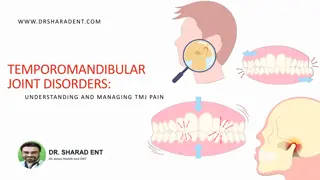
Understanding the Temporomandibular Joint (TMJ) Anatomy

GROSS ANATOMY:
The temporomandibular joint (TMJ) is formed by the
articulation between the articular eminence and the anterior
part of the glenoid fossa of the squamous part of temporal
bone above and the condylar head of the mandible below.
Like most diarthrodial synovial joints, the TMJ contains a
fibrous intra-articular disk that is interposed between the
articular surfaces and functions as a shock absorber.
The disk also increases the type and range of movements and
probably contributes to the stability of the joint. It is an oval,
fibrous, avascular, noninnervated plate that is firmly attached
to the medial and lateral poles of the condyle by medial and
lat eral collateral ligaments.The disk is biconcave in sagittal
section, with a thin intermediate zone, a thick
anterior
band, and a thick posterior
band.
The anterior band continues into loose fibroelastic
connective tissue, which is avascular and innervated, known
as the
anterior foot extension
or anterior ligament. The
latter attaches to the ascending slope of the articular
eminence and becomes contiguous with the anterior capsule
laterally and the superior head of the lat- eral pterygoid
muscle medially.
Some fibers of the superior head of the lateral pterygoid
muscle attach to the anterior band.
The posterior band is
continuous with a loose connective tissue rich in elastic fibers
called the bilaminar zone, which is highly vascular and richly
innervated. The superior stratum, or lamina, of the bilaminar
zone attaches to the posterior wall of the glenoid fossa and
the squamotympanic suture, and the inferior stratum attaches
to the posterior aspect of the mandibular condyle.
The joint capsule is a fibroelastic sac that attaches to the
ascending slope of the articular eminence anteriorly and to
the lips of the squamotympanic fissure posteriorly. Between
these two attachments it is attached to the margins of the
glenoid fossa superiorly and to the neck of the condyle
inferiorly. The posterior capsule is highly vascular and
sometimes is called
pis vasculosa
Parotid gland tissue is usually found in the posterior portion
of the glenoid fossa between the posterior capsule of the joint
and the postglenoid tubercle. The lateral aspect of the capsule
is strengthened by the temporomandibular ligament. The
anterior surface of the capsule, unlike the other surfaces, is
usually ill defined. The inner surface of the capsule is smooth
and glistening because of the presence of a synovial
membrane lining.
The latter does not extend over the articular surfaces of the
disk, the articular eminence, or the condyle. The disk divides
the joint space into two compartments: 1-a lower one
between the condyle and the disk (condylodiskal)
2- and an upper one between the disk and the temporal bone
(temporodiskal).
The disk acts as a third bone and provides a movable
articulation for the condyle. In the lower joint space
rotational movement about an axis through the heads of the
condyles permits opening of the jaws; this is designated as a
hinge movement
. In the upper joint space, because of the
firm attachment of the disk to the lateral and medial poles of
the condyle and the contraction of the inferior head of the
lateral pterygoid muscle, a translatory movement occurs as
the disks and the condyles traverse anteriorly along the
descending slopes of the articular eminences to produce an
anterior and inferior movement of the mandible.
In a healthy joint the condyle, a disk, and temporal
components are contiguous with each other during all
movements, and the superior and inferior joint spaces are
reduced to the thickness of a synovial film except for small
pockets or recesses at the most anterior, posterior, medial,
and lateral limits of the joint spaces.
The posterior part of the disk gets compressed and the blood
is squeezed out when the disk moves posteriorly as in closure
of jaws. When the disk is drawn forward during mouth
opening, the posterior part of the disk gets filled up with
blood. The presence of elastic fibers in the superior lamellae,
is said to recoil and help in the posterior movement of the
disk.
Except for the avascular disk, the joint tissues are innervated
by branches of the auriculotemporal branch of the
mandibular nerve of the fifth cranial or trigeminal nerve.
Proprioceptor fibers from the joint are carried by masseteric
nerves and per- haps other muscular branches of the
mandibular nerve. There are four types of nerve endings in
the TM joint.
1-The
Ruffini’s corpuscles
, present in the capsule are the
proprioceptors
and sense the changes in the joint when
the joint is static.
2-The
pacinian corpuscles
, also present in the capsule,
act as
mechano- receptors
to signal the rapidity and
slowness of the joint movement.
3-The
Golgi tendon,
present in the TM joint ligament,
functions as a
mechanoreceptor
to protect the joint when
joint movements become excessive.
4-The free nerve endings which are
nociceptors
(receptors for pain),
are the most numerous and widely
distributed; protect the joint from excessive movements, by
causing pain and curtailing the movement.
The arterial supply to the joint is through branches of the
maxillary and superficial temporal arteries. Large venules are
consistently seen close to the anterior ligament of the disk,
the bilaminar zone, and the posterior capsule.
DEVELOPMENT OF THE JOINT
At approximately 10 weeks the components of the fetus’
future joint become evident in the mesenchyme between the
condylar cartilage of the mandible and the developing
temporal bone. Two slit-like joint cavities and an intervening
disk make their appearance in this region by 12 weeks.
The mesenchyme around the joint begins to form the fibrous
joint capsule. The developing superior head of the lateral
pterygoid muscle attaches to the anterior portion of the fetal
disk. The disk also continues posteriorly through the
petrotympanic fissure and attaches to the
malleus of the
middle
ear. This connection is usually obliterated by the
growth of the lips of the petrotym- panic fissure and does not
exist in the adult joint.
HISTOLOGY
a-Bony structures
The condyle of the mandible is composed of cancellous bone
covered by a thin layer of compact bone .The trabeculae are
grouped in such a way that they radiate from the neck of the
mandible and reach the cortex at right angles, thus giving
maximal strength to the condyle. The large marrow spaces
decrease in size with progressing age as a result of noticeable
thickening of the trabeculae.
The red marrow in the condyle is of the
myeloid or
cellular
type. In older individuals it is sometimes replaced
by
fatty marrow
. During the period of growth a layer of
hyaline cartilage lies underneath the fibrous covering of the
condyle. This cartilaginous plate grows by apposition from
the deepest layers of the covering connective tissue. At the
same time its deep surface is replaced by bone Remnants of
this cartilage may persist into old age .Unlike
metaphyseal
primary cartilage
of long bones, the hyaline cartilage of
the condyle is not organized in parallel rows of cells at the
interface between the forming bone and the cartilage.
Therefore the cartilage is usually referred to
as secondary
cartilage
. In addition to the appositional subperiosteal
mandibular bone growth, the growth of the secondary
cartilage and its replacement with bone contribute to the
downward and for- ward growth of the mandible.
The roof of the glenoid fossa consists of a thin, compact layer
of bone. The articular eminence is composed of spongy bone
covered with a thin layer of compact bone. Areas of
chondroid bone are commonly seen in the articular
eminence, and in rare cases islands of hyaline cartilage are
found.
2-Articular fibrous covering:
In synovial joints, the articular surfaces are covered with hya-
line cartilage. TM joint, unlike other synovial joints, the
articular surfaces are covered with fibrous tissue. This is
because the mandible is formed from membranous
ossification and there are no cartilages present to cover the
articular surface. The condyle and the articular eminence are
covered by a rather thick layer of fibroelastic tissue
containing fibroblasts and a variable number of chondrocytes.
The fibrous covering of the mandibular condyle is of fairly
even thickness Its superficial layers consist of a network of
strong collagenous fibers.
Chondrocytes may be present and have a tendency to
increase in number with age. They can be recognized by their
thin capsule, which stains heavily with basic dyes. The deepest
layer of the fibrocartilage is rich in small undifferentiated
cells as long as growing hyaline cartilage is present in the
condyle. It contains only a few thin collagenous fibers.
In this zone, called the
reserve cell zone
, appositional
growth of the hyaline cartilage of the condyle takes place
.The presence of fibrocartilage increases the capacity to
withstand the mechanical stress and this type of cartilage
does not tend to calcify, thus it resists ageing process to some
extent. The fibrous layer covering the articulating surface of
the temporal bone .is thin in the articular fossa and thickens
rapidly on the posterior slope of the articular eminence
In this region the fibrous tissue shows a definite arrangement
in two layers, with a small transitional zone between them.
The two layers are characterized by the different courses of
the constituent fibrous bundles. In the inner zone the fibers
are at right angles to the bony surface. In the outer zone they
run parallel to that surface. As in the fibrous covering of the
mandibular condyle, a variable number of chondrocytes are
found in the tissue on the temporal surface. In adults only the
deepest layer shows a thin zone of calcification.
No continuous cellular lining is on the free surface of the
fibrocartilage. Only isolated fibroblasts are situated on the
surface itself. They are characterized by the formation of
long, flat cytoplasmic processes.
The presence of wavy collagen fibers both in the articular
disk and in the fibrous tissue covering the articular surface,
are suggested to be due to compression and are cited in
support of weight bearing nature of the joint.
The finding of chondroitin sulfate as the principal
glycosaminoglycans (GAG) in articular disk of TM joint, as
seen in cartilage, suggests that the disk is subjected to
compressional loads, as in weight bearing joints. However,
the absence of cartilage covering the articular surface is cited
as a point against the weight bearing nature of the joint
3-Articular disk:
In young individuals the articular disk is composed of dense
fibrous tissue. The interlacing fibers are straight and tightly
packed.Elastic fibers are found only in relatively small
numbers, although their number markedly present in the
upper lamina of bilaminar region which probably helps
during retraction or during initial phase of elevation of the
mandible by means of elastic recoiling. The fibroblasts in the
disk are elon- gated and send flat cytoplasmic processes into
the interstices between the adjacent
bundles.With advancing age and in areas of the disk subjected
to excessive mechanical stress, some cells appear rounded
and arranged in pairs similar to chondroid cells. The presence
of rounded cells in the articular disk are not true
chondrocytes, because they lack a capsule, surrounding
them. However the presence of chondrocytes is considered
to be a pathological change Chondrocytes, with typical
territorial matrices that stain heavily with basic dyes, can be
observed in the articular disk of many species, including
humans.
The presence of chondrocytes may increase the resistance
and resilience of the fibrous tissue. The fibrous tissue
covering the articular eminence, the mandibular condyle, and
the large central area of the disk, is devoid of blood vessels
and nerves and thus has limited reparative ability.
4-Synovial membrane:
As in other synovial joints, the articular capsule is lined with
a synovial membrane that folds to form synovial villi.
Synovial villi project into the joint spaces.The synovial
membrane is stretched and flattened during joint
movements, while at rest it shows foldings.
The synovial membrane consists of internal cells, which do
not form a continuous layer but show gaps between the cells,
and the subintimal connective tissue layer, rich in blood
capillaries. The intimal cells are of three types. The first is
rich in rough endoplasmic reticulum (RER) and is called the
fibroblast-like, or B cell. It is sometimes called a secretory S-
cell.
The second type is rich in Golgi complex and lysosomes and
contains little or no RER. It is called the macrophage-like, or
A cell. The third type has a cellular morphology between cell
types A and B. A small amount of a clear, straw-colored
viscous fluid (synovial fluid) is found in the articular spaces.
It is a lubricant and also a nutrient fluid for the avascular
tissues covering the condyle and the articular eminence and
for the disk.
It is elaborated by diffusion from the rich capillary network
of the synovial membrane that is augmented by mucin,
possibly secreted by the synovial cells. As an age change, the
amount of synovial fluid decreases and villous projections are
seen to increase in number. The synovial membrane has
considerable ability to regenerate.
CLINICAL CONSIDERATIONS
The thinness of the bone in the articular fossa is responsible
for fractures if the mandibular head is driven into the fossa by
a heavy blow. In such cases injuries of the dura mater and the
brain have been reported.
A change in force or direction of stress, especially after loss
of posterior teeth, may cause structural changes. These
changes may include fibrillation (separation between collagen
bundles) of the fibrous covering of the articulating surfaces
and of the disk.
Abnormal functional activity may also produce injury to the
articular bones. Compensation and partial repair may be
accomplished by the development of cartilage on the
condylar surface and in the disk. In severe trauma the
articular bone is destroyed, and cartilage and new bone
develop in the marrow spaces and at the periphery of the
condyle. When this occurs, the function of the joint is
severely impaired.
The articular surface is capable of remodeling due to
functional demands. It shows changes due to loss of teeth or
due to attrition of teeth. It also gets remodeled due to
orthodontic treatment. In approximately 35% of the
population the TMJ produces sounds during opening
movements. The joint has palpable irregularities and
produces popping and clicking noises.
The term myofacial pain dysfunction syndrome is used to
indicate a dysfunction of the TMJ. It is characterized by:
(1) masticatory muscle tenderness (most frequently, the
lateral pterygoid and then, in order, the temporalis, medial
pterygoid, and masseter);
(2) limited opening of the mandible (< 37 mm); and
(
3) joint sounds. This symptom complex is seen more often in
females than in males. Its cause is usually spasm of the
masticatory muscles. Since the condition may be related to
stress, treatment should be as conservative as possible.
Dislocation of the TMJ may take place without the impact of
an external force. The dislocation of the jaw is usually
bilateral, and the displacement is anterior.
When the mouth is opened unusually wide during yawning,
the head of the man- dible may slip forward into the
infratemporal fossa, causing articular dislocation of the joint.
The disk, for reasons not yet determined, becomes displaced
anteromedially and creates one or more of the following
signs and symptoms: pain, clicking, limitation of jaw
movement, deviation of the jaw or opening, and locking. If
the condition remains untreated, it could lead to
osteoarthrosis. Diagnosis of cases of the TMJ disk perforation
is also on the increase.
The temporomandibular joint (TMJ) is a complex joint formed by the articulation between the temporal bone and the mandible. It consists of a fibrous intra-articular disk that aids in movement and stability. The disk is biconcave with anterior and posterior bands, and attachments to ligaments and muscles. The joint capsule surrounds the TMJ, contributing to its structure. This detailed anatomy helps in comprehending the function and potential issues related to the TMJ.

Download Presentation

Please find below an Image/Link to download the presentation.
The content on the website is provided AS IS for your information and personal use only. It may not be sold, licensed, or shared on other websites without obtaining consent from the author. Download presentation by click this link. If you encounter any issues during the download, it is possible that the publisher has removed the file from their server.
E N D
Presentation Transcript
GROSS ANATOMY: GROSS ANATOMY: The temporomandibular joint (TMJ) is formed by the articulation between the articular eminence and the anterior part of the glenoid fossa of the squamous part of temporal bone above and the condylar head of the mandible below. Like most diarthrodial synovial joints, the TMJ contains a fibrous intra-articular disk that is interposed between the articular surfaces and functions as a shock absorber.
The disk also increases the type and range of movements and probably contributes to the stability of the joint. It is an oval, fibrous, avascular, noninnervated plate that is firmly attached to the medial and lateral poles of the condyle by medial and lat eral collateral ligaments.The disk is biconcave in sagittal section, with a thin intermediate zone, a thick anterior band, and a thick posterior band.
The anterior band continues into loose fibroelastic connective tissue, which is avascular and innervated, known as the anterior foot extension or anterior ligament. The latter attaches to the ascending slope of the articular eminence and becomes contiguous with the anterior capsule laterally and the superior head of the lat- eral pterygoid muscle medially.
Some fibers of the superior head of the lateral pterygoid muscle attach to the anterior band. The posterior band is continuous with a loose connective tissue rich in elastic fibers called the bilaminar zone, which is highly vascular and richly innervated. The superior stratum, or lamina, of the bilaminar zone attaches to the posterior wall of the glenoid fossa and the squamotympanic suture, and the inferior stratum attaches to the posterior aspect of the mandibular condyle.
The joint capsule is a fibroelastic sac that attaches to the ascending slope of the articular eminence anteriorly and to the lips of the squamotympanic fissure posteriorly. Between these two attachments it is attached to the margins of the glenoid fossa superiorly and to the neck of the condyle inferiorly. The posterior capsule is highly vascular and sometimes is called pis vasculosa
Parotid gland tissue is usually found in the posterior portion of the glenoid fossa between the posterior capsule of the joint and the postglenoid tubercle. The lateral aspect of the capsule is strengthened by the temporomandibular ligament. The anterior surface of the capsule, unlike the other surfaces, is usually ill defined. The inner surface of the capsule is smooth and glistening because of the presence of a synovial membrane lining.
The latter does not extend over the articular surfaces of the disk, the articular eminence, or the condyle. The disk divides the joint space into two compartments: 1-a lower one between the condyle and the disk (condylodiskal) 2- and an upper one between the disk and the temporal bone (temporodiskal).
The disk acts as a third bone and provides a movable articulation for the condyle. In the lower joint space rotational movement about an axis through the heads of the condyles permits opening of the jaws; this is designated as a hinge movement. In the upper joint space, because of the firm attachment of the disk to the lateral and medial poles of the condyle and the contraction of the inferior head of the lateral pterygoid muscle, a translatory movement occurs as the disks and the condyles traverse anteriorly along the descending slopes of the articular eminences to produce an anterior and inferior movement of the mandible.
In a healthy joint the condyle, a disk, and temporal components are contiguous with each other during all movements, and the superior and inferior joint spaces are reduced to the thickness of a synovial film except for small pockets or recesses at the most anterior, posterior, medial, and lateral limits of the joint spaces.
The posterior part of the disk gets compressed and the blood is squeezed out when the disk moves posteriorly as in closure of jaws. When the disk is drawn forward during mouth opening, the posterior part of the disk gets filled up with blood. The presence of elastic fibers in the superior lamellae, is said to recoil and help in the posterior movement of the disk.
Except for the avascular disk, the joint tissues are innervated by branches of the auriculotemporal branch of the mandibular nerve of the fifth cranial or trigeminal nerve. Proprioceptor fibers from the joint are carried by masseteric nerves and per- haps other muscular branches of the mandibular nerve. There are four types of nerve endings in the TM joint.
1-The Ruffinis corpuscles, present in the capsule are the proprioceptors and sense the changes in the joint when the joint is static. 2-The pacinian corpuscles, also present in the capsule, act as mechano- receptors to signal the rapidity and slowness of the joint movement. 3-The Golgi tendon, present in the TM joint ligament, functions as a mechanoreceptor to protect the joint when joint movements become excessive.
4-The free nerve endings which are nociceptors (receptors for pain), are the most numerous and widely distributed; protect the joint from excessive movements, by causing pain and curtailing the movement. The arterial supply to the joint is through branches of the maxillary and superficial temporal arteries. Large venules are consistently seen close to the anterior ligament of the disk, the bilaminar zone, and the posterior capsule.
DEVELOPMENT OF THE JOINT At approximately 10 weeks the components of the fetus future joint become evident in the mesenchyme between the condylar cartilage of the mandible and the developing temporal bone. Two slit-like joint cavities and an intervening disk make their appearance in this region by 12 weeks.
The mesenchyme around the joint begins to form the fibrous joint capsule. The developing superior head of the lateral pterygoid muscle attaches to the anterior portion of the fetal disk. The disk also continues posteriorly through the petrotympanic fissure and attaches to the malleus of the middle ear. This connection is usually obliterated by the growth of the lips of the petrotym- panic fissure and does not exist in the adult joint.
HISTOLOGY a-Bony structures The condyle of the mandible is composed of cancellous bone covered by a thin layer of compact bone .The trabeculae are grouped in such a way that they radiate from the neck of the mandible and reach the cortex at right angles, thus giving maximal strength to the condyle. The large marrow spaces decrease in size with progressing age as a result of noticeable thickening of the trabeculae.
The red marrow in the condyle is of the myeloid or cellular type. In older individuals it is sometimes replaced by fatty marrow. During the period of growth a layer of hyaline cartilage lies underneath the fibrous covering of the condyle. This cartilaginous plate grows by apposition from the deepest layers of the covering connective tissue. At the same time its deep surface is replaced by bone Remnants of this cartilage may persist into old age .Unlike metaphyseal primary cartilage of long bones, the hyaline cartilage of the condyle is not organized in parallel rows of cells at the interface between the forming bone and the cartilage.
Therefore the cartilage is usually referred to as secondary cartilage. In addition to the appositional subperiosteal mandibular bone growth, the growth of the secondary cartilage and its replacement with bone contribute to the downward and for- ward growth of the mandible. The roof of the glenoid fossa consists of a thin, compact layer of bone. The articular eminence is composed of spongy bone covered with a thin layer of compact bone. Areas of chondroid bone are commonly seen in the articular eminence, and in rare cases islands of hyaline cartilage are found.
2-Articular fibrous covering: In synovial joints, the articular surfaces are covered with hya- line cartilage. TM joint, unlike other synovial joints, the articular surfaces are covered with fibrous tissue. This is because the mandible is formed from membranous ossification and there are no cartilages present to cover the articular surface. The condyle and the articular eminence are covered by a rather thick layer of fibroelastic tissue containing fibroblasts and a variable number of chondrocytes. The fibrous covering of the mandibular condyle is of fairly even thickness Its superficial layers consist of a network of strong collagenous fibers.
Chondrocytes may be present and have a tendency to increase in number with age. They can be recognized by their thin capsule, which stains heavily with basic dyes. The deepest layer of the fibrocartilage is rich in small undifferentiated cells as long as growing hyaline cartilage is present in the condyle. It contains only a few thin collagenous fibers.
In this zone, called the reserve cell zone, appositional growth of the hyaline cartilage of the condyle takes place .The presence of fibrocartilage increases the capacity to withstand the mechanical stress and this type of cartilage does not tend to calcify, thus it resists ageing process to some extent. The fibrous layer covering the articulating surface of the temporal bone .is thin in the articular fossa and thickens rapidly on the posterior slope of the articular eminence
In this region the fibrous tissue shows a definite arrangement in two layers, with a small transitional zone between them. The two layers are characterized by the different courses of the constituent fibrous bundles. In the inner zone the fibers are at right angles to the bony surface. In the outer zone they run parallel to that surface. As in the fibrous covering of the mandibular condyle, a variable number of chondrocytes are found in the tissue on the temporal surface. In adults only the deepest layer shows a thin zone of calcification.
No continuous cellular lining is on the free surface of the fibrocartilage. Only isolated fibroblasts are situated on the surface itself. They are characterized by the formation of long, flat cytoplasmic processes. The presence of wavy collagen fibers both in the articular disk and in the fibrous tissue covering the articular surface, are suggested to be due to compression and are cited in support of weight bearing nature of the joint.
The finding of chondroitin sulfate as the principal glycosaminoglycans (GAG) in articular disk of TM joint, as seen in cartilage, suggests that the disk is subjected to compressional loads, as in weight bearing joints. However, the absence of cartilage covering the articular surface is cited as a point against the weight bearing nature of the joint
3-Articular disk: In young individuals the articular disk is composed of dense fibrous tissue. The interlacing fibers are straight and tightly packed.Elastic fibers are found only in relatively small numbers, although their number markedly present in the upper lamina of bilaminar region which probably helps during retraction or during initial phase of elevation of the mandible by means of elastic recoiling. The fibroblasts in the disk are elon- gated and send flat cytoplasmic processes into the interstices between the adjacent
bundles.With advancing age and in areas of the disk subjected to excessive mechanical stress, some cells appear rounded and arranged in pairs similar to chondroid cells. The presence of rounded cells in the articular disk are not true chondrocytes, because they lack a capsule, surrounding them. However the presence of chondrocytes is considered to be a pathological change Chondrocytes, with typical territorial matrices that stain heavily with basic dyes, can be observed in the articular disk of many species, including humans.
The presence of chondrocytes may increase the resistance and resilience of the fibrous tissue. The fibrous tissue covering the articular eminence, the mandibular condyle, and the large central area of the disk, is devoid of blood vessels and nerves and thus has limited reparative ability.
4-Synovial membrane: As in other synovial joints, the articular capsule is lined with a synovial membrane that folds to form synovial villi. Synovial villi project into the joint spaces.The synovial membrane is stretched and flattened during joint movements, while at rest it shows foldings.
The synovial membrane consists of internal cells, which do not form a continuous layer but show gaps between the cells, and the subintimal connective tissue layer, rich in blood capillaries. The intimal cells are of three types. The first is rich in rough endoplasmic reticulum (RER) and is called the fibroblast-like, or B cell. It is sometimes called a secretory S- cell.
The second type is rich in Golgi complex and lysosomes and contains little or no RER. It is called the macrophage-like, or A cell. The third type has a cellular morphology between cell types A and B. A small amount of a clear, straw-colored viscous fluid (synovial fluid) is found in the articular spaces. It is a lubricant and also a nutrient fluid for the avascular tissues covering the condyle and the articular eminence and for the disk.
It is elaborated by diffusion from the rich capillary network of the synovial membrane that is augmented by mucin, possibly secreted by the synovial cells. As an age change, the amount of synovial fluid decreases and villous projections are seen to increase in number. The synovial membrane has considerable ability to regenerate.
CLINICAL CONSIDERATIONS The thinness of the bone in the articular fossa is responsible for fractures if the mandibular head is driven into the fossa by a heavy blow. In such cases injuries of the dura mater and the brain have been reported. A change in force or direction of stress, especially after loss of posterior teeth, may cause structural changes. These changes may include fibrillation (separation between collagen bundles) of the fibrous covering of the articulating surfaces and of the disk.
Abnormal functional activity may also produce injury to the articular bones. Compensation and partial repair may be accomplished by the development of cartilage on the condylar surface and in the disk. In severe trauma the articular bone is destroyed, and cartilage and new bone develop in the marrow spaces and at the periphery of the condyle. When this occurs, the function of the joint is severely impaired.
The articular surface is capable of remodeling due to functional demands. It shows changes due to loss of teeth or due to attrition of teeth. It also gets remodeled due to orthodontic treatment. In approximately 35% of the population the TMJ produces sounds during opening movements. The joint has palpable irregularities and produces popping and clicking noises.
The term myofacial pain dysfunction syndrome is used to indicate a dysfunction of the TMJ. It is characterized by: (1) masticatory muscle tenderness (most frequently, the lateral pterygoid and then, in order, the temporalis, medial pterygoid, and masseter); (2) limited opening of the mandible (< 37 mm); and (
3) joint sounds. This symptom complex is seen more often in females than in males. Its cause is usually spasm of the masticatory muscles. Since the condition may be related to stress, treatment should be as conservative as possible. Dislocation of the TMJ may take place without the impact of an external force. The dislocation of the jaw is usually bilateral, and the displacement is anterior.
When the mouth is opened unusually wide during yawning, the head of the man- dible may slip forward into the infratemporal fossa, causing articular dislocation of the joint. The disk, for reasons not yet determined, becomes displaced anteromedially and creates one or more of the following signs and symptoms: pain, clicking, limitation of jaw movement, deviation of the jaw or opening, and locking. If the condition remains untreated, it could lead to osteoarthrosis. Diagnosis of cases of the TMJ disk perforation is also on the increase.


 joint sounds. This symptom complex is seen more often")