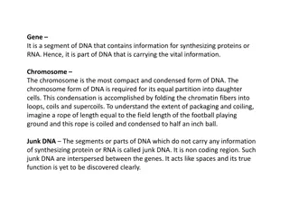
Understanding the Structural Elements of DNA in Molecular Biology
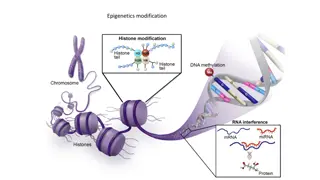
DNA, the genetic material of living organisms, consists of two antiparallel polynucleotide chains forming a double helical structure. Each chain is composed of nucleotide monomers, consisting of deoxyribose sugar, phosphate group, and nitrogenous bases (purines and pyrimidines). The backbone of DNA is held by phosphodiester bonds, while hydrogen bonds link base pairs, ensuring the integrity of the double helix. This insightful exploration sheds light on the fundamental building blocks and bonds that constitute DNA's intricate structure.

Download Presentation

Please find below an Image/Link to download the presentation.
The content on the website is provided AS IS for your information and personal use only. It may not be sold, licensed, or shared on other websites without obtaining consent from the author. Download presentation by click this link. If you encounter any issues during the download, it is possible that the publisher has removed the file from their server.
E N D
Presentation Transcript
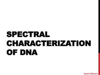
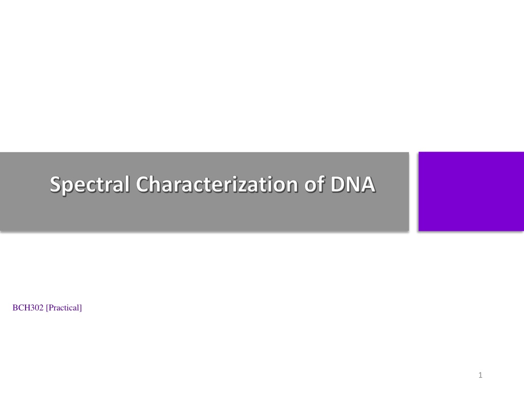
Spectral Characterization of DNA BCH302 [Practical] 1
What DNA made up of : DNA is made of 2 polynucleotide chains which run in opposite direction antiparallel . DNA has a double helical structure. Each polynucleotide chain of DNA consists of monomer units of nucleotides. A monomer unit (nucleotide) consists of 3 main components that are: 1. Pentose sugar. 2. Phosphate. 3. Nitrogenous base. 3
DNA double helical structure: [antiparallel] 4
Nucleotide (DNA building block): Monomer 5
DNA structure: 1. Deoxyribose sugar: Is a monosaccharide 5-Carbon Sugar, Its name indicates that it is a deoxy sugar, meaning that [ it is derived from the sugar ribose by loss of an oxygen atom ]. 6
DNA structure: 2. Phosphate Group: The sugars are joined together by phosphate groups that form phosphodiester bonds between the third and fifth carbon atoms of adjacent sugar rings. 7
DNA structure: 3. Nitrogenous bases: is a nitrogen-containing organic molecule having the chemical properties of a base. They are classified as the derivatives of two parent compounds: 1. Purine: [ Adenine, Guanine ] 2. Pyrimidine : [ Cytosine, Thymine ] 8
DNA structure: 4. Hydrogen bond: The H-bonds form between base pairs of the antiparallel strands. The base in the first strand forms an H-bond only with a complementary base in the second strand. Those two bases form a base-pair (H-bond interaction that keeps strands together and form double helical structure). Sugars and phosphates are located outside of the double helical structure. 9
Types of bonds in DNA: The backbone of the DNA (sugars and phosphate) is held by covalent bond phosphodiester bond . The bases in the two strands are linked togather by hydrogen bond (and hydrophobic effect between the complementary bases). Held by covalent bond Hydrogen bond 10
Denaturation of DNA : Denaturation is a process by which nucleic acids, such as DNA, lose their three- dimensional structures and consequently their primary functions. Many different substances or environmental conditions can denature nucleic acids, such as: 1. 2. 3. Strong acids, organic solvent. Heating. Exposure to Radiation/ UV light. 11
Optical density of DNA : Nucleic acid have maximum absorbance at 260 nm, It absorbs at this wavelength because of the nitrogenous bases (A, G, C and T) of DNA. Absorbance 12 Wave length (nm)
Hyperchromicity: In general : It is the increase of absorbance (optical density) of a material. The hyperchromicity of DNA that occurs when the DNA duplex is denatured. When DNA denatures [e.g. by heat ], it's strands separate, allowing more light to be absorbed by the non-stacked bases[single DNA strands]. Due to denaturation of DNA the bases become exposed to the surface and able to absorb more light at 260 nm. This action is calling the hyperchromic effect. Note: The opposite, a decrease of absorbance is called hypochromicity 13
Experiment 1 : Spectral characterization of DNA Objective: To determine the wave length that represent the maximum absorbance for DNA (the optimum wave length for DNA). To establish the effect of temperature on the absorbance of DNA or [hyperchromic effect]. Principle: 1- The double helix of DNA are bound together mainly by hydrogen bonds and hydrophobic effect between the complementary bases. When DNA in solution is heated above its melting temperature (usually more than 80 C), the double-stranded DNA unwinds to form single-stranded DNA. 2-In single stranded DNA the bases become unstacked and can thus absorb more light. In their native state, the bases of DNA absorb light at the 260 nm wavelength region. When the bases become unstacked, the wavelength of maximum absorbance does not change, but the amount absorbed increases by 30-40%. 15
Experiment 1 : Spectral characterization of DNA Method: 1. Measure the absorbance at the following wavelengths:(240,245,250,255,260,265,270,275 and 280 nm). Using distal water as a blank. Cover the tube and put it in boiling water bath for 15 min. Immediately measure the absorbance at same wave lengths. Plot the absorption spectra of the native DNA solution and the denatured DNA against wavelengths. 2. 3. 4. Results: Wave length (nm) Absorbance of isolated DNA Absorbance of heated DNA Effect of temperature on the absorbance of DNA [hyperchromic ] 240 1.6 245 1.4 250 1.2 Absorbance 1 255 0.8 native DNA 0.6 260 denatured DNA 0.4 265 0.2 0 270 220 240 260 280 300 Wavelenghth (nm) 275 17 280


:")