Practical Bacteriology Laboratory Diagnosis and Sample Collection
 undefined


Laboratory diagnosis
Smear preparation, Simple and Gram stains
4/6/2024
4/6/2024
4/6/2024
4/6/2024
4/6/2024
4/6/2024
4/6/2024
4/6/2024

1
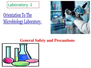
Practical Bacteriology
lab. 2
By:
Asst. Prof. Shiama'a Al-Salihy


L
e
a
r
n
i
n
g
o
b
j
e
c
t
i
v
e
s
:
4/6/2024
4/6/2024
4/6/2024
4/6/2024
4/6/2024
4/6/2024
4/6/2024
4/6/2024

2
After this lab, you must be able to:
1.
Identify laboratory diagnosis steps.
2.
Understand staining techniques
3.
Illustrate the purpose of each stain
4.
Interpret laboratory tests results
Laboratory diagnosis steps:
Specimen:
according to type of infection
Microscopic examination
:
stained or wet
Culture and antibiotic susceptibility test:
inoculation of M.Os in or/and on culture media,
and testing their sensitivity to antibiotic.
Biochemical tests:
catalase, coagulase, sugar
fermentation tests…etc.
Serological and immunological test
Molecular tests
Sample collection (specimens):
Urine
,blood, stool ,sputum , pus, CSF, plural fluid
,peritoneal fluid, different types of swabs.
A. Urine sample:
Collection: Mid stream urine
(M.S.U.) sample to avoid
contamination with normal flora
(
E.coli
),
Staphylococcus
saprophyticus.
The sample of urine must be no
delay more than 1-2hrs.
B. Stool sample:
In sterile container or by rectal
swab. The collected stool should be
processed within 2hrs or inoculated
in transport media
urine collection container
C. Swabs:
Very important methods for
collect the following:
1.
Urethral swabs:
for urethritis
diagnosis in men and vaginal
swab
2.
Throat swab:
culture on
chocolate agar
3.
Nasopharyngeal swab
4.
Ear swab
.
5.
Eye swab.
6.
wound swab.
D. Sputum:
Material from lower RTI or ( a
mixture of bronchial secretion and
inflammatory exudates).
Collection steps:
•
Make the collection in a
disposables wide mouth
screw-capped plastic
container
•
Homogenize the sample.
•
Collect the sample before
antibiotic therapy is began.
Sputum
collection
container
E. Cerebrospinal fluid (CSF):
•
The examination of CSF is an essential step in
the diagnosis of bacterial and fungal
meningitis. Normal CSF is sterile, clear, with
no RBCs.
•
Collection:
5-10 ml of CSF should be collected
in 2 sterile tube by lumber puncture
F. Blood
:
Samples of blood should be taken before antibiotic
administration
skin antisepsis: Iodine, Povidone –Iodine, Alcohol 70% or
Chlorhexidine
10 ml from adult, 2 - 5 ml from children, 1-2 ml from infants
and neonates
Microscopic examination:
Smear:
A thin film of material containing M.Os
which is spread over the surface of slide (stained
or wet)
- Wet preparation
are used to demonstrate
:
•
Blood cells and microbes in fluid specimens such as
urine, feces, CSF
•
Cysts, eggs or parasite in feces.
•
Fungi in skin.
•
Protozoa in blood and tissues.
- Stained smear:
if bacteria are seen in gram stained
smear, their shape, size, arrangement and reaction should
be observed.
S
t
e
p
s
o
f
p
r
e
p
a
r
a
t
i
o
n
s
m
e
a
r
Staining procedure can also be classified
into:
1)
Simple staining:
It is the use of single basic
dye to color the bacterial organism. Such as
methylene blue, crystal violet, safranin.
2)
Differential
staining:
uses more than one dyes
and it is also used to differentiate the organisms
into one of two groups such as gram stain and
acid fast stain. Also it can give the shape of
bacteria
3)
Structural stain:
such as capsule stain,
endospore and flagella
M
e
t
h
o
d
s
o
f
s
i
m
p
l
e
s
t
a
i
n
i
n
g
G
r
a
m
s
t
a
i
n
It is a differential stain uses two or more types of dye to
differentiate between organism according to their response
to these dye
Gram Stain
Gram Stain
a)
Gram positive:
A
p
p
e
a
r
s
v
i
o
l
e
t
a
f
t
e
r
G
r
a
m
’
s
s
t
a
i
n
b)
Gram negative:
Appears
red
after Gram’s
stain
Gram stain procedure
Z
i
e
h
l
-
N
e
e
l
s
e
n
s
t
a
i
n
(
A
c
i
d
f
a
s
t
B
a
c
i
l
l
i
)
To stain Mycobacterium species especially M.
tuberculosis
High lipid content- make decolorization very difficult
Principle:
Acid fast (resist): once the bacteria stained with primary
dye- difficult to decolorize with acid due to high contain of
lipid substances within their cell walls called mycolic
acids.
Basic carbol fuchsin in combination with phenol (the
staining solution) penetrates the cell wall and stain the
bacilli bright red. they resist decolorization with strong
acid.
M
y
c
o
b
a
c
t
e
r
i
u
m
t
u
b
e
r
c
u
l
o
s
i
s
under microscope stained
with AFB stain
Steps of AFB stain
A
l
b
e
r
t
S
t
a
i
n
Used for staining and demonstrated of
Corynebacterium
diphtheriae
,
They are thin gram positive bacilli which resemble Chinese
letters when stained by simple stain.
In albert stain another characteristic is presence of
metachromatic granules (RNA or polymetaphosphate) the
granules are bluish-purple by methylene blue and dark purple
with albert stain
Capsule
Endospore
This practical guide focuses on laboratory diagnosis steps in bacteriology, covering specimen collection, microscopic examination, culture tests, biochemical tests, serological tests, and molecular tests. Learn about various sample collection methods for urine, stool, swabs, sputum, pus, CSF, and more. Understand the importance of proper collection and processing techniques to ensure accurate test results.

Download Presentation

Please find below an Image/Link to download the presentation.
The content on the website is provided AS IS for your information and personal use only. It may not be sold, licensed, or shared on other websites without obtaining consent from the author. Download presentation by click this link. If you encounter any issues during the download, it is possible that the publisher has removed the file from their server.
E N D
Presentation Transcript
Practical Bacteriology Practical Bacteriology lab. lab. 2 2 Laboratory diagnosis Smear preparation, Simple and Gram stains By: Asst. Prof. Shiama'a Al-Salihy 1 4/6/2024
Learning objectives: After this lab, you must be able to: 1. Identify laboratory diagnosis steps. 2. Understand staining techniques 3. Illustrate the purpose of each stain 4. Interpret laboratory tests results 2 4/6/2024
Laboratorydiagnosissteps: Specimen: according to type of infection Microscopicexamination : stained orwet Culture and antibiotic susceptibility test: inoculation of M.Os in or/and on culture media, and testing theirsensitivity to antibiotic. Biochemical tests: catalase, coagulase, sugar fermentation tests etc. Serological and immunological test Moleculartests
Sample ,blood, stool ,sputum , pus, CSF, plural fluid ,peritoneal fluid, different types of swabs. collection (specimens): Urine
A. Urine sample: Collection: (M.S.U.) contamination with normal flora (E.coli), saprophyticus. The sample of urine must be no delay more than 1-2hrs. B. Stool sample: In sterile container or by rectal swab. The collected stool should be processed within 2hrs or inoculated in transport media Mid sample stream to urine avoid Staphylococcus urine collection container
C. Swabs: collect the following: 1. Urethral swabs: for urethritis diagnosis in men and vaginal swab 2. Throat swab: chocolate agar 3. Nasopharyngeal swab 4. Ear swab. 5. Eye swab. 6. wound swab. Very important methods for culture on
D. Sputum: Material from lower RTI or ( a mixture of bronchial secretion and inflammatory exudates). Collection steps: Make the collection in a disposables screw-capped container Homogenize the sample. Collect the sample before antibiotic therapy is began. wide mouth plastic Sputum collection container
E. Cerebrospinal fluid (CSF): The examination of CSF is an essential step in the diagnosis of meningitis. Normal CSF is sterile, clear, with no RBCs. Collection: 5-10 ml of CSF should be collected in 2 sterile tube by lumber puncture bacterial and fungal
F. Blood : Samples administration skin antisepsis: Iodine, Povidone Iodine, Alcohol 70% or Chlorhexidine 10 ml from adult, 2 - 5 ml from children, 1-2 ml from infants and neonates of blood should be taken before antibiotic
Microscopic examination: Smear: A thin film of material containing M.Os which is spread over the surface of slide (stained or wet) - Wet preparation are used to demonstrate: Blood cells and microbes in fluid specimens such as urine, feces, CSF Cysts, eggs or parasite in feces. Fungi in skin. Protozoa in blood and tissues. - Stained smear: if bacteria are seen in gram stained smear, their shape, size, arrangement and reaction should be observed.
Staining procedure can also be classified into: 1) Simple staining: It is the use of single basic dye to color the bacterial organism. Such as methylene blue, crystal violet, safranin. 2) Differential staining: uses more than one dyes and it is also used to differentiate the organisms into one of two groups such as gram stain and acid fast stain. Also it can give the shape of bacteria 3) Structural stain: such as capsule stain, endospore and flagella
Gram stain It is a differential stain uses two or more types of dye to differentiate between organism according to their response to these dye Gram Stain a) Gram positive: b)Gram negative: Appears violet after Gram s stain Appears red after Gram s stain
Ziehl-Neelsen stain (Acid fast Bacilli) To tuberculosis High lipid content- makedecolorizationvery difficult Principle: Acid fast (resist): once the bacteria stained with primary dye- difficult to decolorize with acid due to high contain of lipid substances within their cell walls called mycolic acids. Basic carbol fuchsin in combination with phenol (the staining solution) penetrates the cell wall and stain the bacilli bright red. they resist decolorization with strong acid. stain Mycobacterium species especially M.
Steps of AFB stain Mycobacterium tuberculosis under microscope stained with AFB stain
Albert Stain Used for staining and demonstrated of Corynebacterium diphtheriae, They are thin gram positive bacilli which resemble Chinese letters when stained by simple stain. In albert stain another characteristic metachromatic granules (RNA or polymetaphosphate) the granules are bluish-purple by methylene blue and dark purple with albert stain is presence of
Endospore Capsule


:")
:")


")