Anesthetic Management of Bronchoscopy for Airway Foreign Body
This article discusses the anesthetic management of bronchoscopy for airway foreign body, including presentation, preoperative work-up, intraoperative management, and postoperative care. It highlights the incidence of foreign body inhalation, immediate and delayed presentations, findings on chest X-ray, and additional testing such as CT scans. The article emphasizes the importance of prompt recognition and intervention in cases of airway foreign bodies to prevent complications.

Download Presentation

Please find below an Image/Link to download the presentation.
The content on the website is provided AS IS for your information and personal use only. It may not be sold, licensed, or shared on other websites without obtaining consent from the author. Download presentation by click this link. If you encounter any issues during the download, it is possible that the publisher has removed the file from their server.
E N D
Presentation Transcript
Bronchoscopy for Airway Foreign Body: Anesthetic Management Saeedah Asaf, MD Arkansas Children s Hospital Little Rock, AR The Children s Hospital Lahore, Pakistan
Disclosures No relevant financial relationships
Learning Objectives: Presentation Incidence of foreign body (FB) in the airway Preoperative work up Intraoperative management Postoperative care
15 years old female inhaled scarf pin 6 hours earlier: Persistent Cough
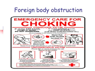
Immediate Presentation Immediate presentation: coughing, dyspnea, wheezing, cyanosis, or stridor Witnessed choking- high predictability
8 years old male with 1 year history of wheezing: Chest X-Ray shows spring from a pen in the right mainstem bronchus
Delayed Presentation Unilateral decreased breath sounds and rhonchi Recurrent or persistent pneumonia May be misdiagnosed as asthma (persistent cough or wheezing)
Incidence of FB Inhalation Leading cause of accidental death under 4 years of age Majority are under 3 years of age Organic foreign body are the most common (e.g. nuts, seeds) Sharp objects (e. g. pins) are the most common in adolescents Site: Bronchial tree 88%, Tracheal 12% Right side 52%, Left 33%, Bilateral 15%
Findings on Chest X-ray Majority of Foreign bodies are radiolucent (89%) Normal CXR (17%)1 Other findings: - Air trapping, atelectasis, localized hyperinflation - Infiltrate - Mediastinal shift - Pneumothorax
Additional Testing: CT Scans Excellent correlation and can aid decision making Can provide virtual bronchoscopy via 3D CT reconstruction ( 6th bronchial generation) Disadvantages - Radiation exposure - Cost - Limited availability
Flexible vs Rigid Bronchoscopy Rigid bronchoscopy- most often utilized - Allows ventilation & airway control through side port - Graspers through main port for FB removal Flexible bronchoscopy - Diagnostic - Possibly therapeutic for small FB removal in distal airways
Rigid Bronchoscopy: Oxygenation and Ventilation through side port1 Anesthesia circuit is attached to the side port of the rigid bronchoscope
Preoperative Assessment Degree of distress urgency of retrieval - Oxygen requirement - SPO2 94% or less - Tachypnea and retractions NPO status - No distress: standard ASA guidelines - Distress: proceed as emergency, OG suction Site of obstruction: tracheal vs bronchial - Unilateral wheeze, decreased breath sounds, CXR findings - Tracheal: usually emergent case
Preoperative Assessment Nature of foreign body - Nuts: inflammation and airway edema Time since inhalation - Recent: coughing, possible dislodgement - Remote: Airway edema and infection
Induction IV induction is preferable Inhalational induction possible if patient is in minimal respiratory distress Consider lidocaine to vocal cords prior to instrumenting airway with bronchoscope: Max dose: 4mg/kg
Anesthetic Plan for Rigid Bronchoscopy Spontaneous vs Controlled Ventilation: Retrospective review of 94 pediatric cases noted no difference in adverse outcomes3 Meta-analysis (423 controlled ventilation and 441 spontaneous ventilation)4 - No difference in desaturation - Lower incidence of laryngospasm and shorter operating time with controlled ventilation
Rigid Bronchoscopy: Spontaneous Ventilation Advantages: - Avoids positive pressure ventilation which can theoretically push foreign body deeper into airway - No muscle relaxant and no reversal Disadvantages: - Difficult to have patient deep enough to avoid coughing and movement and still ventilating adequately - Longer operative time
Rigid Bronchoscopy: Controlled Ventilation Advantages: - Ensures immobility and prevents coughing, gagging or movement during the procedure - Shorter operative time Disadvantages: - Need to monitor and reverse neuromuscular blockade - Need to carefully monitor positive pressure ventilation to prevent air trapping
Rigid Bronchoscopy: Spontaneous Ventilation Preserve spontaneous ventilation Induce with sevoflurane or propofol Maintain anesthesia with TIVA: - Propofol infusion - Fentanyl bolus or remifentanil infusion - +/- dexmedetomidine Lidocaine to vocal cords
Rigid Bronchoscopy: Controlled Ventilation Induce with sevoflurane or propofol Neuromuscular blockade with rocuronium and controlled ventilation via side arm of bronchoscope Maintain anesthesia with TIVA: - Propofol infusion - Fentanyl bolus or remifentanil infusion - +/- dexmedetomidine
Anesthetic Plan for Rigid Bronchoscopy Spontaneous vs Controlled Ventilation. Personal preference of anesthesiologist and surgeon TIVA is optimal approach as minimizes operative team s exposure to volatile agents
Rigid Bronch: Intraop Dexamethasone 0.5 mg/kg for airway edema prophylaxis to a maximum of 10 -16 mg Albuterol to minimize/treat bronchospasm from foreign body removal and airway manipulation
Rigid Bronchoscopy: Challenges Shared airway Must coordinate ventilation with position of bronchoscope (e.g. smaller breaths when bronchoscope is more distal) Difficult to monitor ETCO2 when ventilating through side arm of bronchoscope Risk of airway injury from rigid bronchoscope from coughing, bucking or movement
Bronchoscopy for FB: Common Intraop Problems Hypoxia/hypercarbia during procedure Difficult to monitor ETCO2 when ventilating trough side-arm of rigid bronchoscope Monitor chest rise Significant circuit leak Adjust ventilator, increase flows or ventilate by hand Hypoxia and desaturation when bronchoscope is distal as only ventilating/oxygenating one lung or a fraction of one lung (shunt) Communicate with surgeon to withdraw bronchoscope into trachea Communication with surgeon is important!
Bronchoscopy for FB: Common Intraop Problems Complete airway obstruction Can occur when FB is retrieved into the trachea or lodges in larynx just below the vocal cords during retrieval attempt Inability to ventilate or oxygenate Management: Instruct surgeon to push FB back into bronchus to allow oxygenation/ventilation via one lung
Bronchoscopy for FB: Common Intraop Problems FB fragments during retrieval Most commonly occurs with organic FBs (nuts or beans) May require multiple retrievals and prolongs case consider controlled ventilation May require 2nd or 3rd bronchoscopy to remove all fragments consider leaving patient intubated Betel nut that fragmented during retrieval
Complications from Airway FB Removal Greater than one bronchoscopy required for foreign body extraction ICU admission Hospital length of stay greater than 24 hours Time of surgery greater than 1 hour Sjogren PP, Mills TJ, Pollak AD, Muntz HR, Meier JD, Grimmer JF. Predictors of complicated airway foreign body extraction. Laryngoscope. 2018;128(2):490 495.
Predictors of Complicated Postoperative Course Hyper-lucency on CXR Unwitnessed aspiration Inability to completely remove all of the fragments of a FB (e.g. peanut)
Initial Airway Management After FB removal Face Mask or LMA ETT - + Airway Edema - + FB Fragments still in Airway - + Significant O2 Requirement - + Full Stomach
Postoperative Disposition Majority are admitted a minimum of 4 hours for monitoring Consider ICU admission - Prolonged duration of bronchoscopy - Significant airway edema or bleeding - Inability to remove all the FB fragments on initial bronchoscopy
Conclusions: FB in airway a major source of preventable accidental mortality & morbidity in children Anticipate complications Have a clear, shared airway plan Communicate with surgical colleagues!
References: 1. Fidkowski CW, Zheng H, Firth PG. The Anesthetic Considerations of Tracheobronchial Foreign Bodies in Children. Anesthesia & Analgesia. 2010;111(4):1016 1025. doi: 10.1213/ANE.0b013e3181ef3e9c. 2. Sjogren, P.P., Mills, T.J., Pollak, A.D., Muntz, H.R., Meier, J.D. and Grimmer, J.F. (2018), Predictors of complicated airway foreign body extraction. The Laryngoscope, 128: 490-495. doi:10.1002/lary.26814 3. Litman RS, Ponnuri J, Trogan I. Anesthesia for tracheal or bronchial foreign body removal in children: an analysis of ninety-four cases. Anesth Analg. 2000;91(6):1389-91. 4. Liu, Y., Chen, L. and Li, S. (2014), Controlled ventilation or spontaneous respiration in anesthesia for tracheobronchial foreign body removal: a meta analysis. Paediatr Anaesth, 24: 1023-1030. doi:10.1111/pan.12469 5. Foltran, F., Ballali, S., Rodriguez, H., (Sebastian) van As, A.B., Passali, D., Gulati, A. and Gregori, D. (2013), Inhaled foreign bodies in children: A global perspective on their epidemiological, clinical, and preventive aspects. Pediatr. Pulmonol., 48: 344-351. doi:10.1002/ppul.22701 6. Kendigelen, Pinar .The anaesthetic consideration of tracheobronchial foreign body aspiration in children. Journal of thoracic disease 2016; (2072-1439), 8 (12), p. 3803. 7. Baram, Aram .Scarf pin-related hijab syndrome: A new name for an unusual type of foreign body aspiration.2017. Journal of international medical research (0300-0605), 45 (6), p. 2078. 8. P.S.N. Murthy, V.S. Ingle, Edicula George, S. Ramakrishna, Fahim A. Shah.Sharp foreign bodies in the tracheobronchial tree. 2001.American Journal of Otolaryngology, Volume 22, Issue 2,pages 154- 156,ISSN 0196-0709 9. Divisi, D. "Foreign bodies aspirated in children: role of bronchoscopy.". The Thoracic and cardiovascular surgeon 2007.(0171-6425), 55 (4), p. 249. 10. TOMASKE, M., GERBER, A.C. and WEISS, M. Anesthesia and periinterventional morbidity of rigid bronchoscopy for tracheobronchial foreign body diagnosis and removal. Pediatric Anesthesia, (2006), 16: 123-129. doi:10.1111/j.1460-9592.2005.01714.x 11. https://www.cdc.gov/injury/wisqars/pdf/leading_causes_of_injury_deaths_highlighting_unintentiona l_injury_2011-a.pdf