Overview of Acute Gingival Diseases
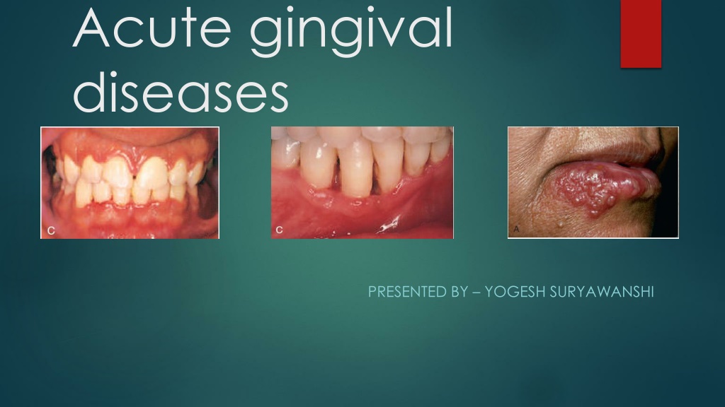


Acute gingival
diseases
PRESENTED BY –
YOGESH SURYAWANSHI
Contents
Introduction
Necrotizing ulcerative gingivitis
Necrotizing ulcerative periodontitis
Necrotizing ulcerative stomatitis
Primary herpetic gingivostomatitis
Periodontal abscess
Pericoronitis
Among these conditions, the following diseases
have been listed:
a)
Necrotizing periodontal diseases –
1.
Necrotizing ulcerative gingivitis
2.
Necrotizing ulcerative periodontitis
3.
Necrotizing ulcerative stomatitis
b) Gingival abscess
c) Periodontal abscess
d) Herpetic gingivostomatitis
e) Pericoronitis,
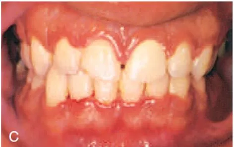
Necrotizing ulcerative gingivitis
Necrotizing ulcerative gingivitis (NUG) is a
microbial disease of the gingiva that most often
occurs in an impaired host.
It manifests with the characteristic clinical signs
of necrosis and sloughing of the gingival tissues
and may be accompanied by systemic
symptoms.
Clinical features
NUG has historically been identified as an acute disease.
However, the term
acute
in this case is used as a clinical
descriptor and not a diagnosis because
chronic forms of
the disease do not exist
.
Although the acronym
ANUG
is frequently used, it is a
misnomer.
A summary of acute periodontal lesions advocated the more simplified
term,
a)
Necrotizing gingivitis
b)
Necrotizing ulcerative gingivitis.
Involvement
can be limited to a single tooth or group of teeth, or it can
be
widespread throughout the mouth.
Oral signs
Characteristic lesions are of punched-out, craterlike
depressions at the crest of the interdental papillae
that subsequently extend to the marginal gingiva
and rarely to the attached gingiva and oral mucosa.
The surface of the gingival crater is covered by a
grey, pseudomembranous slough that is demarcated
from the remainder of the gingival mucosa by a
pronounced linear erythema.
In some cases, the lesions are denuded of the surface
pseudomembrane, thereby exposing the gingival
margin, which is red, shiny, and hemorrhagic.
The characteristic lesions can progressively destroy the
gingiva and the underlying periodontal tissues.
Spontaneous gingival hemorrhage and pronounced
bleeding after the slightest stimulation are additional
characteristic clinical signs.
Other common signs include a fetid odor
and
increased salivation.
etiology
NUG is an multifactorial disease and it include the following etiological
factors-
a)
Role of bacteria
b)
Role of host response
c)
Local predisposing factors
d)
Systemic predisposing factors-
1.
Nutritional deficiency
2.
Debilitating disease
3.
Psychosomatic factors
Differential diagnosis
a) Apthous stomatitis
b) Traumatic ulcers
c) Desquamative gingivitis
d) Erythema multiforme
e) Agranulocytosis
f) Infectious mononucleosis
g) Acute leukemia
h) Allergic stomatitis
i) Secondary syphilis.
treatment
The treatment of NUG consists of –
1. Alleviation of the acute inflammation by reduction of the microbial load
and removal of necrotic tissue.
2. Treatment of chronic disease either underlying the acute involvement or
elsewhere in the oral cavity
3. Alleviation of generalized symptoms such as fever and malaise
4. Correction of systemic conditions or factors that contribute to the
initiation or progression of gingival changes.
Treatment of NUG should follow an orderly sequence, according to
specific steps, at three clinical visits.
First visit
At the first visit, the clinician should conduct a comprehensive
evaluation of the patient, including –
A thorough medical history, with special attention to –
a)
Recent illness g) Type of employment
b)
Living conditions
h) Hours of rest
c)
Dietary background
d)
Cigarette smoking
e)
Risk factors for human immunodeficiency virus (HIV) infection
f)
Psychosocial parameters (e.g., stress, depression).
The patient is questioned regarding the history of the acute disease,
including its onset and duration, as follows:
Is the disease recurrent?
Are the recurrences associated with specific factors such as –
a.
Menstruation
b.
Particular foods
c.
Exhaustion
d.
Mental stress
Has there been any previous treatment? When and for how
long?
The clinician should also inquire as to the type of treatment received
and the patient’s impression regarding the effectiveness of
previous
treatment.
The initial physical examination should include an assessment of general
appearance, presence of halitosis, presence of skin lesions, vital signs
including temperature, and palpation for the presence of enlarged
lymph nodes, especially submaxillary and submental nodes.
The oral cavity is examined for the characteristic NUG lesions distribution,
and the possible involvement of the oropharyngeal region.
The goals of initial therapy are reduction of the microbial load and
removal of necrotic tissue to the degree that repair and regeneration
of normal tissue barriers can be reestablished.
Treatment during the initial visit is continued to the acutely involved
areas, which are isolated with cotton rolls and dried.
A topical anesthetic is applied, and after 2 or 3 minutes the areas are
gently swabbed with a moistened cotton pellet to remove the
pseudomembrane and nonattached surface debris.
Antibiotics are effective in the treatment of patients with NUG.
Patients with moderate or severe NUG and local lymphadenopathy or
other systemic signs or symptoms are placed on an antibiotic regimen
of amoxicillin 500 mg orally every 6 hours for 10 days.
For amoxicillin-sensitive patients, other antibiotics are prescribed, such
as erythromycin (500 mg every 6 hours) or metronidazole (500 mg twice
daily for 7 days). Systemic complications should subside in 1 to 3 days.
Antibiotics are not recommended in NUG patients who do not have
systemic complications.
Instructions to the patients
The patient is discharged with the following instructions:
1. Avoid tobacco, alcohol, and condiments.
2. Rinse with a glassful of an equal mixture of 3% hydrogen peroxide and
warm water every 2 hours and/or 0.12%
chlorhexidine solution twice daily.
3. Get adequate rest. Pursue usual activities, but avoid excessive physical
exertion or prolonged exposure to the sun, as in golfing, playing tennis,
swimming, or sunbathing.
Second visit
At the second visit, 1 or 2 days after the first visit, the patient is evaluated
for amelioration of signs and symptoms.
The patient’s condition is usually improved; the pain is diminished or is no
longer present.
The gingival margins of the involved areas are erythematous but without
a superficial pseudomembrane.
Scaling is performed if it is necessary and sensitivity permits.
Shrinkage of the gingiva may expose previously covered calculus, which
is gently removed.
Third visit
At the next visit, approximately 5 days after the second visit, the patient
is evaluated for resolution of symptoms, and a comprehensive plan for
management of the patient’s periodontal condition is formulated.
The patient should be essentially symptom-free at this time.
Some erythema may still be present in the involved areas, and the
gingiva may be slightly painful on tactile stimulation.
The patient is instructed in biofilm control procedures, which are
essential for the success of the treatment and the maintenance of
periodontal health.
The patient is further counseled on nutrition, smoking cessation, and
other conditions or habits associated with potential recurrence.
The hydrogen peroxide rinses are discontinued, but chlorhexidine
rinses may be maintained for an additional 2 or 3 weeks.
Scaling and root planing is repeated if necessary.
Unfortunately, patients often discontinue treatment because the acute
condition has subsided; however, this is when comprehensive treatment
of the chronic periodontal problem should begin.
Patients need to be educated about the importance of
comprehensive periodontal treatment and encouraged to complete it.
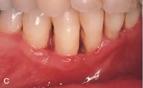
Necrotizing ulcerative periodontitis
In the 1999 classification, necrotizing ulcerative gingivitis (NUG) and
necrotizing ulcerative periodontitis (NUP) were included among
Necrotizing diseases of Periodontium.
Studies have suggested that they may represent different stages of the
same disease, because they have similar etiology, clinical
characteristics, and treatment, and may even progress to more severe
forms such as necrotizing stomatitis (NS) and noma.
Clinical features
Clinical cases of NUP are defined by necrosis and
ulceration of the coronal portion of the interdental
papillae and gingival margin, with a painful, bright-
red marginal gingiva that bleeds easily.
This is similar to the clinical features of NUG.
The distinguishing feature of NUP is the
destructive
progression
of the disease, which includes
periodontal attachment and bone loss.
Deep interdental osseous craters typify periodontal
lesions of NUP.
However, “conventional” periodontal pockets with
deep probing depth are not found, because the
ulcerative and necrotizing nature of the gingival lesion
destroys the marginal epithelium and connective tissue,
resulting in gingival
recession.
etiology
The etiology of NUP has not been determined, although a mixed fusiform–
spirochete bacterial flora appears to play a key role.
Because bacterial pathogens are not solely responsible for causing the
disease, some predisposing “host” factors may be necessary.
Numerous predisposing factors have been attributed to NUG, including
poor oral hygiene, preexisting periodontal disease, smoking, viral
infections, immunocompromised status, psychosocial stress, and
malnutrition.
One case report attributed heavy tobacco use as the most significant
contributing factor associated with NUP in a 21-year-old who had been
smoking (>20 cigarettes/day) and chewing tobacco since the
age of 7
years.
Smoking, malnutrition, and high plaque levels all increase the risk of NUP
and need to be changed so that treatment success is
obtained.
treatment
Necrotizing ulcerative periodontitis (NUP) is a rare disease, especially
in
developed countries.
Often, NUP is diagnosed in individuals with a compromised host immune
response.
The incidence of NUP in specific populations, such as patients who are
positive for human immunodeficiency virus (HIV) infection or have
acquired immunodeficiency syndrome (AIDS), has been reported to be
between 0 and 6%.
Most patients diagnosed with NUP have diseases or conditions that
impair their host immune response.
These patients often have an underlying predisposing systemic factor
that renders them susceptible to NUP disease.
For this reason, patients presenting with NUP should be treated in
consultation with their physician.
A comprehensive medical evaluation and diagnosis of
any condition that may be contributing to an altered host
immune response should be completed.
It is also important to rule out any hematologic disease
(e.g., leukemia) before initiating treatment of any case
that has a similar presentation to NUP.
Treatment can be initiated only after a thorough medical
history and examination to identify the existence of any
systemic diseases.
Treatment for NUP includes local debridement of lesions with scaling and
root planing, lavage, and instructions for good oral hygiene.
It may be necessary to use local anesthesia during the debridement
because lesions are frequently painful.
The use of ultrasonic instrumentation with profuse irrigation may
enhance debridement and flushing of deep lesions.
Achieving good oral hygiene may also be challenging until the lesions
and associated pain resolve.
Antimicrobial adjuncts, such as chlorhexidine, added to the oral hygiene
regimen may contribute to the daily reduction of bacterial loads.
Locally applied topical antimicrobials and systemic antibiotics, as well as
systemic analgesics, should be used as indicated by signs and symptoms.
Patients with NUP often harbor bacteria, fungi, viruses, and other non oral
microorganisms, complicating the selection of antimicrobial therapy.
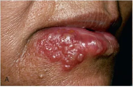
Primary herpetic gingivostomatitis
Primary herpetic gingivostomatitis is an infection of the oral cavity
caused by herpes simplex virus (HSV) type 1.
It occurs most often among infants and children who are younger
than 6 years of age, but it is also seen in adolescents and adults.
It occurs with equal frequency in male and female patients.
The incubation period is 1–26 days and can occur throughout the
year.
In most individuals, however, the primary infection is asymptomatic.
The primary HSV infection is usually acquired through direct contact with
affected area or through secretions.
The virus once attached to the cells at inoculation site through specific
receptors; it replicates too many virions to the maximum number and
discharges to neighboring cells.
Subsequently it affects adjacent cells, spreads to distant sensory nerve
endings and autonomic axons, further to adjacent related ganglia, and
remains latent there.
The lymph node is involved through viral proteins by mobile dendritic
cells and begins its primary immune response.
The reason for the wide anatomical distribution and multiple crops may
be due to descending spread of virions back to periphery from various
neuronal
site.
Secondary manifestations include herpes labialis,
herpetic stomatitis, herpes genitalis, ocular herpes, and
herpetic encephalitis.
Secondary herpetic stomatitis can occur on the
palate, on the gingiva, or on the mucosa as a result of
dental treatment.
Clinical features
The disease occurring in
children is frequently the primary
attack and is characterized by the development of
fever, irritability, headache, pain upon swallowing, and
regional lymphadenopathy.
Within a few days, the mouth becomes painful and the
gingiva which is intensely inflamed appears
erythematous and edematous.
The lips, tongue, buccal mucosa, palate, pharynx, and tonsils may
also be involved.
Shortly, yellowish, fluid-filled vesicles develop.
These vesicles rupture and form shallow, ragged, extremely painful
ulcers covered by a grey membrane and surrounded by an
erythematous halo.
It is important to recognize that the gingival inflammation precedes the
formation of the ulcers by several days.
The ulcers vary considerably in size, ranging from very tiny lesions to
lesions measuring several millimeters or even a centimeter in diameter.
They heal spontaneously within 7–14 days and
leave no scar.
After approximately 24 hours, the vesicles rupture and form painful
small ulcers with red, elevated, halo-like margins and depressed
yellowish or grayish white central portions.
They occur in widely separated areas or in clusters where
confluence occurs.
The disease is accompanied by generalized soreness of the oral
cavity, which interferes with eating, drinking, and oral hygiene.
The ruptured vesicles are the focal sites of pain; they are particularly
sensitive to touch, thermal changes, foods such as condiments and
fruit juices, and the action of coarse foods.
In infants, the disease is marked by irritability and refusal to take food.
diagnosis
It is critical to arrive at a diagnosis as early as possible for a patient with
a primary herpetic infection.
Treatment with antiviral medications can dramatically alter the course
of the disease by reducing symptoms and potentially reducing
recurrences.
The diagnosis is usually established from the patient’s history and the
clinical findings.
It can be diagnosed by laboratory procedures as well.
Scrapings obtained from the base of the lesions are stained with
Wright’s, and Giemsa stain.
Pap stain demonstrates balloon cells, multinucleated giant cells and
intranuclear inclusions.
Though cytological procedures give a quick result but it will not
differentiate between HSV and
varicella zoster virus (VZV).
treatment
It consists of –
a)
Symptomatic treatment
b)
Supportive care
c)
Systemic treatment
Symptomatic treatment
Pain control measures – Topical anesthetic like 2% lignocaine, 0.5%
benzocaine hydrochloride are used.
In some cases systemic analgesics are used.
Topical anti infective agents –
Agents used are 0.2 % chlorhexidine, tetracycline mouthwash.
Supportive care
Fluids are given to maintain proper hydration and
electrolyte balance.
Antipyretics are given to reduce fever.
Avoid spicy food, hard bristles for tooth brushing during
acute stage.
Systemic treatment
Acyclovir suspension 15 mg/kg or acyclovir tablets 200 mg 5 times a
day for 5 days initiated within first 3 days of onset can reduce the
duration of lesion.
For primary herpes labialis, antiviral agents like acyclovir may reduce
the pain and time of healing.
Valvacyclovir 500 mg or Famciclovir 250 mg 2 times a day for 5 days
can be used as an alternative.
pericoronitis
Pericoronitis or operculitis is an acute or chronic periodontal inflammation
around the occlusal surface of partially or completely erupted
mandibular third molar, which is seen most commonly around 20 to 29
years of age, rarely seen before 20 or after 40.
Acute pericoronitis is characterized by cardinal signs of swelling , namely
rubor, dolor, calor , tumor.
Chronic pericoronitis is due to soft tissue trauma covering the occlusal
surface of partially or completely erupted mandibular third molar which
is traumatized by the cusps of the opposing maxillary third molar, done
repeatedly and for a long duration.
The symptoms associated with chronic pericoronitis are swelling, pus
discharge, which may progress to mandibular space infection.
etiology
The most common cause of pericoronitis is entrapment of food debris
and plaque between the crown and the overlying operculum.
This constant entrapment of plaque and food debris leads to
inflammation, which aggravates pain, redness, occlusal trauma and
also leads to release of inflammatory fluid and cellular exudate.
The microflora of pericoronitis is diverse and differs from pathogens that
cause periodontitis.
In a study of microbiota of pericoronitis, Actinomyces oris,
Fusobacterium nucleatum, Treponema denticola were present in high
levels.
In short, pericoronitis results from bacterial overgrowth in a confined
space that is exacerbated by poor cleansability.
Clinical features
Acute pericoronitis is characterized by pain, swollen, red, suppurating
lesion , tender & throbbing in nature and radiates to ear,
temporomandibular joint and posterior submandibular region, making
the patient unable to sleep.
Patient also complains of dysphagia, halitosis, putrescent odor and
inability to close the jaws.
Trismus is seen along the swelling of cheek in the angle of jaw.
Signs of trauma in operculum such as indentations of cusps of upper
teeth leading to ulceration.
This leads to systemic conditions like pyrexia, leukocytosis, regional
lymphadenopathy with a diffuse spread to tissue spaces.
treatment
Various treatment options for treating pericoronitis are:-
1.
Non surgical –
a.
Local debridement
b.
Pain management
c.
Systemic antibiotics
2. Surgical removal of overlying flap
Local debridement
This method involves irrigation of the pericoronal space with warm
saline to flush away the food debris using a 10cc syringe of 20 gauge
needle.
The other irrigating solutions are:-
Phenol 5%, tincture of iodine.
Elevate the flap gently from the tooth with scaler and curette and
swab the area with an antiseptic.
If pericoronal abscess is present, make an incision anteroposteriorly with
a no.15 BP blade to establish drainage.
Pain management
Pain can greatly limit chewing and taking in good oral intake for patients
with pericoronitis.
Pain management, in addition to resolving infection, is crucial in
pericoronitis management.
There are multiple modalities for pain management: local anesthetic
injections, topical analgesics, and oral analgesics.
Oral analgesia with NSAID should be the primary method of pain
management.
Systemic antibiotics
If pericoronitis is severe or systemic symptoms are
present, antibiotics , namely, either metronidazole
400mg twice a day for five days or penicillin 500mg
thrice a day for five days.
Patients allergic to penicillin are advised to take
erythromycin 500mg thrice a day for five days is suitable.
Surgical removal of overlying flap
Soft tissue surgery: Removing the infected soft tissue can help resolve
pericoronitis as it is the soft tissue disease overlying an erupting third
molar.
The operculum, soft tissue covering erupting third molar, can be
removed to eliminate the deep pocket formed between the gingiva
and the tooth.
Removal of the soft tissue covering the third molar can also help with the
tooth eruption.
This treatment option is limited to third molars with a favorable eruption
position.
For example, if a tooth is erupting vertically with adequate space from
the distal aspect to the tooth to the anterior border of the ramus,
operculectomy can help resolve pericoronitis.
However, if a tooth is horizontally impacted or there is inadequate space
for eruption, the areas that are hard to clean will likely persist, which may
cause pericoronitis to recur.
Gingival abscess
The gingival abscess is a localized acute inflammatory
lesion that may arise from a variety of sources,
including microbial plaque infection, trauma, and
foreign body impaction.
Clinical features include a red, smooth, sometimes
painful, often fluctuant swelling
Periodontal abscess
Periodontal abscesses are typically found in patients with
untreated periodontitis and in association with moderate
to deep periodontal
pockets.
1
Periodontal abscesses often arise as acute
exacerbations
of preexisting pockets.
2
Primarily related to incomplete calculus removal,
periodontal abscesses have been linked to several
clinical situations.
3
They have been identified in patients after periodontal
surgery,
1
after preventive maintenance, after systemic
antibiotic therapy,
2
and as the result of recurrent disease.
Conditions in which periodontal abscesses are not related
to inflammatory periodontal disease include tooth
perforation
3
or and foreign body impaction.
Poorly controlled diabetes mellitus has been considered
a predisposing factor for periodontal abscess formation.
Formation of periodontal abscess has been reported as
a major cause of tooth loss; however, with proper
treatment followed by consistent preventive periodontal
maintenance, teeth with significant bone loss may be
retained for many years.
Acute abscesses are characterized by painful, red,
edematous, smooth, and ovoid swelling of the gingival
tissues.
Exudate may be expressed with gentle pressure; the tooth
may be percussion sensitive and feel elevated in the socket.
Fever and regional lymphadenopathy are occasional
findings.
Treatment
The principles for
the management of simple dental infections are as
follows:
1. Local measures
i. Drainage
ii. Maintain drainage
iii. Eliminate cause
2. Systemic measures in conjunction with the local measures
The management of a patient with periodontal abscess
can divided into three stages:
i. Immediate management
ii. Initial management
iii. Definitive therapy
Acute gingival diseases encompass various conditions such as Necrotizing Ulcerative Gingivitis, Necrotizing Ulcerative Periodontitis, Necrotizing Ulcerative Stomatitis, and others. Necrotizing Ulcerative Gingivitis (NUG) is a microbial disease primarily affecting the gingiva, characterized by necrosis and sloughing of gingival tissues. Clinical features include acute presentation with punched-out lesions and pseudomembranous slough. Management typically involves addressing the microbial imbalance and promoting oral hygiene. Early detection and prompt treatment are essential to prevent progression and systemic implications.
- Gingival Diseases
- Necrotizing Ulcerative Gingivitis
- Oral Health
- Periodontal Conditions
- Acute Symptoms

Download Presentation

Please find below an Image/Link to download the presentation.
The content on the website is provided AS IS for your information and personal use only. It may not be sold, licensed, or shared on other websites without obtaining consent from the author. Download presentation by click this link. If you encounter any issues during the download, it is possible that the publisher has removed the file from their server.
E N D
Presentation Transcript
Acute gingival diseases PRESENTED BY YOGESH SURYAWANSHI
Contents Introduction Necrotizing ulcerative gingivitis Necrotizing ulcerative periodontitis Necrotizing ulcerative stomatitis Primary herpetic gingivostomatitis Periodontal abscess Pericoronitis
Among these conditions, the following diseases have been listed: a) Necrotizing periodontal diseases 1. Necrotizing ulcerative gingivitis 2. Necrotizing ulcerative periodontitis 3. Necrotizing ulcerative stomatitis b) Gingival abscess c) Periodontal abscess d) Herpetic gingivostomatitis e) Pericoronitis,
Necrotizing ulcerative gingivitis Necrotizing ulcerative gingivitis (NUG) is a microbial disease of the gingiva that most often occurs in an impaired host. It manifests with the characteristic clinical signs of necrosis and sloughing of the gingival tissues and may be accompanied by systemic symptoms.
Clinical features NUG has historically been identified as an acute disease. However, the term acutein this case is used as a clinical descriptor and not a diagnosis because chronic forms of the disease do not exist. Although the acronym ANUGis frequently used, it is a misnomer.
A summary of acute periodontal lesions advocated the more simplified term, a) Necrotizing gingivitis b) Necrotizing ulcerative gingivitis. Involvement can be limited to a single tooth or group of teeth, or it can be widespread throughout the mouth.
Oral signs Characteristic lesions are of punched-out, craterlike depressions at the crest of the interdental papillae that subsequently extend to the marginal gingiva and rarely to the attached gingiva and oral mucosa. The surface of the gingival crater is covered by a grey, pseudomembranous slough that is demarcated from the remainder of the gingival mucosa by a pronounced linear erythema.
In some cases, the lesions are denuded of the surface pseudomembrane, thereby exposing the gingival margin, which is red, shiny, and hemorrhagic. The characteristic lesions can progressively destroy the gingiva and the underlying periodontal tissues. Spontaneous gingival hemorrhage and pronounced bleeding after the slightest stimulation are additional characteristic clinical signs. Other common signs include a fetid odor and increased salivation.
etiology NUG is an multifactorial disease and it include the following etiological factors- a) Role of bacteria b) Role of host response c) Local predisposing factors d) Systemic predisposing factors- 1. Nutritional deficiency 2. Debilitating disease 3. Psychosomatic factors
Differential diagnosis a) Apthous stomatitis b) Traumatic ulcers c) Desquamative gingivitis d) Erythema multiforme e) Agranulocytosis g) Acute leukemia h) Allergic stomatitis i) Secondary syphilis. f) Infectious mononucleosis
treatment The treatment of NUG consists of 1. Alleviation of the acute inflammation by reduction of the microbial load and removal of necrotic tissue. 2. Treatment of chronic disease either underlying the acute involvement or elsewhere in the oral cavity
3. Alleviation of generalized symptoms such as fever and malaise 4. Correction of systemic conditions or factors that contribute to the initiation or progression of gingival changes. Treatment of NUG should follow an orderly sequence, according to specific steps, at three clinical visits.
First visit At the first visit, the clinician should conduct a comprehensive evaluation of the patient, including A thorough medical history, with special attention to a) Recent illness g) Type of employment b) Living conditions h) Hours of rest c) Dietary background d) Cigarette smoking e) Risk factors for human immunodeficiency virus (HIV) infection Psychosocial parameters (e.g., stress, depression). f)
The patient is questioned regarding the history of the acute disease, including its onset and duration, as follows: Is the disease recurrent? Are the recurrences associated with specific factors such as a. Menstruation b. Particular foods c. Exhaustion d. Mental stress Has there been any previous treatment? When and for how long?
The clinician should also inquire as to the type of treatment received and the patient s impression regarding the effectiveness of previous treatment. The initial physical examination should include an assessment of general appearance, presence of halitosis, presence of skin lesions, vital signs including temperature, and palpation for the presence of enlarged lymph nodes, especially submaxillary and submental nodes. The oral cavity is examined for the characteristic NUG lesions distribution, and the possible involvement of the oropharyngeal region.
The goals of initial therapy are reduction of the microbial load and removal of necrotic tissue to the degree that repair and regeneration of normal tissue barriers can be reestablished. Treatment during the initial visit is continued to the acutely involved areas, which are isolated with cotton rolls and dried. A topical anesthetic is applied, and after 2 or 3 minutes the areas are gently swabbed with a moistened cotton pellet to remove the pseudomembrane and nonattached surface debris.
Antibiotics are effective in the treatment of patients with NUG. Patients with moderate or severe NUG and local lymphadenopathy or other systemic signs or symptoms are placed on an antibiotic regimen of amoxicillin 500 mg orally every 6 hours for 10 days. For amoxicillin-sensitive patients, other antibiotics are prescribed, such as erythromycin (500 mg every 6 hours) or metronidazole (500 mg twice daily for 7 days). Systemic complications should subside in 1 to 3 days. Antibiotics are not recommended in NUG patients who do not have systemic complications.
Instructions to the patients The patient is discharged with the following instructions: 1. Avoid tobacco, alcohol, and condiments. 2. Rinse with a glassful of an equal mixture of 3% hydrogen peroxide and warm water every 2 hours and/or 0.12% chlorhexidine solution twice daily. 3. Get adequate rest. Pursue usual activities, but avoid excessive physical exertion or prolonged exposure to the sun, as in golfing, playing tennis, swimming, or sunbathing.
Second visit At the second visit, 1 or 2 days after the first visit, the patient is evaluated for amelioration of signs and symptoms. The patient s condition is usually improved; the pain is diminished or is no longer present. The gingival margins of the involved areas are erythematous but without a superficial pseudomembrane. Scaling is performed if it is necessary and sensitivity permits. Shrinkage of the gingiva may expose previously covered calculus, which is gently removed.
Third visit At the next visit, approximately 5 days after the second visit, the patient is evaluated for resolution of symptoms, and a comprehensive plan for management of the patient s periodontal condition is formulated. The patient should be essentially symptom-free at this time. Some erythema may still be present in the involved areas, and the gingiva may be slightly painful on tactile stimulation. The patient is instructed in biofilm control procedures, which are essential for the success of the treatment and the maintenance of periodontal health.
The patient is further counseled on nutrition, smoking cessation, and other conditions or habits associated with potential recurrence. The hydrogen peroxide rinses are discontinued, but chlorhexidine rinses may be maintained for an additional 2 or 3 weeks. Scaling and root planing is repeated if necessary.
Unfortunately, patients often discontinue treatment because the acute condition has subsided; however, this is when comprehensive treatment of the chronic periodontal problem should begin. Patients need to be educated comprehensive periodontal treatment and encouraged to complete it. about the importance of
Necrotizing ulcerative periodontitis In the 1999 classification, necrotizing ulcerative gingivitis (NUG) and necrotizing ulcerative periodontitis (NUP) were included among Necrotizing diseases of Periodontium. Studies have suggested that they may represent different stages of the same disease, because they characteristics, and treatment, and may even progress to more severe forms such as necrotizing stomatitis (NS) and noma. have similar etiology, clinical
Clinical features Clinical cases of NUP are defined by necrosis and ulceration of the coronal portion of the interdental papillae and gingival margin, with a painful, bright- red marginal gingiva that bleeds easily. This is similar to the clinical features of NUG. The distinguishing feature of NUP is the destructive progression of the disease, periodontal attachment and bone loss. which includes
Deep interdental osseous craters typify periodontal lesions of NUP. However, conventional periodontal pockets with deep probing depth are not found, because the ulcerative and necrotizing nature of the gingival lesion destroys the marginal epithelium and connective tissue, resulting in gingival recession.
etiology The etiology of NUP has not been determined, although a mixed fusiform spirochete bacterial flora appears to play a key role. Because bacterial pathogens are not solely responsible for causing the disease, some predisposing host factors may be necessary. Numerous predisposing factors have been attributed to NUG, including poor oral hygiene, preexisting periodontal disease, smoking, viral infections, immunocompromised malnutrition. status, psychosocial stress, and
One case report attributed heavy tobacco use as the most significant contributing factor associated with NUP in a 21-year-old who had been smoking (>20 cigarettes/day) and chewing tobacco since the age of 7 years. Smoking, malnutrition, and high plaque levels all increase the risk of NUP and need to be changed so that treatment success is obtained.
treatment Necrotizing ulcerative periodontitis (NUP) is a rare disease, especially in developed countries. Often, NUP is diagnosed in individuals with a compromised host immune response. The incidence of NUP in specific populations, such as patients who are positive for human immunodeficiency virus (HIV) infection or have acquired immunodeficiency syndrome (AIDS), has been reported to be between 0 and 6%.
Most patients diagnosed with NUP have diseases or conditions that impair their host immune response. These patients often have an underlying predisposing systemic factor that renders them susceptible to NUP disease. For this reason, patients presenting with NUP should be treated in consultation with their physician.
A comprehensive medical evaluation and diagnosis of any condition that may be contributing to an altered host immune response should be completed. It is also important to rule out any hematologic disease (e.g., leukemia) before initiating treatment of any case that has a similar presentation to NUP. Treatment can be initiated only after a thorough medical history and examination to identify the existence of any systemic diseases.
Treatment for NUP includes local debridement of lesions with scaling and root planing, lavage, and instructions for good oral hygiene. It may be necessary to use local anesthesia during the debridement because lesions are frequently painful. The use of ultrasonic instrumentation with profuse irrigation may enhance debridement and flushing of deep lesions. Achieving good oral hygiene may also be challenging until the lesions and associated pain resolve.
Antimicrobial adjuncts, such as chlorhexidine, added to the oral hygiene regimen may contribute to the daily reduction of bacterial loads. Locally applied topical antimicrobials and systemic antibiotics, as well as systemic analgesics, should be used as indicated by signs and symptoms. Patients with NUP often harbor bacteria, fungi, viruses, and other non oral microorganisms, complicating the selection of antimicrobial therapy.
Primary herpetic gingivostomatitis Primary herpetic gingivostomatitis is an infection of the oral cavity caused by herpes simplex virus (HSV) type 1. It occurs most often among infants and children who are younger than 6 years of age, but it is also seen in adolescents and adults. It occurs with equal frequency in male and female patients. The incubation period is 1 26 days and can occur throughout the year.
In most individuals, however, the primary infection is asymptomatic. The primary HSV infection is usually acquired through direct contact with affected area or through secretions. The virus once attached to the cells at inoculation site through specific receptors; it replicates too many virions to the maximum number and discharges to neighboring cells.
Subsequently it affects adjacent cells, spreads to distant sensory nerve endings and autonomic axons, further to adjacent related ganglia, and remains latent there. The lymph node is involved through viral proteins by mobile dendritic cells and begins its primary immune response. The reason for the wide anatomical distribution and multiple crops may be due to descending spread of virions back to periphery from various neuronal site.
Secondary manifestations include herpes labialis, herpetic stomatitis, herpes genitalis, ocular herpes, and herpetic encephalitis. Secondary herpetic stomatitis can occur on the palate, on the gingiva, or on the mucosa as a result of dental treatment.
Clinical features The disease occurring in children is frequently the primary attack and is characterized by the development of fever, irritability, headache, pain upon swallowing, and regional lymphadenopathy. Within a few days, the mouth becomes painful and the gingiva which is intensely erythematous and edematous. inflamed appears
The lips, tongue, buccal mucosa, palate, pharynx, and tonsils may also be involved. Shortly, yellowish, fluid-filled vesicles develop. These vesicles rupture and form shallow, ragged, extremely painful ulcers covered by a grey membrane and surrounded by an erythematous halo.
It is important to recognize that the gingival inflammation precedes the formation of the ulcers by several days. The ulcers vary considerably in size, ranging from very tiny lesions to lesions measuring several millimeters or even a centimeter in diameter. They heal spontaneously within 7 14 days and leave no scar.
After approximately 24 hours, the vesicles rupture and form painful small ulcers with red, elevated, halo-like margins and depressed yellowish or grayish white central portions. They occur in widely separated areas or in clusters where confluence occurs.
The disease is accompanied by generalized soreness of the oral cavity, which interferes with eating, drinking, and oral hygiene. The ruptured vesicles are the focal sites of pain; they are particularly sensitive to touch, thermal changes, foods such as condiments and fruit juices, and the action of coarse foods. In infants, the disease is marked by irritability and refusal to take food.
diagnosis It is critical to arrive at a diagnosis as early as possible for a patient with a primary herpetic infection. Treatment with antiviral medications can dramatically alter the course of the disease by reducing symptoms and potentially reducing recurrences. The diagnosis is usually established from the patient s history and the clinical findings.
It can be diagnosed by laboratory procedures as well. Scrapings obtained from the base of the lesions are stained with Wright s, and Giemsa stain. Pap stain demonstrates balloon cells, multinucleated giant cells and intranuclear inclusions. Though cytological procedures give a quick result but it will not differentiate between HSV and varicella zoster virus (VZV).
treatment It consists of a) Symptomatic treatment b) Supportive care c) Systemic treatment
Symptomatic treatment Pain control measures Topical anesthetic like 2% lignocaine, 0.5% benzocaine hydrochloride are used. In some cases systemic analgesics are used. Topical anti infective agents Agents used are 0.2 % chlorhexidine, tetracycline mouthwash.
Supportive care Fluids are given to maintain proper hydration and electrolyte balance. Antipyretics are given to reduce fever. Avoid spicy food, hard bristles for tooth brushing during acute stage.
Systemic treatment Acyclovir suspension 15 mg/kg or acyclovir tablets 200 mg 5 times a day for 5 days initiated within first 3 days of onset can reduce the duration of lesion. For primary herpes labialis, antiviral agents like acyclovir may reduce the pain and time of healing. Valvacyclovir 500 mg or Famciclovir 250 mg 2 times a day for 5 days can be used as an alternative.
pericoronitis Pericoronitis or operculitis is an acute or chronic periodontal inflammation around the occlusal surface of partially or completely erupted mandibular third molar, which is seen most commonly around 20 to 29 years of age, rarely seen before 20 or after 40. Acute pericoronitis is characterized by cardinal signs of swelling , namely rubor, dolor, calor , tumor.
Chronic pericoronitis is due to soft tissue trauma covering the occlusal surface of partially or completely erupted mandibular third molar which is traumatized by the cusps of the opposing maxillary third molar, done repeatedly and for a long duration. The symptoms associated with chronic pericoronitis are swelling, pus discharge, which may progress to mandibular space infection.