Evaluation and Management of Pediatric Intoeing: A Comprehensive Guide
Evaluation and treatment of pediatric intoeing involves a detailed history, screening examinations, and special tests to assess rotational profiles and identify underlying pathologies such as femoral anteversion, tibial torsion, and forefoot adduction. Proper diagnosis and education of parents are crucial for the successful management of intoeing in children.

Download Presentation

Please find below an Image/Link to download the presentation.
The content on the website is provided AS IS for your information and personal use only. It may not be sold, licensed, or shared on other websites without obtaining consent from the author. Download presentation by click this link. If you encounter any issues during the download, it is possible that the publisher has removed the file from their server.
E N D
Presentation Transcript
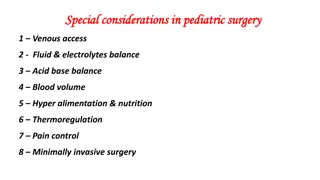
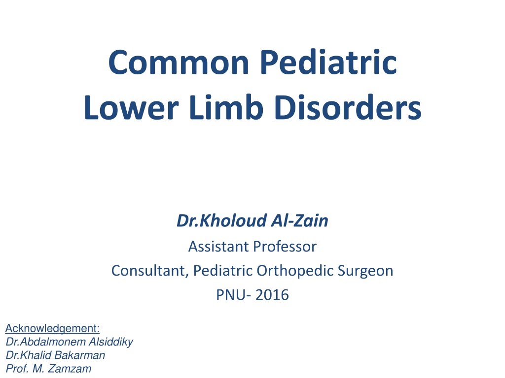
Common Pediatric Lower Limb Disorders Dr.Kholoud Al-Zain Assistant Professor Consultant, Pediatric Orthopedic Surgeon PNU- 2016 Acknowledgement: Dr.Abdalmonem Alsiddiky Dr.Khalid Bakarman Prof. M. Zamzam
Topics to Cover 1. In-toeing 2. Genu (varus & valgus), & proximal tibia vara 3. Club foot 4. L.L deformities in C.P patients 5. Limping & leg length inequality 6. Leg aches
Intoeing- Evaluation Detailed history Onset, who noticed it, progression Fall a lot How sits on the ground Screening examination (head to toe) Pathology at the level of: Femoral anteversion Tibial torsion Forefoot adduction Wandering big toe
Intoeing- Asses rotational profile Pathology Level Femoral anteversion Special Test Hips rotational profile: Supine Prone Inter-malleolus axis: Supine Prone Foot thigh axis Heel bisector line Tibial torsion Forefoot adduction Wandering big toe
Intoeing- Special Test Foot Propagation Angle normal is (-10 ) to (+15 )
Intoeing- Femoral Anteversion Hips rotational profile, supine IR/ER normal = 40-45/45-50
Intoeing- Tibial Torsion Inter-malleolus axis Supine position Sitting position
Intoeing- Tibial Torsion Foot Thigh Axis normal (0 ) to (-10 )
Intoeing- Forefoot Adduction Heel bisector line normal along 2 toe
Intoeing- Treatment Establish correct diagnosis Parents education Annual clinic F/U asses degree of deformity Femoral anti-version sit cross legged Tibial torsion spontaneous improvement Forefoot adduction anti-version shoes, or proper shoes reversal Adducted big toe spontaneous improvement
Intoeing- Treatment Operative correction indicated for children: (> 8) years of age With significant cosmetic and functional deformity <1%
Genu Varum and Genu Valgum Types: Physiological is usually bilateral Pathological can be unilateral
Genu Varum and Genu Valgum Types: Physiologic Pathologic
Genu Varum and Genu Valgum Evaluation History (detailed) Examination (signs of Rickets) Laboratory
Genu Varum and Genu Valgum Evaluation: Imaging Rickets
Genu Varum and Genu Valgum Management principles: Non-operative: Physiological usually Pathological must treat underlying cause, as rickets Epiphysiodesis Corrective osteotomies
Proximal Tibia Vara Blount disease : damage of proximal medial tibial growth plate of unknown cause Usually: Overweight Dark skinned Types: Infantile < 3y of age, usually Bil & early walkers Juvenile 3 -10 y, combination Adolescent > 10y, usually unilateral
Blount Disease- Investigation MRI is mandatory: When: Sever cases Recurrence Why?
Blount Disease- Treatment Unilateral Bilateral Types: Infantile Adolescent
Clubfoot Etiology Postural fully correctable, needs only intensive P.T Idiopathic (CTEV) partially correctable Secondary (Spina Bifida) rigid deformity, pt needs workup
Clubfoot Clinical examination Characteristic Deformity : Hind foot: Equinus (Ankle joint, tight A.T) Varus (Subtalar joint) Mid & fore foot: Forefoot Adduction Cavus (pronation)
Clubfoot Clinical examination: Deformities don t prevent walking Calf muscles wasting Foot is smaller in unilateral affection Callosities at abnormal pressure areas Abnormal cavus crease in middle of the foot
Clubfoot Management: The goal of treatment for is to obtain a foot that is plantigrade, functional, painless, and stable over time A cosmetically pleasing appearance is also an important goal sought by surgeon and family
Clubfoot Manipulation and serial casts: Technique Ponseti serial casting weekly (usually 6-8w) Validity up to 12-months soft tissue becomes more tight
Clubfoot Manipulation and serial casts: Maintaining correction Dennis Brown Splint 3-4y old
Clubfoot Indications of surgical treatment: Late presentation (>12m old) Complementary to conservative treatment, as residual forefoot adduction (also > 12m) Failure of conservative treatment (>9m old) Recurrence after conservative treatment (>9m old)
Clubfoot Types of surgery: Soft tissue > 9-12 m old Bony > 3-4 y old If severe & rigid arthrodesis (types), >10y old
4) L.L Deformities in C.P Patients
Lower Limb Deformities in CP Child C.P is a non-progressive brain insult that occurred during the peri-natal period. Causes skeletal muscles imbalance that affects joint s movements. Can be associated with: Mental retardation (various degrees) Hydrocephalus and V.P shunt Convulsions Its not-un-common
Cerebral Palsy- Types Physiological classification: Spastic Athetosis Ataxia Rigidity Mixed Topographic classification: Monoplegia Diplegia Paraplegia Hemiplegia Triplegia Quadriplegia or tetraplegia
Cerebral Palsy- Clinical Picture Hip Flexion Adduction Internal rotation Knee Flexion Ankle Equinus Varus or valgus Gait In-toeing Scissoring Crouch
Cerebral Palsy- Clinical Picture Right hemiplegia classic appearance: Flexed elbow Flexed wrist Foot equines
Cerebral Palsy- Examination Assessment: Hips Thomas test
Cerebral Palsy- Examination Assessment: Knees popliteal angle
Cerebral Palsy- Examination Assessment: Ankles Achilles tendon shorting
Cerebral Palsy- Treatment Is multidisciplinary Parents education Pediatric neurology diagnosis, F/U, treat fits P.T (home & center) joints R.O.M, gait training Orthotics maintain correction, aid in gait Social / Government aid Others: Neurosurgery (V.P shunt), Ophthalmology (eyes sequent), etc.
Cerebral Palsy- Treatment P.T should be as fun & games Being a quadriplegic dose not mean they can not walk or can not get a colleague degree
Cerebral Palsy- Treatment Give them a chance, support them, let them enjoy their lives
Cerebral Palsy- Treatment Indications of Orthopedic surgery: Sever contractures preventing P.T P.T plateaued due to contractures Perennial hygiene (sever hips adduction) In a non-walker to sit comfortable in wheelchair Prevent: Neuropathic skin ulceration (as feet) Joint dislocation (as hip)
Cerebral Palsy- Treatment Options of Surgery: Tendon elongation Tendon Transfer Tenotomy Neurectomy Bony surgery Osteotomy/Fusion


 Intoeing")


 Genu Varus & Valgus")


 Club Foot")


 L.L Deformities in")


 Limping")