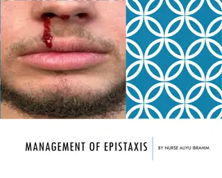
Understanding Epistaxis: Causes, Sites of Bleeding, and Management
Epistaxis, commonly known as nosebleed, can range from trivial to life-threatening. It can result from various local and general causes, including idiopathic bleeds, trauma, inflammatory conditions, neoplastic growths, and systemic diseases. Understanding the vascular anatomy and sites of bleeding in the nasal region is crucial for effective management of epistaxis. Treatment strategies may involve simple measures like pressure application to more complex interventions, depending on the severity and underlying cause.

Download Presentation

Please find below an Image/Link to download the presentation.
The content on the website is provided AS IS for your information and personal use only. It may not be sold, licensed, or shared on other websites without obtaining consent from the author. Download presentation by click this link. If you encounter any issues during the download, it is possible that the publisher has removed the file from their server.
E N D
Presentation Transcript
EPISTAXIS By Dr.Abdullhussein Kareem
Epistaxis is bleeding from the nose ,could be trivial or life threatining. Vascular anatomy. -Arterial supply,the nose is supplied by the following arteries. 1.Branches of external carotid artery. A.Maxilarry art by the sphenopalatine and greater palatine arteries. B.Facial art by the superior labial ,lateral nasal and ascending palatine arteries
2.Branches of internal carotid artery. A.Anterior ethmoidal art. B.Posterior ethmoidal art. Venous drainage. The veins of the lateral wall drain through the sphenopalatine foramen into the ptergoid venous plexus to the internal jugular vein.Anteriorly ,drainage is via superior labial and greater palatine veins to the facialvein and then to the external jugular system.
Aetiology. A.Local causes. 1.Idiopathic.Spontaneous arterial bleeding from the nasal septum especially in Little s area is the commonest in adult while in children and youngs ,retrocollumelar vein is frequent cause of epistaxis. 2.Traumatic with abrasion of the mucosa due to direct blow on the nose,or may be due to compound fractures of the bones or cartilages of the nasal cavity or skull base fractures or may be due to foreign body ,after nasal surgery or nose picking.
3.Inflammatory like acute and chronic rhinitis ,atrophic rhinitis and rhinitis sicca. 4.Neoplastic, benign and malignant tumours can cause epistaxis .Juvenile angiofibroma is the most common cause of severe epistaxis which may be fatal. 5.Environmental.High altitude ,air- conditioned rooms may cause dryness of the nasal mucosa and exposure to dust and caustic fumes.
B.General causes. 1.Hypertension doesn t cause epistaxis but epistaxis is more severe and prolonged in hypertensives. 2.Raised venous pressure as in cardiac and pulmonary disorders. 3.Diseases of blood and blood vessels such as leukaemia ,haemophilia,vitamine K deficiency.
Sites of bleeding. 1.Nasal septum ,Little s area (Kiesselbach s plexus) is the commonest site or bleeding can occur behind septal spur . 2.Inferior turbinate . 3.Nasal floor. 4.Above the middle turbinate ,in cases of hypertension. 5.The middle meatus and the sinuses. Anterior epistaxis is anterior to the pyriform aperture and posterior one is posterior to it
Clinical features. Bleeding is variable from trivial to lethal ,usually occurs from the anterior nares but may flow back into the pharynx and appears at the apposite nostril. Sometimes the bleeding is entirely into the pharynx and inhaled or swallowed to present later as haematemesis and haemoptysis respectively.
Treatment. When we face a patient with epistaxis ,we have to do proper assessment(amount of bleeding ,vital signs ,find IV line ,resuscitate and stop the bleeding immediately). We should wear gown.gloves,mask and googles . Instrument and equipment to deal with bleeding like suction system and nasal packs. Decide whether the patient needs admission to hospital or not ,it is unwise to send home an old man with chronic disease and nasal pack in.
Treatment . 1.DIRECT;means identification of the bleeding point and deal with it like cauterization. 2.INDIRECT; means treatment without identification of bleeding point like packing. 1.Immediate treatment. 1.Pressure on the nostrils ,the patient is instructed to breathe quietly through the mouth with the head leant forward.
2.Ice or cold packs applied to the bridge of the nose. 3.Packing of the nose. A.Anterior packing by using gauze ot cotton- wool impregnated with paraffin or using poly vinyl acetate pack(merocel). B.Posterior packing by inserting Foley s catheter under local anaesthesia or postnasal space pack under GA . 4.Sedatives. 5.Systemic antibiotics to prevent secondary infection from packing.
Anterior pack should be kept for 48 hours with observation of the patient . If the patient still has bleeding after removal of the anterior pack ,posterior pack should be inserted and it is removed after 72 hours. Complications of the pack. 1.Nasal mucosal abrasions. 2.Dry mouth and pharyngitis. 3.Snoring and sleep apnoea. 4.Otitis media due to Eustachian tube obstruction. 5.Toxic shock syndrome if kept for long time due to staphylococcal toxins.
Curative and preventive treatment. These measures are needed when immediate treatment fails or repeated bleeding occurs. 1.Cauterization of the bleeding point either electrical (galvanocautery) or chemical by using silver nitrate ,trichloroacetic acid and chromic acid after using local anaesthesia for nasal mucosa. 2.Examination under GA tom allow better identification of the bleeding point.
3.Septal surgery to correct septal deviation and dealing with the bleeding point. 4.Arterial ligation is indicated when the bleeding is not controlled by packing or cauterization.The following arteries may be ligated A.External carotid artery distal to the lingual art. B.Maxillary art. C.Ethmoidal art D.Sphenopalatine art ligation by using endoscope (ESPAL),this is the most effective method to control epistaxis.
5.Embolization under angiographic guidance using transfemoral catheter and particles(polyvinyl alcohol or steel microcoils) ,can be done for internal maxillart art and facial art. 6.Blood transfusion. 7.Drugs like aminocaproic acid,vitamin K and vitamineC.
References. 1.Scott-Brown otolaryngology. 2.Synopsis of otolaryngology.